How Big Is a Cell? Most Are a Hair's Width or Far Narrower

Slide the ladder sideways to read its labels, or open it full size.
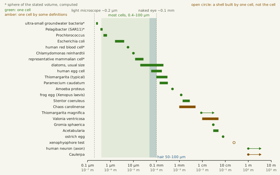
Most cells: about 0.4–100 µm across [10−7–10−4 m]; single cells run from about 0.2 µm to an 8 cm ostrich egg, and past 1 m along a nerve cell's axon or with many nuclei.
Log scale from 10 cm to 0.1 nm; each rung is ten times smaller than the one to its left. Dashed lines: naked eye about 0.1 mm and light microscope about 0.2 µm, explained on the Zoom ladder page.
"Zoom ladder: how big is a cell" by Ants, Cells & Atoms, https://antscellsandatoms.com/scale/how-big-is-a-cell/ — CC BY 4.0 (https://creativecommons.org/licenses/by/4.0/)
Data sources for this figure
Data: Marshall WF et al. 2012, What determines cell size?, BMC Biology 10:101, CC BY 2.0, https://doi.org/10.1186/1741-7007-10-101 (essays by Young KD: SAR11 0.2 µm, most known bacteria 0.4–2 µm in diameter; Frankel J: ostrich egg 8 cm, Caulerpa 1 m or more; retrieved 2026-10-25); Roy D, Tedeschi A 2021, Cells 10(5):1078, CC BY 4.0, https://doi.org/10.3390/cells10051078 (human axons up to one metre or more; retrieved 2026-10-25); subject band 0.4–100 µm from the article front matter zoom, same value as the Bench card (lower end: Young's most known bacteria, 0.4–2 µm in diameter); ladder computed 2026-10-25 for this page
Bench card
- Question
- How big is a cell?
- Answer range
- most cells about 0.4–100 µm [10−7–10−4 m]; measured single cells run from about 0.2 µm (SAR11 bacteria) to about 8 cm (an ostrich egg); a human nerve cell's axon reaches 1 m or more, and so does Caulerpa if a cell with many nuclei counts as one cellSource: cell-sizes.csv on this page
- Method
- compiled measurements
- Inputs
- 62 rows from 22 sources (55 as stated by the sources, 7 computed as spheres of a stated volume)Source: cell-sizes.csv on this page
how-big-is-a-cell-bench.csv (CSV) · where each value comes from, and its license (JSON)
Data: Marshall WF et al. 2012, What determines cell size?, BMC Biology 10:101, CC BY 2.0, https://doi.org/10.1186/1741-7007-10-101 (retrieved 2026-10-25); Gooday AJ, Durden JM, Smith CR 2020, Communicative & Integrative Biology 13(1):189-197, CC BY 4.0, https://doi.org/10.1080/19420889.2020.1843818 (retrieved 2026-10-25); row and source counts computed 2026-10-25 for this page from cell-sizes.csv
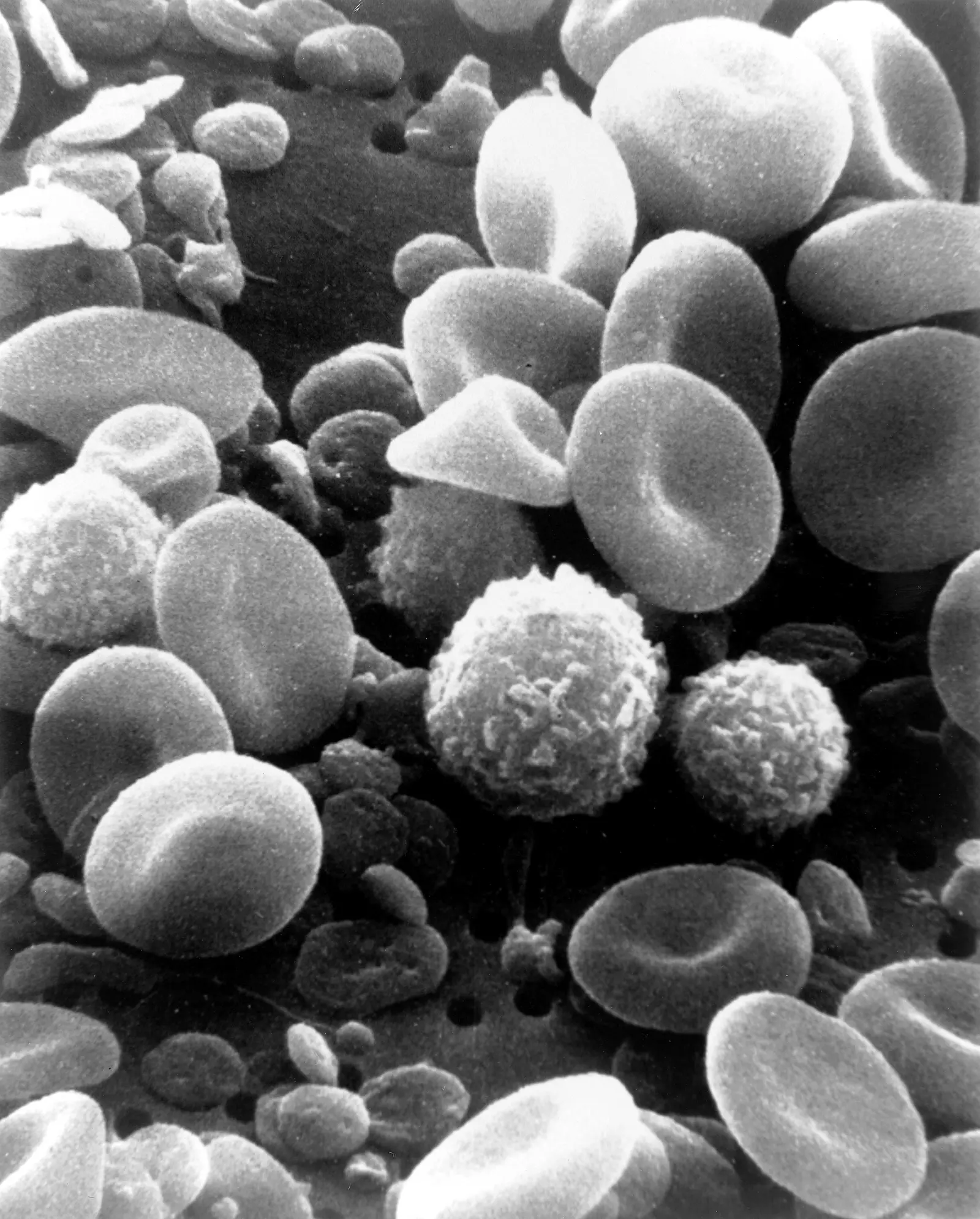
Human blood: red cells of about 100 µm3, the volume of a sphere about 6 µm across [10−6 m], with white blood cells and platelets.
Color: native (electron micrograph, no color)
“SEM blood cells” by Bruce Wetzel and Harry Schaefer, National Cancer Institute (1982) · Public domain
Full credit
"SEM blood cells" by Bruce Wetzel and Harry Schaefer, National Cancer Institute (1982) — https://commons.wikimedia.org/wiki/File:SEM_blood_cells.jpg — Public domain (work of United States federal employees at the National Cancer Institute (NIH), public domain in the United States under 17 U.S.C. §105; Commons tag PD-USGov; the NCI Visuals Online source states that all images are in the public domain and may be reproduced without permission, with credit to the listed source or author; dated February 1982; used in the EU on the source's explicit public-domain statement)

The downloadable table behind this page holds 51 sizes from 18 sources. The 23 that are plotted are listed under the chart. Each row names what was measured and where the figure comes from, and marks the sources whose abstract alone was available. A size computed for this page, such as the width of a sphere that holds a measured volume, says computed.
How big are cells, on one log scale?
Put every cell in the table on an ordinary ruler 1 m long, and anything under 1 mm crowds into the first 0.1% of it. A log scale gives each factor of ten the same room, so a 0.2 µm bacterium and a metre-long alga fit on one line. For sizes spread this wide, a logarithmic axis is the only honest one.
On that axis the cells bunch up. Kevin Young, writing in a 2012 BMC Biology forum on cell size, puts most known bacteria at 0.4–2 µm in diameter and 0.5–5 µm long. The human cells counted by Sender, Fuchs and Milo in 2016 include red blood cells of about 100 µm3, among the smallest human cell types, and fat and muscle cells usually over 10,000 µm3. Spheres of those volumes would be about 6 µm and about 27 µm or more across, computed for this page.
Slide the diagram sideways to read its labels, or open it full size.
Cell sizes from 0.24 µm to over 1 m [10−7–100 m]; the shaded band marks most cells, about 0.4–100 µm; amber rows count as one cell only by some definitions, and the open circle marks a shell, not a cell.
Data: cell-sizes.csv (sources, version and license, JSON) (62 rows; the table below lists the 23 plotted).
"Cell sizes on one log scale, from 0.2 µm bacteria to metre-long cells" by Ants, Cells & Atoms, https://antscellsandatoms.com/scale/how-big-is-a-cell/ — CC BY 4.0 (https://creativecommons.org/licenses/by/4.0/)
Data sources for this figure
Data: sizes from the 23 plotted rows of cell-sizes.csv; sources named in each row (retrieved 2026-10-25); most-cells band 0.4–100 µm from the article front matter zoom; lower end from Marshall WF et al. 2012, What determines cell size?, BMC Biology 10:101, CC BY 2.0, https://doi.org/10.1186/1741-7007-10-101 (essay by Young KD: most known bacteria 0.4–2 µm in diameter; retrieved 2026-10-25); Li C et al. 2023, Journal of Biomedical Optics 28(11):116501, CC BY 4.0, https://doi.org/10.1117/1.JBO.28.11.116501 (human hair, typically 50–100 µm; retrieved 2026-10-25); chart computed 2026-10-25 for this page
Data sources for this figure
Data: Marshall WF et al. 2012, What determines cell size?, BMC Biology 10:101, CC BY 2.0, https://doi.org/10.1186/1741-7007-10-101 (essays by Young KD, Frankel J and Wallingford J; retrieved 2026-10-25); Luef B et al. 2015, Nature Communications 6:6372 (abstract), https://doi.org/10.1038/ncomms7372 (retrieved 2026-10-25); Zhao X et al. 2017, Applied and Environmental Microbiology 83(3):e02807-16 (abstract), https://doi.org/10.1128/AEM.02807-16 (retrieved 2026-10-25); Partensky F, Hess WR, Vaulot D 1999, Microbiology and Molecular Biology Reviews 63(1):106-127 (abstract), https://doi.org/10.1128/MMBR.63.1.106-127.1999 (retrieved 2026-10-25); Volland JM et al. 2022, Science 376(6600):1453-1458 (abstract), https://doi.org/10.1126/science.abb3634 (retrieved 2026-10-25); Volkmer B, Heinemann M 2011, PLoS ONE 6(7):e23126, CC BY 4.0, https://doi.org/10.1371/journal.pone.0023126 (Table 1; retrieved 2026-10-25); Gooday AJ et al. 2018, Scientific Reports 8:12103, CC BY 4.0, https://doi.org/10.1038/s41598-018-30186-2 (retrieved 2026-10-25); Gooday AJ, Durden JM, Smith CR 2020, Communicative & Integrative Biology 13(1):189-197, CC BY 4.0, https://doi.org/10.1080/19420889.2020.1843818 (retrieved 2026-10-25); Lisicki M et al. 2019, eLife 8:e44907, CC BY 4.0, https://doi.org/10.7554/eLife.44907 (Appendix 1 flagellates, Appendix 2 ciliates; retrieved 2026-10-25); Velho Rodrigues MF, Lisicki M, Lauga E 2021, PLOS ONE 16(6):e0252291, CC BY 4.0, https://doi.org/10.1371/journal.pone.0252291 (Table 9; retrieved 2026-10-25); Moore ER et al. 2017, PLOS ONE 12(7):e0181098, CC BY 4.0, https://doi.org/10.1371/journal.pone.0181098 (retrieved 2026-10-25); Piegari G et al. 2019, Frontiers in Veterinary Science 6:404, CC BY 4.0, https://doi.org/10.3389/fvets.2019.00404 (retrieved 2026-10-25); Terracciano M, De Stefano L, Rea I 2018, Pharmaceutics 10(4):242, CC BY 4.0, https://doi.org/10.3390/pharmaceutics10040242 (retrieved 2026-10-25); Gastineau R et al. 2021, Biology 10(4):328, CC BY 4.0, https://doi.org/10.3390/biology10040328 (retrieved 2026-10-25); Razavi M, Saberi Fathi SM, Tuszynski JA 2022, Entropy 24(1):94, CC BY 4.0, https://doi.org/10.3390/e24010094 (retrieved 2026-10-25); Sender R, Fuchs S, Milo R 2016, PLoS Biology 14(8):e1002533, CC BY 4.0, https://doi.org/10.1371/journal.pbio.1002533 (retrieved 2026-10-25); Pors SE et al. 2022, Journal of Assisted Reproduction and Genetics 39(10):2209-2214, CC BY 4.0, https://doi.org/10.1007/s10815-022-02602-0 (retrieved 2026-10-25); Roy D, Tedeschi A 2021, Cells 10(5):1078, CC BY 4.0, https://doi.org/10.3390/cells10051078 (retrieved 2026-10-25); GBIF Secretariat, GBIF Backbone Taxonomy, checklist dataset https://doi.org/10.15468/39omei, CC BY 4.0 (taxon keys, accessed 2026-10-25); equivalent-sphere diameters computed 2026-10-25 for this page with units carried at every step; Rillo MC, Miller CG, Kucera M, Ezard THG 2020, Ecology and Evolution 10(20):11579-11590, CC BY 4.0, https://doi.org/10.1002/ece3.6792 (adult Globorotalia menardii shells; retrieved 2026-10-25); Hohenegger J, Kinoshita S, Briguglio A, Eder W, Wöger J 2019, Scientific Reports 9:8286, CC BY 4.0, https://doi.org/10.1038/s41598-019-44646-w (Nummulites gizehensis tests of 5 and 30 mm; largest larger benthic foraminifera 13 cm living and 19 cm fossil; retrieved 2026-10-25); Gerber T et al. 2022, eLife 11:e69745, CC BY 4.0, https://doi.org/10.7554/eLife.69745 (Physarum polycephalum plasmodium extent, area and nucleus size; retrieved 2026-10-25); Janssens GE, Veenhoff LM 2016, PLOS ONE 11(12):e0167394 and its S1 File, CC BY 4.0, https://doi.org/10.1371/journal.pone.0167394 (baker's yeast mother cell diameters; equivalent diameters computed 2026-10-25 for this page; retrieved 2026-10-25)
| Cell | Size | What was measured | Source |
|---|---|---|---|
| ultra-small groundwater bacteria | 0.24–0.28 µm | sphere of the measured volume | Luef 2015 (abstract), computed |
| Pelagibacter (SAR11) | 0.37–0.45 µm | sphere of the measured volume | Zhao 2017 (abstract), computed |
| Prochlorococcus | 0.5–0.7 µm | diameter | Partensky 1999 (abstract) |
| Escherichia coli | 1.6–3.9 µm | mean length, 22 growth conditions | Volkmer 2011 |
| human red blood cell | 5.8 µm | sphere of about 100 µm3 | Sender 2016, computed |
| Chlamydomonas reinhardtii | 10 µm | typical size | Lisicki 2019 |
| representative mammalian cell | 12–27 µm | sphere of 1,000–10,000 µm3 (assumed) | Sender 2016, computed |
| diatoms, usual size | 20–200 µm | size | Piegari 2019 |
| human egg cell | 60–171 µm | diameter, 1,563 eggs | Pors 2022 |
| Thiomargarita (typical) | 100–300 µm | diameter | Gooday 2018 |
| Paramecium caudatum | 140–311 µm | length | Velho Rodrigues 2021 |
| Amoeba proteus | about 500 µm | length, moving | Frankel 2012 |
| frog egg (Xenopus laevis) | about 1.2 mm | diameter | Frankel 2012 |
| Stentor coeruleus | 1.4–2.8 mm | length, extended | Frankel 2012 |
| Chaos carolinense (Chaos chaos in the source) | 1–5 mm | length, extended | Frankel 2012 |
| Thiomargarita magnifica | over 9,000 µm | average length | Volland 2022 (abstract) |
| Valonia ventricosa | 1–5 cm | diameter | Razavi 2022 |
| Gromia sphaerica | about 3 cm | diameter | Frankel 2012 |
| Acetabularia | 3–5 cm | length | Frankel 2012 |
| ostrich egg | about 8 cm | size | Frankel 2012 |
| xenophyophore test | up to 24 cm | test (shell) size | Gooday 2020 |
| human neuron (axon) | 1 m or more | axon length | Roy & Tedeschi 2021 |
| Caulerpa | 1 m or more | stolon length | Frankel 2012 |
What is the smallest cell?
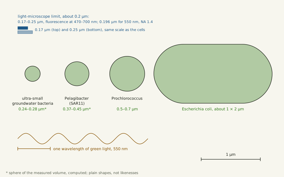
The smallest cells in these sources are bacteria. In 2015 Luef and colleagues reported cells from groundwater that had passed through a filter of about 0.2 µm, with volumes of 0.009 ± 0.002 µm3 (abstract). A sphere of 0.009 µm3 would be about 0.26 µm across, computed for this page.
Young calls the marine SAR11 bacteria "the tiniest 0.2 µm cells". Yet one measured SAR11 strain is larger by volume than the groundwater cells. Frozen-hydrated cells of Candidatus Pelagibacter ubique (Pelagibacter ubique in GBIF), strain HTCC1062, averaged 0.037 µm3, by Zhao and colleagues' 2017 account (abstract). That is about four times the groundwater cells' volume, and a sphere that size is about 0.41 µm across, both computed.
Zhao and colleagues describe SAR11 as bacterioplankton found throughout the oceans, part of the plankton. Prochlorococcus, 0.5–0.7 µm in diameter, was the smallest known photosynthetic organism as of a 1999 review by Partensky, Hess and Vaulot (abstract). Young describes Escherichia coli as a rod normally about 1 µm wide and 2 µm long.
Slide the diagram sideways to read its labels, or open it full size.
The smallest cells drawn to one scale, 0.24 to 2 µm [10−7–10−6 m], beside the light-microscope limit of about 0.2 µm; spheres hold the measured volumes.
Data: cell-sizes.csv (sources, version and license, JSON) and reference-objects.csv (sources, version and license, JSON).
"The smallest cells to scale, beside the light-microscope limit" by Ants, Cells & Atoms, https://antscellsandatoms.com/scale/how-big-is-a-cell/ — CC BY 4.0 (https://creativecommons.org/licenses/by/4.0/)
Data sources for this figure
Data: Luef B et al. 2015, Nature Communications 6:6372 (abstract), https://doi.org/10.1038/ncomms7372 (cell volume 0.009 ± 0.002 µm³; sphere width computed 2026-10-25); Zhao X et al. 2017, Applied and Environmental Microbiology 83(3):e02807-16 (abstract), https://doi.org/10.1128/AEM.02807-16 (cell volume 0.037 ± 0.011 µm³; sphere width computed 2026-10-25); Partensky F, Hess WR, Vaulot D 1999, Microbiology and Molecular Biology Reviews 63(1):106-127 (abstract), https://doi.org/10.1128/MMBR.63.1.106-127.1999 (Prochlorococcus 0.5–0.7 µm; retrieved 2026-10-25); Marshall WF et al. 2012, BMC Biology 10:101, CC BY 2.0, https://doi.org/10.1186/1741-7007-10-101 (essay by Young KD: E. coli about 1 µm by 2 µm; retrieved 2026-10-25); Valli J et al. 2021, Journal of Biological Chemistry 297(1):100791, CC BY 4.0, https://doi.org/10.1016/j.jbc.2021.100791 (lateral limit 170–250 nm for a standard fluorescence microscope; 0.196 µm computed 2026-10-25 for 550 nm and NA 1.4); geometric shapes computed 2026-10-25 for this page, not likenesses of the cells
Data sources for this figure
Data: Marshall WF et al. 2012, What determines cell size?, BMC Biology 10:101, CC BY 2.0, https://doi.org/10.1186/1741-7007-10-101 (essays by Young KD, Frankel J and Wallingford J; retrieved 2026-10-25); Luef B et al. 2015, Nature Communications 6:6372 (abstract), https://doi.org/10.1038/ncomms7372 (retrieved 2026-10-25); Zhao X et al. 2017, Applied and Environmental Microbiology 83(3):e02807-16 (abstract), https://doi.org/10.1128/AEM.02807-16 (retrieved 2026-10-25); Partensky F, Hess WR, Vaulot D 1999, Microbiology and Molecular Biology Reviews 63(1):106-127 (abstract), https://doi.org/10.1128/MMBR.63.1.106-127.1999 (retrieved 2026-10-25); Volland JM et al. 2022, Science 376(6600):1453-1458 (abstract), https://doi.org/10.1126/science.abb3634 (retrieved 2026-10-25); Volkmer B, Heinemann M 2011, PLoS ONE 6(7):e23126, CC BY 4.0, https://doi.org/10.1371/journal.pone.0023126 (Table 1; retrieved 2026-10-25); Gooday AJ et al. 2018, Scientific Reports 8:12103, CC BY 4.0, https://doi.org/10.1038/s41598-018-30186-2 (retrieved 2026-10-25); Gooday AJ, Durden JM, Smith CR 2020, Communicative & Integrative Biology 13(1):189-197, CC BY 4.0, https://doi.org/10.1080/19420889.2020.1843818 (retrieved 2026-10-25); Lisicki M et al. 2019, eLife 8:e44907, CC BY 4.0, https://doi.org/10.7554/eLife.44907 (Appendix 1 flagellates, Appendix 2 ciliates; retrieved 2026-10-25); Velho Rodrigues MF, Lisicki M, Lauga E 2021, PLOS ONE 16(6):e0252291, CC BY 4.0, https://doi.org/10.1371/journal.pone.0252291 (Table 9; retrieved 2026-10-25); Moore ER et al. 2017, PLOS ONE 12(7):e0181098, CC BY 4.0, https://doi.org/10.1371/journal.pone.0181098 (retrieved 2026-10-25); Piegari G et al. 2019, Frontiers in Veterinary Science 6:404, CC BY 4.0, https://doi.org/10.3389/fvets.2019.00404 (retrieved 2026-10-25); Terracciano M, De Stefano L, Rea I 2018, Pharmaceutics 10(4):242, CC BY 4.0, https://doi.org/10.3390/pharmaceutics10040242 (retrieved 2026-10-25); Gastineau R et al. 2021, Biology 10(4):328, CC BY 4.0, https://doi.org/10.3390/biology10040328 (retrieved 2026-10-25); Razavi M, Saberi Fathi SM, Tuszynski JA 2022, Entropy 24(1):94, CC BY 4.0, https://doi.org/10.3390/e24010094 (retrieved 2026-10-25); Sender R, Fuchs S, Milo R 2016, PLoS Biology 14(8):e1002533, CC BY 4.0, https://doi.org/10.1371/journal.pbio.1002533 (retrieved 2026-10-25); Pors SE et al. 2022, Journal of Assisted Reproduction and Genetics 39(10):2209-2214, CC BY 4.0, https://doi.org/10.1007/s10815-022-02602-0 (retrieved 2026-10-25); Roy D, Tedeschi A 2021, Cells 10(5):1078, CC BY 4.0, https://doi.org/10.3390/cells10051078 (retrieved 2026-10-25); GBIF Secretariat, GBIF Backbone Taxonomy, checklist dataset https://doi.org/10.15468/39omei, CC BY 4.0 (taxon keys, accessed 2026-10-25); equivalent-sphere diameters computed 2026-10-25 for this page with units carried at every step; Rillo MC, Miller CG, Kucera M, Ezard THG 2020, Ecology and Evolution 10(20):11579-11590, CC BY 4.0, https://doi.org/10.1002/ece3.6792 (adult Globorotalia menardii shells; retrieved 2026-10-25); Hohenegger J, Kinoshita S, Briguglio A, Eder W, Wöger J 2019, Scientific Reports 9:8286, CC BY 4.0, https://doi.org/10.1038/s41598-019-44646-w (Nummulites gizehensis tests of 5 and 30 mm; largest larger benthic foraminifera 13 cm living and 19 cm fossil; retrieved 2026-10-25); Gerber T et al. 2022, eLife 11:e69745, CC BY 4.0, https://doi.org/10.7554/eLife.69745 (Physarum polycephalum plasmodium extent, area and nucleus size; retrieved 2026-10-25); Janssens GE, Veenhoff LM 2016, PLOS ONE 11(12):e0167394 and its S1 File, CC BY 4.0, https://doi.org/10.1371/journal.pone.0167394 (baker's yeast mother cell diameters; equivalent diameters computed 2026-10-25 for this page; retrieved 2026-10-25)
Data sources for this figure
Data: Li C et al. 2023, Journal of Biomedical Optics 28(11):116501, CC BY 4.0, https://doi.org/10.1117/1.JBO.28.11.116501 (human hair; retrieved 2026-10-25); VanDusen BM, Fegley SR, Peterson CH 2012, PLoS ONE 7(12):e52694, CC BY 4.0, https://doi.org/10.1371/journal.pone.0052694 (sand class bounds; retrieved 2026-10-25); Fowler KD et al. 2020, PLOS ONE 15(4):e0231046, CC BY 4.0, https://doi.org/10.1371/journal.pone.0231046 (fingerprint ridge breadth; retrieved 2026-10-25); de Jong PTVM 2024, Eye 38(1):13-24, CC BY 4.0, https://doi.org/10.1038/s41433-022-02180-6 (one minute of arc, quoting Hooke; retrieved 2026-10-25); Valli J et al. 2021, Journal of Biological Chemistry 297(1):100791, CC BY 4.0, https://doi.org/10.1016/j.jbc.2021.100791 (lateral resolution limit, Abbe 1873; retrieved 2026-10-25); naked-eye size and 550 nm Abbe limit computed 2026-10-25 for this page with units carried at every step
Cells this small sit at the edge of what a light microscope can show. Abbe's limit of 1873, as a 2021 methods review by Valli and colleagues gives it, is about the wavelength divided by twice the numerical aperture, a measure of the range of angles the lens accepts. For a standard fluorescence microscope detecting light of 470–700 nm, the review puts it at around 170–250 nm. Two points closer than that blur into one spot.
The first two sizes above were measured with electron microscopes. Luef's study used cryogenic transmission electron microscopy, and Zhao's used cryo-electron tomography. Viruses are not cells and are smaller still: the head of bacteriophage T4, a virus that infects bacteria, is 120 nm long and 86 nm wide, by Rao and Black's 2010 account.
How big is an ordinary cell?
Grown in culture, Escherichia coli cells averaged 1.0–1.5 µm wide and 1.6–3.9 µm long across 22 growth conditions (Volkmer and Heinemann 2011). How a cell that size moves through water is set by its Reynolds number, a ratio of inertia to stickiness that grows with both size and speed.
In a 70 kg reference man, Sender and colleagues count about 3.0×10¹³ human cells, 84% of them red blood cells, and about as many bacteria (How Many Cells in the Human Body?). Red cells have no nucleus, and so no nuclear DNA. Fat and muscle cells are about 0.2% of the count.
Across the whole body, Hatton and colleagues found in 2023 that cell masses span about seven orders of magnitude, from a red blood cell to the largest muscle fibres, each fibre a cell with thousands of nuclei. For cells of the same shape, a million times the mass means only a hundred times the width, computed as a cube root.
Slide the diagram sideways to read its labels, or open it full size.
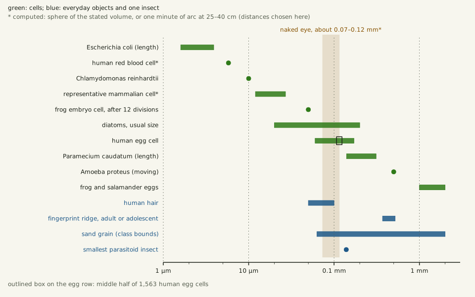
Ordinary cells next to a hair (50–100 µm), a fingerprint ridge and sand grains [10−6–10−3 m]; the human egg cell sits on the naked-eye band.
Data: cell-sizes.csv (sources, version and license, JSON) and reference-objects.csv (sources, version and license, JSON).
"Ordinary cells next to a hair, a sand grain and the naked-eye limit" by Ants, Cells & Atoms, https://antscellsandatoms.com/scale/how-big-is-a-cell/ — CC BY 4.0 (https://creativecommons.org/licenses/by/4.0/)
Data sources for this figure
Data: cell sizes from cell-sizes.csv (Volkmer & Heinemann 2011; Sender et al. 2016; Lisicki et al. 2019; Marshall et al. 2012; Piegari et al. 2019; Pors et al. 2022; Velho Rodrigues et al. 2021), retrieved 2026-10-25; reference objects from reference-objects.csv (Li et al. 2023; Fowler et al. 2020; VanDusen et al. 2012; naked-eye size computed 2026-10-25 from de Jong 2024 at viewing distances chosen for this page); Polilov AA 2015, ZooKeys 526:61-64, CC BY 4.0, https://doi.org/10.3897/zookeys.526.6531 (smallest known parasitoid insect, 139 µm, citing Mockford 1997 and Huber & Noyes 2013; retrieved 2026-10-25); chart computed 2026-10-25 for this page
Data sources for this figure
Data: Marshall WF et al. 2012, What determines cell size?, BMC Biology 10:101, CC BY 2.0, https://doi.org/10.1186/1741-7007-10-101 (essays by Young KD, Frankel J and Wallingford J; retrieved 2026-10-25); Luef B et al. 2015, Nature Communications 6:6372 (abstract), https://doi.org/10.1038/ncomms7372 (retrieved 2026-10-25); Zhao X et al. 2017, Applied and Environmental Microbiology 83(3):e02807-16 (abstract), https://doi.org/10.1128/AEM.02807-16 (retrieved 2026-10-25); Partensky F, Hess WR, Vaulot D 1999, Microbiology and Molecular Biology Reviews 63(1):106-127 (abstract), https://doi.org/10.1128/MMBR.63.1.106-127.1999 (retrieved 2026-10-25); Volland JM et al. 2022, Science 376(6600):1453-1458 (abstract), https://doi.org/10.1126/science.abb3634 (retrieved 2026-10-25); Volkmer B, Heinemann M 2011, PLoS ONE 6(7):e23126, CC BY 4.0, https://doi.org/10.1371/journal.pone.0023126 (Table 1; retrieved 2026-10-25); Gooday AJ et al. 2018, Scientific Reports 8:12103, CC BY 4.0, https://doi.org/10.1038/s41598-018-30186-2 (retrieved 2026-10-25); Gooday AJ, Durden JM, Smith CR 2020, Communicative & Integrative Biology 13(1):189-197, CC BY 4.0, https://doi.org/10.1080/19420889.2020.1843818 (retrieved 2026-10-25); Lisicki M et al. 2019, eLife 8:e44907, CC BY 4.0, https://doi.org/10.7554/eLife.44907 (Appendix 1 flagellates, Appendix 2 ciliates; retrieved 2026-10-25); Velho Rodrigues MF, Lisicki M, Lauga E 2021, PLOS ONE 16(6):e0252291, CC BY 4.0, https://doi.org/10.1371/journal.pone.0252291 (Table 9; retrieved 2026-10-25); Moore ER et al. 2017, PLOS ONE 12(7):e0181098, CC BY 4.0, https://doi.org/10.1371/journal.pone.0181098 (retrieved 2026-10-25); Piegari G et al. 2019, Frontiers in Veterinary Science 6:404, CC BY 4.0, https://doi.org/10.3389/fvets.2019.00404 (retrieved 2026-10-25); Terracciano M, De Stefano L, Rea I 2018, Pharmaceutics 10(4):242, CC BY 4.0, https://doi.org/10.3390/pharmaceutics10040242 (retrieved 2026-10-25); Gastineau R et al. 2021, Biology 10(4):328, CC BY 4.0, https://doi.org/10.3390/biology10040328 (retrieved 2026-10-25); Razavi M, Saberi Fathi SM, Tuszynski JA 2022, Entropy 24(1):94, CC BY 4.0, https://doi.org/10.3390/e24010094 (retrieved 2026-10-25); Sender R, Fuchs S, Milo R 2016, PLoS Biology 14(8):e1002533, CC BY 4.0, https://doi.org/10.1371/journal.pbio.1002533 (retrieved 2026-10-25); Pors SE et al. 2022, Journal of Assisted Reproduction and Genetics 39(10):2209-2214, CC BY 4.0, https://doi.org/10.1007/s10815-022-02602-0 (retrieved 2026-10-25); Roy D, Tedeschi A 2021, Cells 10(5):1078, CC BY 4.0, https://doi.org/10.3390/cells10051078 (retrieved 2026-10-25); GBIF Secretariat, GBIF Backbone Taxonomy, checklist dataset https://doi.org/10.15468/39omei, CC BY 4.0 (taxon keys, accessed 2026-10-25); equivalent-sphere diameters computed 2026-10-25 for this page with units carried at every step; Rillo MC, Miller CG, Kucera M, Ezard THG 2020, Ecology and Evolution 10(20):11579-11590, CC BY 4.0, https://doi.org/10.1002/ece3.6792 (adult Globorotalia menardii shells; retrieved 2026-10-25); Hohenegger J, Kinoshita S, Briguglio A, Eder W, Wöger J 2019, Scientific Reports 9:8286, CC BY 4.0, https://doi.org/10.1038/s41598-019-44646-w (Nummulites gizehensis tests of 5 and 30 mm; largest larger benthic foraminifera 13 cm living and 19 cm fossil; retrieved 2026-10-25); Gerber T et al. 2022, eLife 11:e69745, CC BY 4.0, https://doi.org/10.7554/eLife.69745 (Physarum polycephalum plasmodium extent, area and nucleus size; retrieved 2026-10-25); Janssens GE, Veenhoff LM 2016, PLOS ONE 11(12):e0167394 and its S1 File, CC BY 4.0, https://doi.org/10.1371/journal.pone.0167394 (baker's yeast mother cell diameters; equivalent diameters computed 2026-10-25 for this page; retrieved 2026-10-25)
Data sources for this figure
Data: Li C et al. 2023, Journal of Biomedical Optics 28(11):116501, CC BY 4.0, https://doi.org/10.1117/1.JBO.28.11.116501 (human hair; retrieved 2026-10-25); VanDusen BM, Fegley SR, Peterson CH 2012, PLoS ONE 7(12):e52694, CC BY 4.0, https://doi.org/10.1371/journal.pone.0052694 (sand class bounds; retrieved 2026-10-25); Fowler KD et al. 2020, PLOS ONE 15(4):e0231046, CC BY 4.0, https://doi.org/10.1371/journal.pone.0231046 (fingerprint ridge breadth; retrieved 2026-10-25); de Jong PTVM 2024, Eye 38(1):13-24, CC BY 4.0, https://doi.org/10.1038/s41433-022-02180-6 (one minute of arc, quoting Hooke; retrieved 2026-10-25); Valli J et al. 2021, Journal of Biological Chemistry 297(1):100791, CC BY 4.0, https://doi.org/10.1016/j.jbc.2021.100791 (lateral resolution limit, Abbe 1873; retrieved 2026-10-25); naked-eye size and 550 nm Abbe limit computed 2026-10-25 for this page with units carried at every step
Pors and colleagues measured 1,563 human egg cells in 2022, collected immature and cultured in the laboratory: 60 to 171 µm across, mean 115 µm, not counting the outer coat, the zona pellucida. Hooke reckoned the naked eye could not resolve finer than one minute of arc, a sixtieth of a degree, as quoted in de Jong's 2024 history of visual acuity testing. Seen from 25–40 cm, a viewing distance chosen for this page, that angle spans about 73–116 µm, computed for this page.
Paramecium caudatum cells measure 140–311 µm long across the studies compiled in the BOSO-Micro database by Velho Rodrigues and colleagues in 2021 (Paramecium). A male Dicopomorpha echmepterygis (eschmepterigis in the paper), the smallest known parasitoid insect, is smaller than that whole range: 139 µm in size by Polilov's 2015 account, citing earlier measurements (smallest insect). Budding yeast, Saccharomyces cerevisiae, also listed in GBIF as baker's yeast, commits to its next division only at a set size, as Swaffer, Wood and Nurse describe in the 2012 forum.
What is the largest single cell?
Bacteria hold their own records. Young puts some species of Thiomargarita and Epulopiscium at over 600 to 700 µm, visible to the naked eye. Spherical Thiomargarita cells are typically 100–300 µm and can reach 750 µm, as Gooday and colleagues note in 2018. In 2022 Volland and colleagues described Candidatus Thiomargarita magnifica (Thiomargarita magnifica in GBIF), with an average cell length greater than 9,000 µm (abstract). That is more than 4,000 times the roughly 2 µm they give for most bacterial species, computed for this page.
Several algae and protists reach centimetres as single cells. Joseph Frankel, in the same 2012 forum, lists the stalked alga Acetabularia at 3–5 cm and the deep-sea protist Gromia sphaerica at 3 cm. The bubble alga Valonia ventricosa is typically 1–5 cm across, by the account of Razavi and colleagues in 2022.
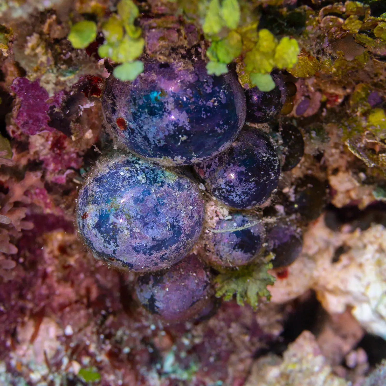
Bubble alga Valonia ventricosa underwater off Zanzibar; its round cells are typically 1–5 cm across [10−2 m].
Color: native (underwater photograph, no stain)
"Alga burbuja (Valonia ventricosa), Zanzíbar, Tanzania, 2024-05-30, DD 59" by Diego Delso — https://commons.wikimedia.org/wiki/File:Alga_burbuja_(Valonia_ventricosa),_Zanz%C3%ADbar,_Tanzania,_2024-05-30,_DD_59.jpg — CC BY-SA 4.0 (https://creativecommons.org/licenses/by-sa/4.0/)

Past this point the answer depends on the definition. Frankel calls the green alga Caulerpa, with many nuclei and a stolon a metre or more long, the champion unicellular organism. Valonia itself, long treated as one giant cell, is built of many cytoplasmic domains with one nucleus each, by the account of Shepherd, Beilby and Bisson in 2004 (abstract), who use the synonym Ventricaria ventricosa.
Gerber and colleagues write that the plasmodium of the slime mold Physarum polycephalum, a single cell with many nuclei, can grow to cover square metres. A human neuron is one cell too, and its axon can run a metre or more, by Roy and Tedeschi's account.
The xenophyophores, giant foraminifera of the deep sea, build tests of foreign particles up to 24 cm in size, among the largest single-celled organisms known, by Gooday and colleagues' 2020 account. The cell itself takes up only a small part of that test. Rough earlier estimates for 12 Pacific specimens, recounted by Gooday's group in 2018, mostly put the cell's branching strands at 1% of the test volume or less; that group's own scans of three species found 1.7–5.1%.
Is an egg one cell?
Frankel writes that in animals the largest dividing cells are eggs. The most familiar frogs and salamanders lay eggs 1 to just over 2 mm across, and some amphibian eggs reach 10 mm. The egg of the frog Xenopus laevis, 1.2 mm across, is a single cell until it divides. After 12 divisions its cells are about 50 µm, John Wallingford notes in the same forum.
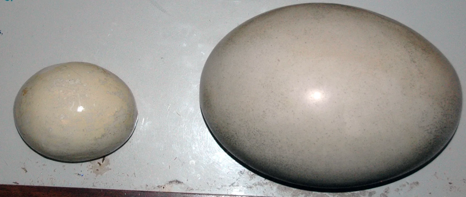
Museum models of an ostrich egg, left, and an elephant bird egg; Frankel lists an ostrich egg, 8 cm [10−2 m], among centimetre-scale cells.
Color: native (photograph of models, not real eggs)
"Ostrich egg (left) & elephant bird egg (right)" by James St. John — https://commons.wikimedia.org/wiki/File:Ostrich_egg_(left)_%26_elephant_bird_egg_(right).jpg — CC BY 2.0 (https://creativecommons.org/licenses/by/2.0/)

 Illustration
A whole egg with a hair beside it.
Illustration — not to scale
Illustration: Ants, Cells & Atoms. Generated schematic illustration — not a photograph, micrograph or specimen record. © Ants, Cells & Atoms, all rights reserved.
Frankel's list of centimetre-scale cells starts with the ostrich egg, measured at 8 cm, a figure he credits to E. B. Wilson. In bird and reptile eggs, he adds, only a small portion of the mass cleaves. The essay does not say which part of the egg the 8 cm describes. The mean human egg cell above, 115 µm, is about 1/700 of that, computed for this page.
Why do most cells stay small?
Young gives one reason for bacteria. Their nutrients reach them by diffusion alone, so the ratio of surface to volume has to stay high: "to maximize this ratio, most bacteria produce cells in the 0.2 to 10 μm size range".
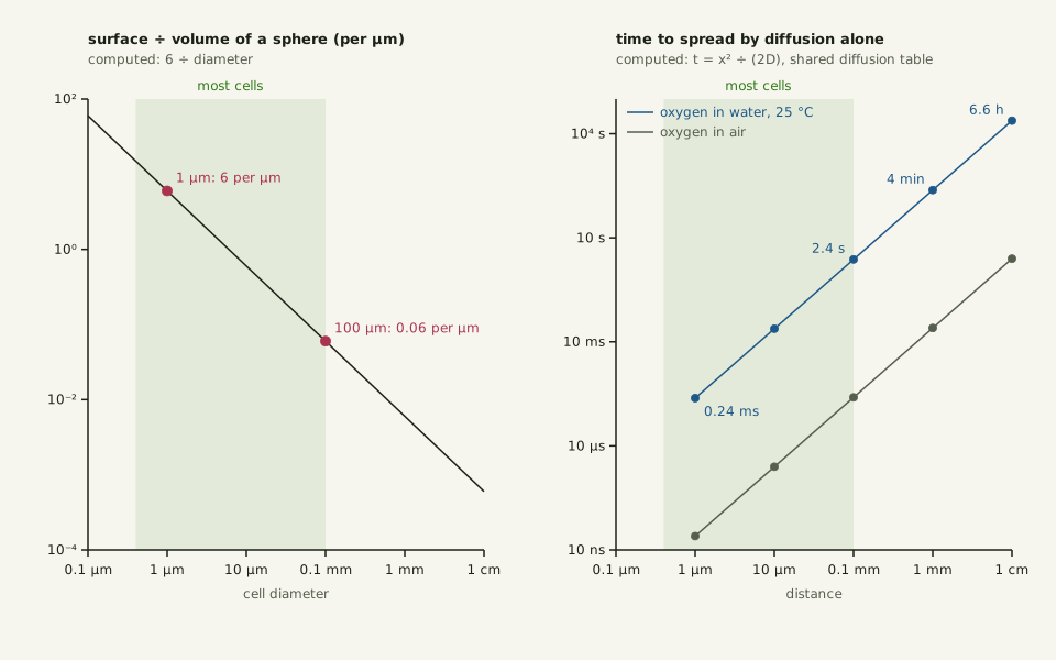
Geometry adds to that. A sphere's surface divided by its volume is 6 divided by its diameter: 6 per µm at 1 µm, 0.06 per µm at 100 µm, computed for this page. Diffusion time grows with the square of distance.
In water at 25 °C, oxygen diffuses about 1 µm in 0.24 ms, 100 µm in 2.4 s, 1 mm in 4 minutes and 1 cm in 6.6 hours. These times come from a table shared with the page on how insects breathe, built on Somerville and Proctor's diffusion coefficient and on t = x2/(2D) (Vestergaard and colleagues).
Slide the diagram sideways to read its labels, or open it full size.
Surface per volume and diffusion time against size, 0.1 µm to 1 cm [10−7–10−2 m]: both favour smaller cells.
Data: computed for this page; diffusion times from the shared diffusion table, oxygen in water at 25 °C and in air.
"Why most cells stay small: surface per volume and diffusion time" by Ants, Cells & Atoms, https://antscellsandatoms.com/scale/how-big-is-a-cell/ — CC BY 4.0 (https://creativecommons.org/licenses/by/4.0/)
Data sources for this figure
Data: surface-to-volume ratio of a sphere, 6/d, computed 2026-10-25 for this page; most-cells band 0.4–100 µm from the article front matter zoom; lower end from Marshall WF et al. 2012, What determines cell size?, BMC Biology 10:101, CC BY 2.0, https://doi.org/10.1186/1741-7007-10-101 (essay by Young KD: most known bacteria 0.4–2 µm in diameter; retrieved 2026-10-25); diffusion times from the shared file diffusion-distance.csv: Somerville GA, Proctor RA 2013, BMC Microbiology 13:9, CC BY 2.0, https://doi.org/10.1186/1471-2180-13-9 (O2 diffusion coefficients in water at 25 °C and in air); t = x²/(2D) from the Gaussian solution given by Vestergaard CL, Flyvbjerg H, Møller IM 2012, Frontiers in Plant Science 3:295, CC BY 3.0, https://doi.org/10.3389/fpls.2012.00295 (retrieved 2026-10-25); chart computed 2026-10-25 for this page
Large cells, colonies and animals work around the limit. In Thiomargarita, a liquid-filled vacuole takes some 98% of the volume, leaving the active cytoplasm a thin outer layer, Gooday and colleagues note. Oxygen diffuses nearly 10,000 times more readily in air than in water, Somerville and Proctor write, and in many tissues an insect's finest air tubes may reach practically every cell, by Maina's 2012 account. Volvox, a spherical shell of many green cells, would outgrow what diffusion alone can feed; its beating flagella stir enough water to get past that limit, by Short and colleagues' account (abstract).
Every tenfold step in width multiplies the diffusion time a hundredfold. A cell 1,000 times wider than a 1 µm bacterium would wait a million times longer for oxygen to cross it by diffusion alone.
Sources
- Li C, Du Y, Chen H, Han X, Wu W, Kong X, Zhang C, Yuan X. "Transmission infrared micro-spectroscopic study of individual human hair." Journal of Biomedical Optics 28(11):116501 (2023). https://doi.org/10.1117/1.JBO.28.11.116501 Read: full text. Text license: CC BY 4.0 (https://creativecommons.org/licenses/by/4.0/).
- Marshall WF, Young KD, Swaffer M, Wood E, Nurse P, Kimura A, Frankel J, Wallingford J, Walbot V, Qu X, Roeder AHK. "What determines cell size?" BMC Biology 10:101 (2012). https://doi.org/10.1186/1741-7007-10-101 Read: full text. Text license: CC BY 2.0 (https://creativecommons.org/licenses/by/2.0/).
- Sender R, Fuchs S, Milo R. "Revised Estimates for the Number of Human and Bacteria Cells in the Body." PLoS Biology 14(8):e1002533 (2016). https://doi.org/10.1371/journal.pbio.1002533 Read: full text. Text license: CC BY 4.0 (https://creativecommons.org/licenses/by/4.0/).
- Luef B, Frischkorn KR, Wrighton KC, Holman H-YN, Birarda G, Thomas BC, Singh A, Williams KH, Siegerist CE, Tringe SG, Downing KH, Comolli LR, Banfield JF. "Diverse uncultivated ultra-small bacterial cells in groundwater." Nature Communications 6:6372 (2015). https://doi.org/10.1038/ncomms7372 Read: abstract.
- Zhao X, Schwartz CL, Pierson J, Giovannoni SJ, McIntosh JR, Nicastro D. "Three-Dimensional Structure of the Ultraoligotrophic Marine Bacterium "Candidatus Pelagibacter ubique"." Applied and Environmental Microbiology 83(3):e02807-16 (2017). https://doi.org/10.1128/AEM.02807-16 Read: abstract.
- Partensky F, Hess WR, Vaulot D. "Prochlorococcus, a Marine Photosynthetic Prokaryote of Global Significance." Microbiology and Molecular Biology Reviews 63(1):106–127 (1999). https://doi.org/10.1128/MMBR.63.1.106-127.1999 Read: abstract.
- Volkmer B, Heinemann M. "Condition-Dependent Cell Volume and Concentration of Escherichia coli to Facilitate Data Conversion for Systems Biology Modeling." PLoS ONE 6(7):e23126 (2011). https://doi.org/10.1371/journal.pone.0023126 Read: full text. Text license: CC BY 4.0 (https://creativecommons.org/licenses/by/4.0/).
- Lisicki M, Velho Rodrigues MF, Goldstein RE, Lauga E. "Swimming eukaryotic microorganisms exhibit a universal speed distribution." eLife 8:e44907 (2019). https://doi.org/10.7554/eLife.44907 Read: full text. Text license: CC BY 4.0 (https://creativecommons.org/licenses/by/4.0/).
- Piegari G, De Biase D, d'Aquino I, Prisco F, Fico R, Ilsami R, Pozzato N, Genovese A, Paciello O. "Diagnosis of Drowning and the Value of the Diatom Test in Veterinary Forensic Pathology." Frontiers in Veterinary Science 6:404 (2019). https://doi.org/10.3389/fvets.2019.00404 Read: full text. Text license: CC BY 4.0 (https://creativecommons.org/licenses/by/4.0/).
- Pors SE, Nikiforov D, Cadenas J, Ghezelayagh Z, Wakimoto Y, Jara LAZ, Cheng J, Dueholm M, Macklon KT, Flachs EM, Mamsen LS, Kristensen SG, Andersen CY. "Oocyte diameter predicts the maturation rate of human immature oocytes collected ex vivo." Journal of Assisted Reproduction and Genetics 39(10):2209–2214 (2022). https://doi.org/10.1007/s10815-022-02602-0 Read: full text. Text license: CC BY 4.0 (https://creativecommons.org/licenses/by/4.0/).
- Gooday AJ, Sykes D, Góral T, Zubkov MV, Glover AG. "Micro-CT 3D imaging reveals the internal structure of three abyssal xenophyophore species (Protista, Foraminifera) from the eastern equatorial Pacific Ocean." Scientific Reports 8:12103 (2018). https://doi.org/10.1038/s41598-018-30186-2 Read: full text. Text license: CC BY 4.0 (https://creativecommons.org/licenses/by/4.0/).
- Velho Rodrigues MF, Lisicki M, Lauga E. "The bank of swimming organisms at the micron scale (BOSO-Micro)." PLOS ONE 16(6):e0252291 (2021). https://doi.org/10.1371/journal.pone.0252291 Read: full text. Text license: CC BY 4.0 (https://creativecommons.org/licenses/by/4.0/).
- Volland JM, Gonzalez-Rizzo S, Gros O, Tyml T, Ivanova N, Schulz F, Goudeau D, Elisabeth NH, Nath N, Udwary D, Malmstrom RR, Guidi-Rontani C, Bolte-Kluge S, Davies KM, Jean MR, Mansot JL, Mouncey NJ, Angert ER, Woyke T, Date SV. "A centimeter-long bacterium with DNA contained in metabolically active, membrane-bound organelles." Science 376(6600):1453–1458 (2022). https://doi.org/10.1126/science.abb3634 Read: abstract.
- Razavi M, Saberi Fathi SM, Tuszynski JA. "The Effect of the Protein Synthesis Entropy Reduction on the Cell Size Regulation and Division Size of Unicellular Organisms." Entropy 24(1):94 (2022). https://doi.org/10.3390/e24010094 Read: full text. Text license: CC BY 4.0 (https://creativecommons.org/licenses/by/4.0/).
- Gooday AJ, Durden JM, Smith CR. "Giant, highly diverse protists in the abyssal Pacific: vulnerability to impacts from seabed mining and potential for recovery." Communicative & Integrative Biology 13(1):189–197 (2020). https://doi.org/10.1080/19420889.2020.1843818 Read: full text. Text license: CC BY 4.0 (https://creativecommons.org/licenses/by/4.0/).
- Roy D, Tedeschi A. "The Role of Lipids, Lipid Metabolism and Ectopic Lipid Accumulation in Axon Growth, Regeneration and Repair after CNS Injury and Disease." Cells 10(5):1078 (2021). https://doi.org/10.3390/cells10051078 Read: full text. Text license: CC BY 4.0 (https://creativecommons.org/licenses/by/4.0/).
- Valli J, Garcia-Burgos A, Rooney LM, Vale de Melo e Oliveira B, Duncan RR, Rickman C. "Seeing beyond the limit: A guide to choosing the right super-resolution microscopy technique." Journal of Biological Chemistry 297(1):100791 (2021). https://doi.org/10.1016/j.jbc.2021.100791 Read: full text. Text license: CC BY 4.0 (https://creativecommons.org/licenses/by/4.0/).
- Hatton IA, Galbraith ED, Merleau NSC, Miettinen TP, Smith BM, Shander JA. "The human cell count and size distribution." Proceedings of the National Academy of Sciences 120(39):e2303077120 (2023). https://doi.org/10.1073/pnas.2303077120 Read: full text. Text license: CC BY 4.0 (https://creativecommons.org/licenses/by/4.0/).
- de Jong PTVM. "A history of visual acuity testing and optotypes." Eye 38(1):13–24 (2024). https://doi.org/10.1038/s41433-022-02180-6 Read: full text. Text license: CC BY 4.0 (https://creativecommons.org/licenses/by/4.0/).
- Polilov AA. "How small is the smallest? New record and remeasuring of Scydosella musawasensis Hall, 1999 (Coleoptera, Ptiliidae), the smallest known free-living insect." ZooKeys 526:61–64 (2015). https://doi.org/10.3897/zookeys.526.6531 Read: full text. Text license: CC BY 4.0 (https://creativecommons.org/licenses/by/4.0/).
- GBIF Secretariat. "GBIF Backbone Taxonomy." Checklist dataset https://doi.org/10.15468/39omei (accessed 2026-10-25). Taxon keys for the organisms on this page, and English vernacular names of Saccharomyces cerevisiae (7440012), including baker's yeast and budding yeast. Read: dataset record. Text license: CC BY 4.0 (https://creativecommons.org/licenses/by/4.0/).
- Shepherd VA, Beilby MJ, Bisson MA. "When is a cell not a cell? A theory relating coenocytic structure to the unusual electrophysiology of Ventricaria ventricosa (Valonia ventricosa)." Protoplasma 223(2–4) (2004). https://doi.org/10.1007/s00709-003-0032-4 Read: abstract.
- Gerber T, Loureiro C, Schramma N, Chen S, Jain A, Weber A, Weigert A, Santel M, Alim K, Treutlein B, Camp JG. "Spatial transcriptomic and single-nucleus analysis reveals heterogeneity in a gigantic single-celled syncytium." eLife 11:e69745 (2022). https://doi.org/10.7554/eLife.69745 Read: full text. Text license: CC BY 4.0 (https://creativecommons.org/licenses/by/4.0/).
- Somerville GA, Proctor RA. "Cultivation conditions and the diffusion of oxygen into culture media: the rationale for the flask-to-medium ratio in microbiology." BMC Microbiology 13:9 (2013). https://doi.org/10.1186/1471-2180-13-9 Read: full text. Text license: CC BY 2.0 (https://creativecommons.org/licenses/by/2.0/).
- Vestergaard CL, Flyvbjerg H, Møller IM. "Intracellular signaling by diffusion: can waves of hydrogen peroxide transmit intracellular information in plant cells?" Frontiers in Plant Science 3:295 (2012). https://doi.org/10.3389/fpls.2012.00295 Read: full text. Text license: CC BY 3.0 (https://creativecommons.org/licenses/by/3.0/).
- Maina JN. "Comparative molecular developmental aspects of the mammalian- and the avian lungs, and the insectan tracheal system by branching morphogenesis: recent advances and future directions." Frontiers in Zoology 9:16 (2012). https://doi.org/10.1186/1742-9994-9-16 Read: full text. Text license: CC BY 2.0 (https://creativecommons.org/licenses/by/2.0/).
- Short MB, Solari CA, Ganguly S, Powers TR, Kessler JO, Goldstein RE. "Flows driven by flagella of multicellular organisms enhance long-range molecular transport." Proceedings of the National Academy of Sciences 103(22):8315–8319 (2006). https://doi.org/10.1073/pnas.0600566103 Read: abstract.
- Gastineau R, et al. "Haslea silbo, A Novel Cosmopolitan Species of Blue Diatoms." Biology 10(4):328 (2021). https://doi.org/10.3390/biology10040328 Read: full text. Text license: CC BY 4.0 (https://creativecommons.org/licenses/by/4.0/).
- Rao VB, Black LW. "Structure and assembly of bacteriophage T4 head." Virology Journal 7:356 (2010). https://doi.org/10.1186/1743-422X-7-356 Read: full text. Text license: CC BY 2.0 (https://creativecommons.org/licenses/by/2.0/).
Cite this page
Ants, Cells & Atoms. “How Big Is a Cell? Most Are a Hair's Width or Far Narrower.” Published 2026-10-25. https://antscellsandatoms.com/scale/how-big-is-a-cell/. Accessed (add the date you read it).
No DOI is issued for entries. Figures made here are CC BY 4.0; see each caption for the license of photographs.
Related entries
How Many Cells in the Human Body? About Half Are Bacteria
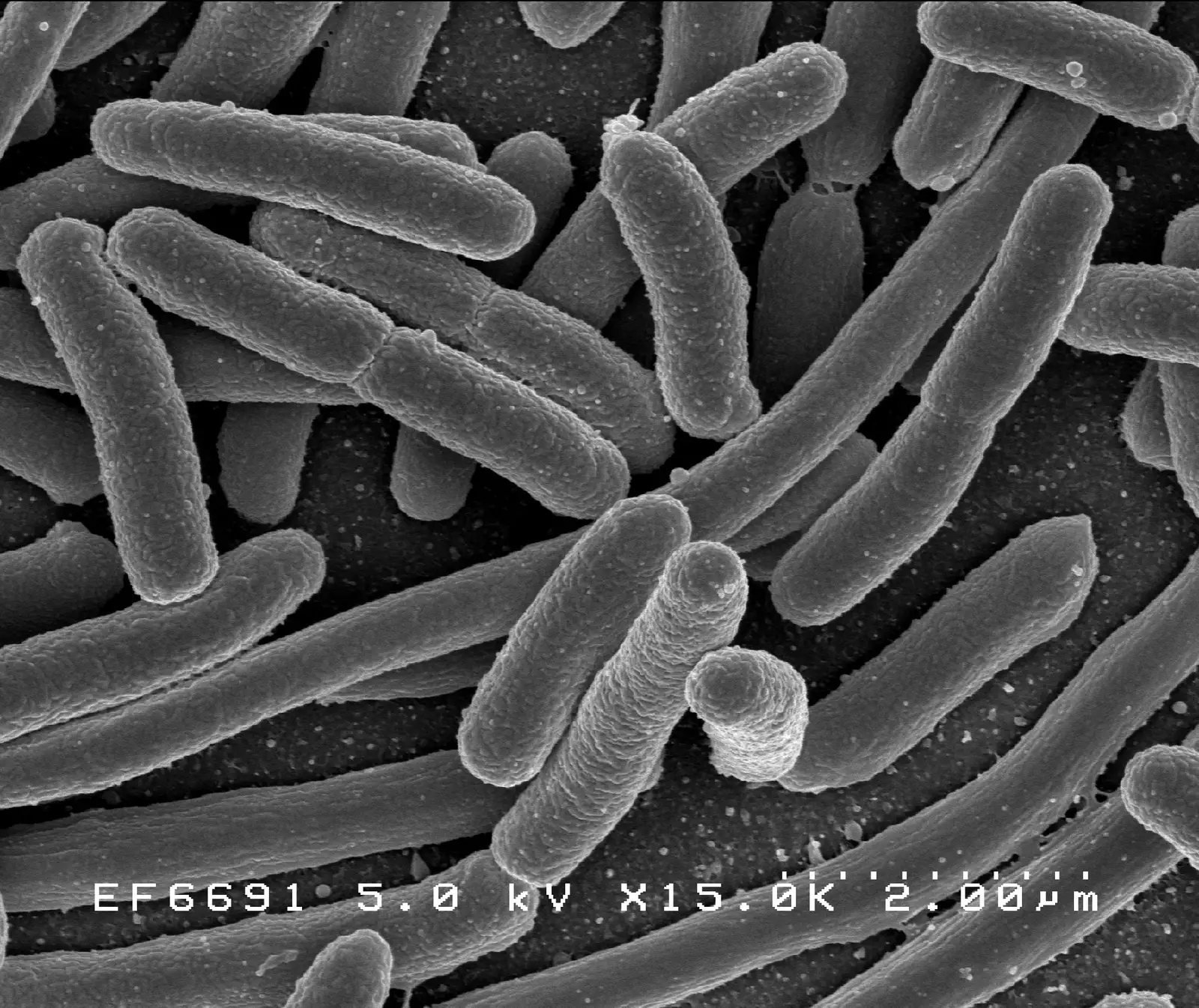
Image: “E. coli Bacteria (7316101966)” by NIAID (2002) · Public domain
"E. coli Bacteria (7316101966)" by NIAID (2002) — https://commons.wikimedia.org/wiki/File:E._coli_Bacteria_(7316101966).jpg — Public domain (work of a United States federal agency (NIAID, part of NIH), public domain in the United States under 17 U.S.C. §105; Commons tag PD-USGov-NIH-NIAID; Flickr source page credits NIAID with no contractor note; dated 2002-11-14 in the file metadata; no copyright term applies in the EU for a US government work released as public domain by its author agency (used as the source's explicit PD mark))

Reynolds Number: Why a Bacterium Can't Coast and a Fish Can
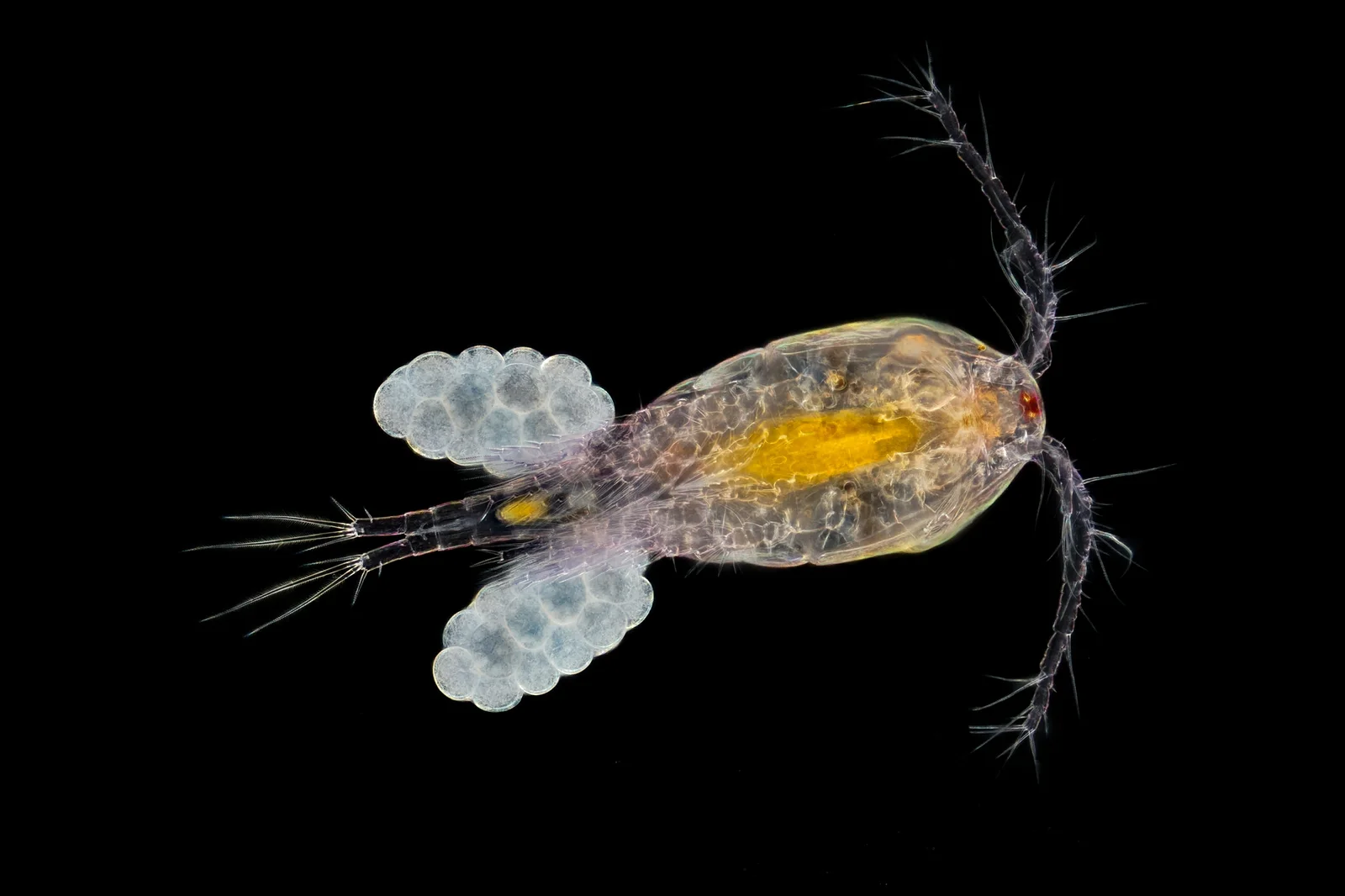
Image: “Oczlik (Cyclops) - samica” by Mikron86 (2025) · CC BY 4.0
"Oczlik (Cyclops) - samica" by Mikron86 — https://commons.wikimedia.org/wiki/File:Oczlik_(Cyclops)_-_samica.jpg — CC BY 4.0 (https://creativecommons.org/licenses/by/4.0/)

 Illustration
How Small Is an Atom? For Most Elements, About 0.1–0.5 nm
Image: Illustration: Ants, Cells & Atoms, all rights reserved
Illustration: Ants, Cells & Atoms. Generated schematic illustration — not a photograph, micrograph or specimen record. © Ants, Cells & Atoms, all rights reserved.