Foraminifera: Single Cells Known for Shells, Some Coin-Sized

Slide the ladder sideways to read its labels, or open it full size.
Foraminiferal tests: adult Globorotalia menardii shells 150–1,500 µm, and under 2 mm the "normal" test size of the smaller kinds [10−4–10−3 m]; fossil Nummulites tests of 5 and 30 mm in one pyramid block.
Log scale from 10 cm to 0.1 nm, each rung ten times smaller; dashed lines: naked eye about 0.1 mm, light microscope about 0.2 µm (How to read the Zoom ladder). Data: foraminifera-sizes.csv (sources, version and license, JSON).
"Zoom ladder: foraminifera" by Ants, Cells & Atoms, https://antscellsandatoms.com/microscopic-life/foraminifera/ — CC BY 4.0 (https://creativecommons.org/licenses/by/4.0/)
Data sources for this figure
Data: Adult Globorotalia menardii shells 150–1,500 µm: Rillo MC, Miller CG, Kucera M, Ezard THG 2020, Ecology and Evolution 10(20):11579–11590, CC BY 4.0, https://doi.org/10.1002/ece3.6792, retrieved 2026-10-25; Normal test size under 2 mm; Nummulites gizehensis tests of 5 and 30 mm in a block of the pyramid of Khufu: Hohenegger J, Kinoshita S, Briguglio A, Eder W, Wöger J 2019, Scientific Reports 9:8286, CC BY 4.0, https://doi.org/10.1038/s41598-019-44646-w, retrieved 2026-10-25; Xenophyophore tests up to 24 cm: Gooday AJ, Durden JM, Smith CR 2020, Communicative & Integrative Biology 13(1):189–197, CC BY 4.0, https://doi.org/10.1080/19420889.2020.1843818, as listed in this site's cell-sizes.csv; ladder computed 2026-10-25
Data sources for this figure
Data: Rillo MC, Miller CG, Kucera M, Ezard THG 2020, Ecology and Evolution 10(20):11579–11590, CC BY 4.0, https://doi.org/10.1002/ece3.6792, retrieved 2026-10-25; Hohenegger J, Kinoshita S, Briguglio A, Eder W, Wöger J 2019, Scientific Reports 9:8286, CC BY 4.0, https://doi.org/10.1038/s41598-019-44646-w, retrieved 2026-10-25; Greco M, Westgård A, Sykes FE, Ezat MM, Meilland J 2023, Journal of Plankton Research 45(4):652–660, CC BY 4.0, https://doi.org/10.1093/plankt/fbad031, retrieved 2026-10-25; one specimen size computed 2026-10-25; Gooday AJ, Durden JM, Smith CR 2020, Communicative & Integrative Biology 13(1):189–197, CC BY 4.0, https://doi.org/10.1080/19420889.2020.1843818, retrieved 2026-10-25; Polilov AA 2015, ZooKeys 526:61–64, CC BY 4.0, https://doi.org/10.3897/zookeys.526.6531, retrieved 2026-10-25; Piegari G et al. 2019, Frontiers in Veterinary Science 6:404, CC BY 4.0, https://doi.org/10.3389/fvets.2019.00404; Li C et al. 2023, Journal of Biomedical Optics 28(11):116501, CC BY 4.0, https://doi.org/10.1117/1.JBO.28.11.116501; VanDusen BM, Fegley SR, Peterson CH 2012, PLoS ONE 7(12):e52694, CC BY 4.0, https://doi.org/10.1371/journal.pone.0052694; as listed in this site's shared cell-sizes.csv and reference-objects.csv, retrieved 2026-10-25; GBIF Secretariat 2023, GBIF Backbone Taxonomy, checklist dataset https://doi.org/10.15468/39omei, CC BY 4.0 (taxon keys), retrieved 2026-10-25
Bench card
- Name
- Foraminifera (foraminifers), single-celled organisms known for the shell they build, called a test
- Actual size
- shells (tests) of adult drifting Globorotalia menardii 150–1,500 µm [10−4–10−3 m]; under 2 mm is the "normal" test size of the smaller seafloor and drifting kinds; tests of larger seafloor kinds reach 13 cm living and 19 cm fossilSource: Rillo et al. 2020, https://doi.org/10.1002/ece3.6792; Hohenegger et al. 2019, https://doi.org/10.1038/s41598-019-44646-w
- Size in this image
- not measurable from this image
- Lives in
- mostly on or in the seafloor; about 50 modern species drift as plankton; some in fresh water and soilSource: Holzmann et al. 2024, https://doi.org/10.1038/s41597-024-04137-8
- Seen with
- naked eye for fossil nummulites of 5 and 30 mm; light microscope, or a stereomicroscope in reflected light, for the smaller kindsSource: Sremac et al. 2024, https://doi.org/10.3934/microbiol.2024030; Hohenegger et al. 2019, https://doi.org/10.1038/s41598-019-44646-w
- Color in images
- native: fossil shells, no stain; shells from a sediment trap, no stain stated; the Haeckel plate is printed in one inkSource: figure captions on this page
foraminifera-bench.csv (CSV) · where each value comes from, and its license (JSON)
Data credits for this card
Data: GBIF Secretariat 2023, GBIF Backbone Taxonomy, Foraminifera (taxon key 8376456), checklist dataset https://doi.org/10.15468/39omei, CC BY 4.0, retrieved 2026-10-25; Rillo MC, Miller CG, Kucera M, Ezard THG 2020, Ecology and Evolution 10(20):11579–11590, CC BY 4.0, https://doi.org/10.1002/ece3.6792, retrieved 2026-10-25; Hohenegger J, Kinoshita S, Briguglio A, Eder W, Wöger J 2019, Scientific Reports 9:8286, CC BY 4.0, https://doi.org/10.1038/s41598-019-44646-w, retrieved 2026-10-25; Holzmann M, Nguyen NL, Angeles IB, Pawlowski J 2024, Scientific Data 11:1292, CC BY 4.0, https://doi.org/10.1038/s41597-024-04137-8, retrieved 2026-10-25; Sremac J, Bošnjak M, Firi KF, Šimičević A, Aščić Š 2024, AIMS Microbiology 10(3):644–673, CC BY 4.0, https://doi.org/10.3934/microbiol.2024030, retrieved 2026-10-25
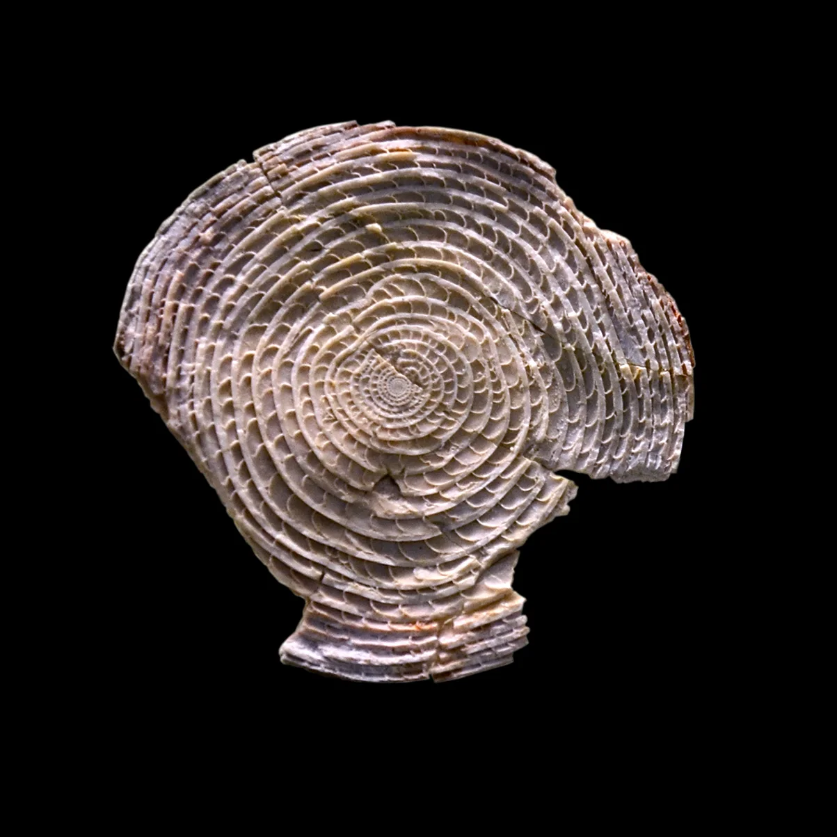
A fossil Nummulites puschi in the Natural History Museum, Basel, showing its spiral of chambers; Nummulites gizehensis tests in a block of the pyramid of Khufu measure 5 and 30 mm [10−3–10−2 m].
Color: native (a fossil shell photographed on black; no stain)
"Nummulite Naturhistorisches Museum Basel 27102013" by Vassil — https://commons.wikimedia.org/wiki/File:Nummulite_Naturhistorisches_Museum_Basel_27102013.jpg — CC0 1.0 (https://creativecommons.org/publicdomain/zero/1.0/)

Are foraminifera animals?
No. Each foraminifer is one cell, and the GBIF Backbone Taxonomy lists Foraminifera as a phylum in the kingdom Chromista, not among animals. In a 2024 reference dataset, Holzmann and colleagues call them protists, or microbial eukaryotes, set apart by net-forming threads of cytoplasm called granuloreticulopodia and by a test that is calcareous, agglutinated from gathered particles, or organic.
The name means "hole bearers", Sremac and colleagues note in a 2024 review of marine microfossils. Tests vary in shape, size, number of chambers and chemistry: commonly calcite, but also aragonite, gathered particles, chitin or, rarely, silica. Holzmann's group counts 8,896 modern and 39,976 fossil species, citing the World Register of Marine Species as of 2024. It adds that DNA surveys have turned up many unknown species, most of them soft-walled or naked (shell-less) single-chambered kinds that shell-based research had largely overlooked.
Most are benthic, living on or in the seafloor; about 50 modern species drift as plankton, and others live in fresh water and soil. Their shells also keep a record: by Sremac and colleagues' account, oxygen isotopes in foraminiferal tests are used to estimate past sea temperatures.
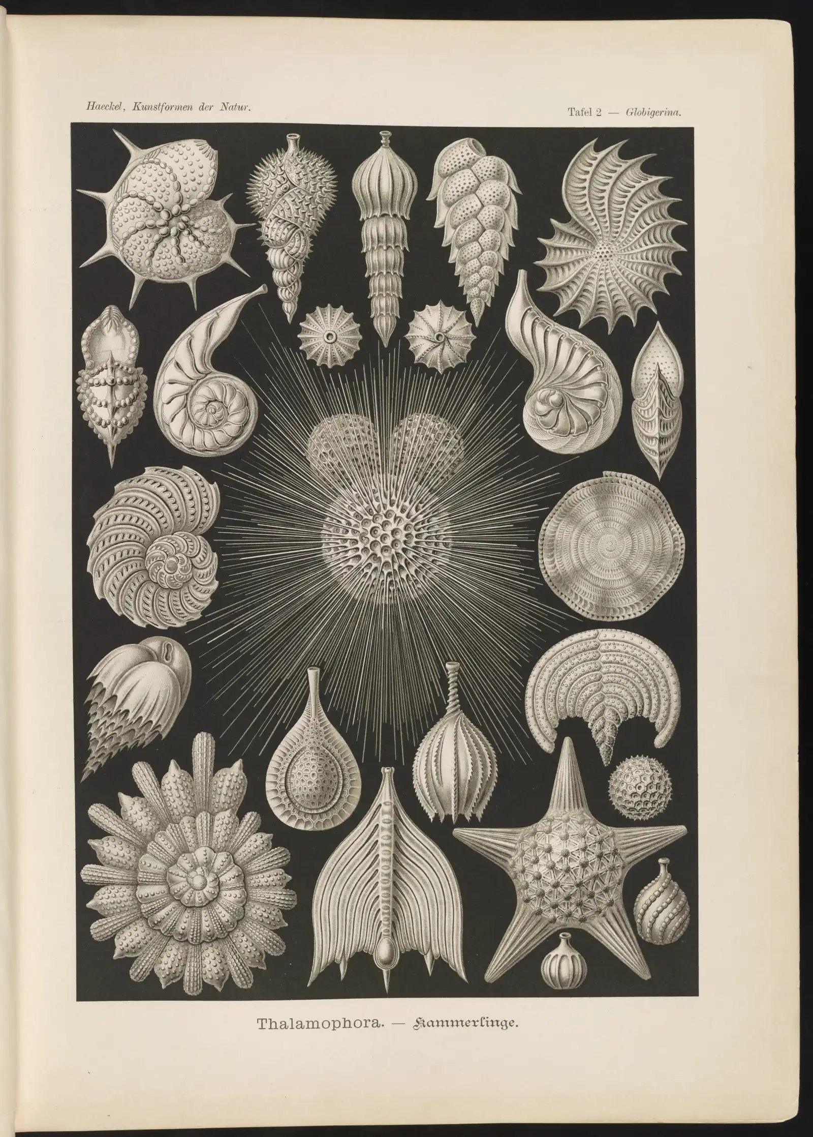
Thalamophora, plate 2 of Ernst Haeckel's Kunstformen der Natur (1904), headed Globigerina: chambered foraminifera shells around a spiny one; in the smaller kinds a test under 2 mm is the "normal" size [10−3 m].
Color: native (the plate's single printed ink; a lithograph, not a micrograph)
“Thalamophora. - Kammerlinge” by Ernst Haeckel (artist) and Adolf Giltsch (lithographer) (1904) · Public domain
Full credit
"Thalamophora. - Kammerlinge" by Ernst Haeckel (artist) and Adolf Giltsch (lithographer) (1904) — https://commons.wikimedia.org/wiki/File:Thalamophora._-_Kammerlinge_LCCN2015647733.jpg — Public domain (artist Ernst Haeckel died 1919 and lithographer Adolf Giltsch died 1911, so the plate is out of copyright in the EU (life plus 70 years) and in the US (published 1904, before 1930); the scan is from the Library of Congress Prints and Photographs division (lccn.loc.gov/2015647733, digital ID ds.07540), which lists no known restrictions on publication)

How big are foraminifera?
The "normal" test size for the smaller seafloor and drifting kinds is under 2 mm, and the larger seafloor kinds surpass it, Hohenegger and colleagues wrote in 2019. In a 2020 study of 3,799 museum shells, Rillo and colleagues found adult shells of the drifting Globorotalia menardii ranging from 150 to 1,500 µm, a tenfold spread within one species.
Those shells sit among familiar small things. A human hair is typically 50–100 µm wide (Li and colleagues, 2023), diatoms are usually 20–200 µm (Piegari and colleagues, 2019), and sand grains run from 63 µm to 2 mm on the grain-size scale VanDusen and colleagues used in 2012. A 150 µm adult G. menardii shell is about the size of a 139 µm male wasp, the smallest known parasitoid insect in Polilov's 2015 account.
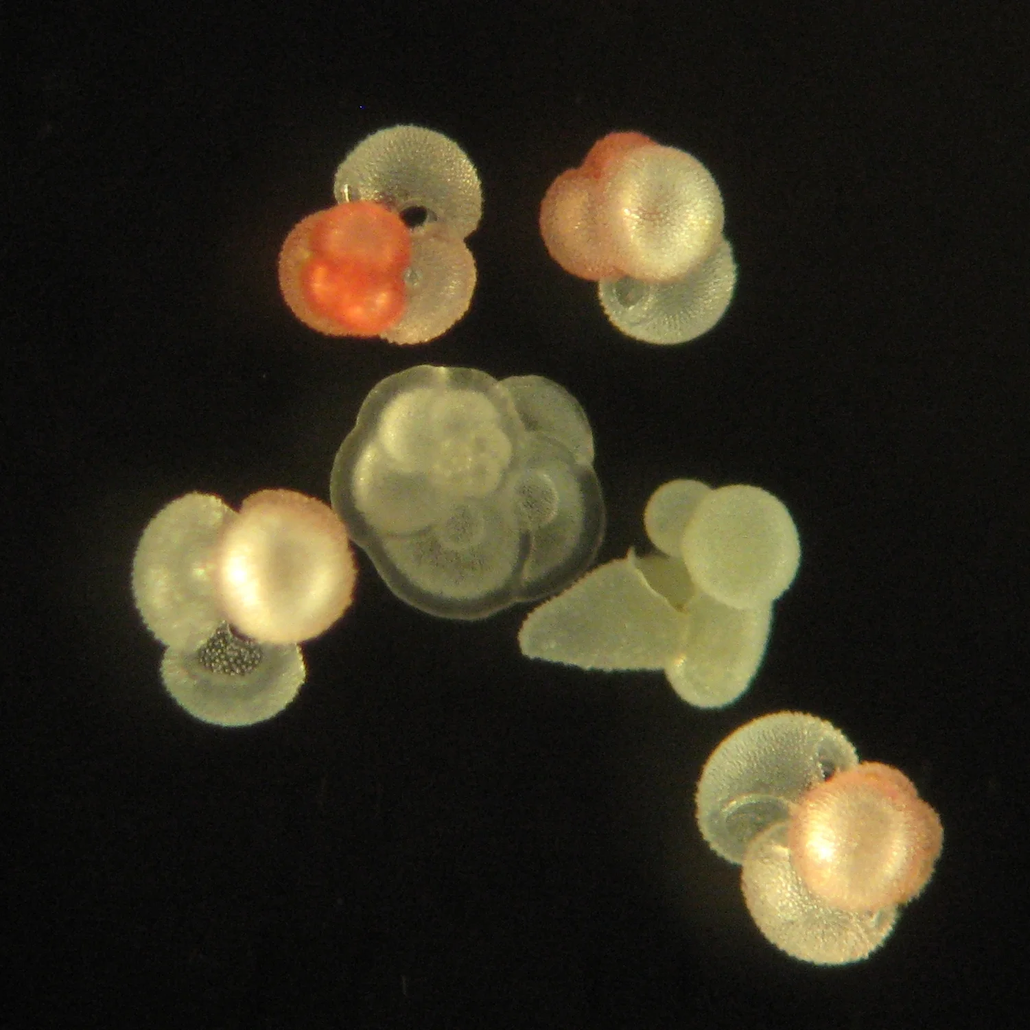
Drifting foraminifera from a sediment trap in the northern Gulf of Mexico, magnified 255×: pink Globigerinoides ruber and, at centre, Globorotalia menardii, whose adult shells measure 150–1,500 µm [10−4–10−3 m].
Color: native (the pink variety of Globigerinoides ruber, as the source names it; no stain stated)
“Planktic Foraminifera of the northern Gulf of Mexico” by Jessica Spear, U.S. Geological Survey (2010) · Public domain
Full credit
"Planktic Foraminifera of the northern Gulf of Mexico" by Jessica Spear, U.S. Geological Survey (2010) — https://commons.wikimedia.org/wiki/File:Planktic_Foraminifera_of_the_northern_Gulf_of_Mexico.jpg — Public domain (photograph by Jessica Spear of the U.S. Geological Survey, a US federal agency, public domain in the United States under 17 U.S.C. §105; Commons tag PD-USGov-USGS; the USGS copyrights and credits page states that USGS-authored or produced data and information are in the US public domain and that third-party material on USGS sites is marked as copyrighted (this photo carries no such mark on Commons); dated 2010-03-15; used in the EU on the source's explicit public-domain statement)

At the other end, the tests of larger seafloor foraminifera, which house algae in warm shallow seas, reach 13 cm in living species and 19 cm as fossils. Hohenegger's group cites both figures from earlier records. Xenophyophores, giant deep-sea foraminifera, build tests up to 24 cm from seafloor particles (Gooday and colleagues, 2020). From a 150 µm shell to a 24 cm test is a factor of 1,600, computed for this page; only a log scale shows both on one chart.
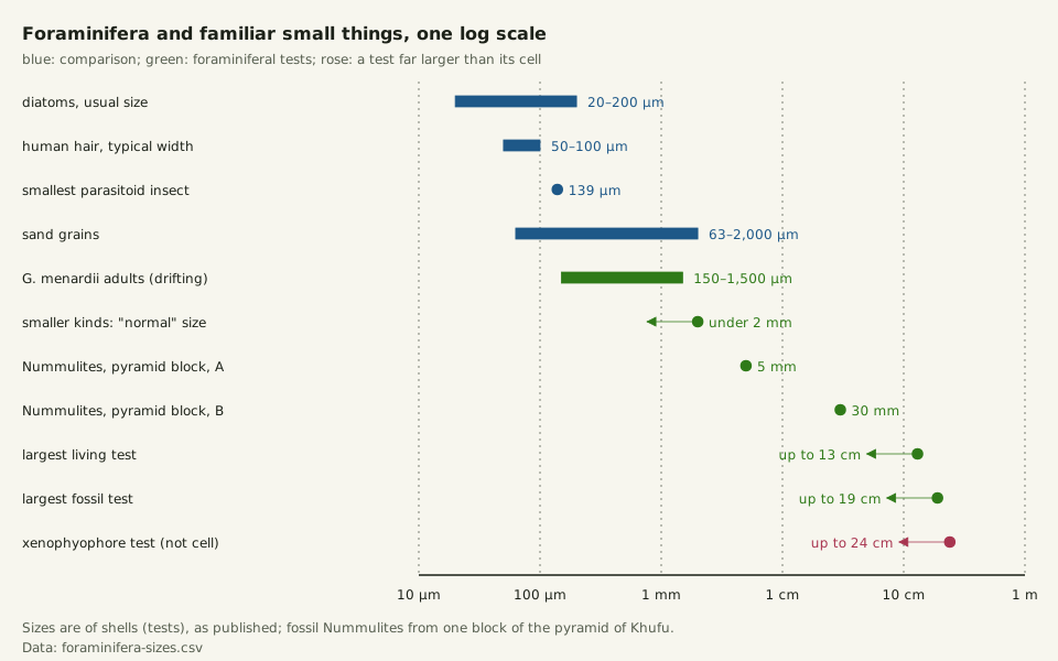
Slide the diagram sideways to read its labels, or open it full size.
Foraminiferal tests on one log scale [10−4–10−1 m]: adult G. menardii 150–1,500 µm and the 2 mm "normal" limit beside a hair, diatoms and sand grains; Nummulites of 5 and 30 mm; tests to 13 cm living, 19 cm fossil and 24 cm in xenophyophores.
Sizes are of shells as published. Data: foraminifera-sizes.csv (sources, version and license, JSON).
"Foraminifera sizes on one log scale, from a 150 µm shell to a 24 cm test" by Ants, Cells & Atoms, https://antscellsandatoms.com/microscopic-life/foraminifera/ — CC BY 4.0 (https://creativecommons.org/licenses/by/4.0/)
Data sources for this figure
Data: Rillo MC, Miller CG, Kucera M, Ezard THG 2020, Ecology and Evolution 10(20):11579–11590, CC BY 4.0, https://doi.org/10.1002/ece3.6792 (Globorotalia menardii adult shells 150–1,500 µm; retrieved 2026-10-25); Hohenegger J, Kinoshita S, Briguglio A, Eder W, Wöger J 2019, Scientific Reports 9:8286, CC BY 4.0, https://doi.org/10.1038/s41598-019-44646-w (normal test size under 2 mm; Nummulites tests of 5 and 30 mm; largest tests 13 cm living and 19 cm fossil; retrieved 2026-10-25); Gooday AJ, Durden JM, Smith CR 2020, Communicative & Integrative Biology 13(1):189–197, CC BY 4.0, https://doi.org/10.1080/19420889.2020.1843818 (xenophyophore tests up to 24 cm; retrieved 2026-10-25); Comparison rows from this site's shared files cell-sizes.csv and reference-objects.csv: Piegari G et al. 2019, Frontiers in Veterinary Science 6:404, CC BY 4.0, https://doi.org/10.3389/fvets.2019.00404 (diatoms 20–200 µm); Li C et al. 2023, Journal of Biomedical Optics 28(11):116501, CC BY 4.0, https://doi.org/10.1117/1.JBO.28.11.116501 (human hair 50–100 µm); VanDusen BM, Fegley SR, Peterson CH 2012, PLoS ONE 7(12):e52694, CC BY 4.0, https://doi.org/10.1371/journal.pone.0052694 (sand grains 63 µm–2 mm); retrieved 2026-10-25; Polilov AA 2015, ZooKeys 526:61–64, CC BY 4.0, https://doi.org/10.3897/zookeys.526.6531 (smallest known parasitoid insect, 139 µm; retrieved 2026-10-25); chart computed 2026-10-25 for this page
Data sources for this figure
Data: Rillo MC, Miller CG, Kucera M, Ezard THG 2020, Ecology and Evolution 10(20):11579–11590, CC BY 4.0, https://doi.org/10.1002/ece3.6792, retrieved 2026-10-25; Hohenegger J, Kinoshita S, Briguglio A, Eder W, Wöger J 2019, Scientific Reports 9:8286, CC BY 4.0, https://doi.org/10.1038/s41598-019-44646-w, retrieved 2026-10-25; Greco M, Westgård A, Sykes FE, Ezat MM, Meilland J 2023, Journal of Plankton Research 45(4):652–660, CC BY 4.0, https://doi.org/10.1093/plankt/fbad031, retrieved 2026-10-25; one specimen size computed 2026-10-25; Gooday AJ, Durden JM, Smith CR 2020, Communicative & Integrative Biology 13(1):189–197, CC BY 4.0, https://doi.org/10.1080/19420889.2020.1843818, retrieved 2026-10-25; Polilov AA 2015, ZooKeys 526:61–64, CC BY 4.0, https://doi.org/10.3897/zookeys.526.6531, retrieved 2026-10-25; Piegari G et al. 2019, Frontiers in Veterinary Science 6:404, CC BY 4.0, https://doi.org/10.3389/fvets.2019.00404; Li C et al. 2023, Journal of Biomedical Optics 28(11):116501, CC BY 4.0, https://doi.org/10.1117/1.JBO.28.11.116501; VanDusen BM, Fegley SR, Peterson CH 2012, PLoS ONE 7(12):e52694, CC BY 4.0, https://doi.org/10.1371/journal.pone.0052694; as listed in this site's shared cell-sizes.csv and reference-objects.csv, retrieved 2026-10-25; GBIF Secretariat 2023, GBIF Backbone Taxonomy, checklist dataset https://doi.org/10.15468/39omei, CC BY 4.0 (taxon keys), retrieved 2026-10-25
What were Strabo's stone lentils?
Strabo, born at Amasia in Pontus, lived under the emperors Augustus and Tiberius. In Book 17 of his Geography he describes the stones:
"One extraordinary thing which I saw at the pyramids must not be omitted. Heaps of stones from the quarries lie in front of the pyramids. Among these are found pieces which in shape and size resemble lentils. Some contain substances like grains half peeled. These, it is said, are the remnants of the workmen's food converted into stone; which is not probable." Strabo, Geography 17.1.34, in W. Falconer's translation (London: Bohn, 1857)
So the petrified-food story was hearsay, and Strabo judged it unlikely; he compared the pieces with lentil-like pebbles on a long hill at home, near Amasia. His translators added a note in 1857: Niebuhr had reported small petrified lentil-shaped bodies in these stones, like shells he collected at Bushir, and to Clarke they looked like the petrified remains of some unknown animal.
The stone can now be named. Nummulites is a genus of large seafloor foraminifera with coin-shaped tests, and nummulitic limestone, made up of them, was the main building stone of the Egyptian pyramids, by the account of Sremac and colleagues, who cite a 2018 magazine article. Members of the genus built up large beds of carbonate rock, Hohenegger and colleagues note, and one species, N. venosus, still lives off Okinawa.
Their 2019 paper shows the surface of a block from the pyramid of Khufu, also called Cheops, built of Nummulites gizehensis tests (spelled ghizehensis in the paper): many 5 mm tests of one generation, called A, and 30 mm tests of another, called B.
Strabo gave no measurements, so the pieces he saw cannot be matched one by one to these shells. The claims line up like this (foraminifera-strabo-claims.csv (sources, version and license, JSON) Data: Strabo, Geography 17.1.34, translated by H. C. Hamilton and W. Falconer, vol. 3 (London: Henry G. Bohn, 1857), public domain, https://www.gutenberg.org/ebooks/44886, retrieved 2026-10-25; Hohenegger J, Kinoshita S, Briguglio A, Eder W, Wöger J 2019, Scientific Reports 9:8286, CC BY 4.0, https://doi.org/10.1038/s41598-019-44646-w, retrieved 2026-10-25; Holzmann M, Nguyen NL, Angeles IB, Pawlowski J 2024, Scientific Data 11:1292, CC BY 4.0, https://doi.org/10.1038/s41597-024-04137-8, retrieved 2026-10-25; Sremac J, Bošnjak M, Firi KF, Šimičević A, Aščić Š 2024, AIMS Microbiology 10(3):644–673, CC BY 4.0, https://doi.org/10.3934/microbiol.2024030, retrieved 2026-10-25; GBIF Secretariat 2023, GBIF Backbone Taxonomy, checklist dataset https://doi.org/10.15468/39omei, CC BY 4.0 (Foraminifera, taxon key 8376456), retrieved 2026-10-25):
| Claim | Who made it | Reading today |
|---|---|---|
| Lentil-shaped pieces lie in the quarry-stone heaps before the pyramids | Strabo, as an eyewitness | One block of the pyramid of Khufu is built of Nummulites tests of 5 and 30 mm (Hohenegger 2019); the pieces Strabo saw are not identified |
| The pieces are the workmen's food turned to stone | Unnamed people, reported by Strabo | Strabo called it "not probable"; that pyramid block is built of fossil shells |
| The stones hold small petrified shells | Niebuhr, in the translators' 1857 note | Consistent: Nummulites tests are shells |
| They are remains of an unknown animal | Clarke, in the same note | The fossils in that block are Nummulites: single cells, not animals |
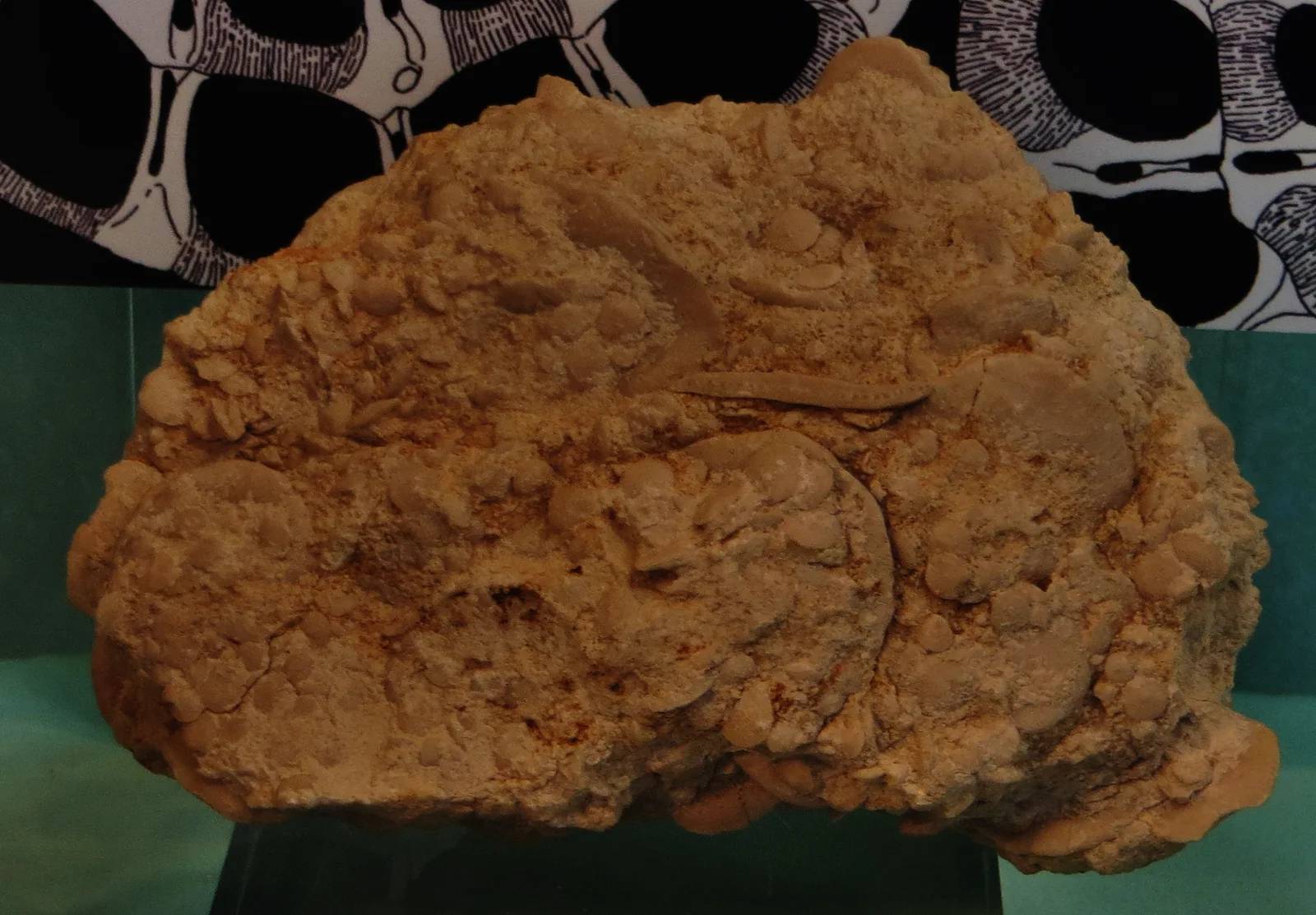
A slab of Nummulites limestone in the Goldfuß Museum, Bonn, labelled Nummulites gizehensis in its file title; in a block of the pyramid of Khufu, this species' tests measure 5 and 30 mm [10−3–10−2 m].
Color: native (fossil shells in rock, photographed in a museum display; no stain)
"Nummulites gizehensis" by Ghedoghedo — https://commons.wikimedia.org/wiki/File:Nummulites_gizehensis.jpg — CC BY-SA 4.0 (https://creativecommons.org/licenses/by-sa/4.0/)

Does a big shell mean a big cell?
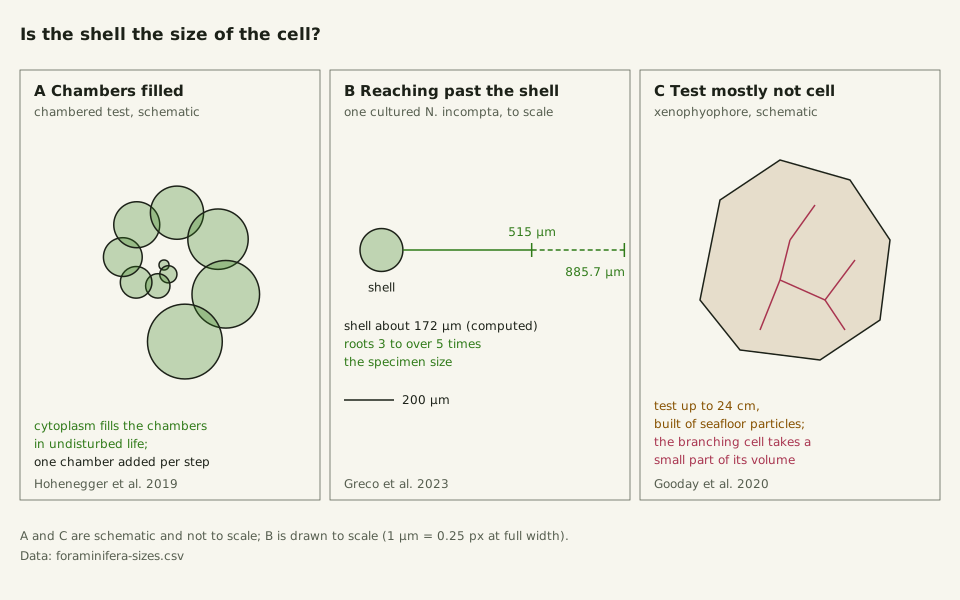
In chambered foraminifera, the cell normally fills its shell. They build the test by adding a new chamber at every growth step, and during undisturbed life the cytoplasm fills the chambers completely, Hohenegger and colleagues write; chamber volumes therefore trace the growth of one cell. For living Nummulites venosus off Okinawa, a species that houses diatoms as partners, they estimated an expected life span of 18 months.
Drifting species grow the same way, one chamber at a time, until they reproduce, when the cell dies (Rillo and colleagues, 2020).
The living cell also reaches past its shell. Foraminifera move on reticulopodia, threads of cytoplasm that join into networks with cytoplasm flowing both ways, Greco and colleagues explain in a 2023 study. In culture, one Neogloboquadrina incompta held itself in place with ectoplasmic roots, thick strands of the cell's outer cytoplasm, 515–885.7 µm long, three to more than five times its own size; twig-like strands of N. pachyderma reached up to four times the size of the shell.
At the giant end the reverse holds. A xenophyophore's test can reach 24 cm, but its branching cell body occupies only a small proportion of the test volume, Gooday and colleagues wrote in 2020, so the 24 cm measures a shell, not a cell. How Big Is a Cell? sets that test beside the largest single cells.
Slide the diagram sideways to read its labels, or open it full size.
Shell and cell in foraminifera [10−4–10−1 m]: chambers filled by cytoplasm; a Neogloboquadrina incompta about 0.17 mm in size, computed, whose roots reached 515–885.7 µm; a xenophyophore test up to 24 cm with a thin branching cell.
A and C are schematic; B is to scale. Data: foraminifera-sizes.csv (sources, version and license, JSON).
"Shell and cell: filled chambers, roots past the shell, a mostly empty giant test" by Ants, Cells & Atoms, https://antscellsandatoms.com/microscopic-life/foraminifera/ — CC BY 4.0 (https://creativecommons.org/licenses/by/4.0/)
Data sources for this figure
Data: Chambers filled by cytoplasm in undisturbed life: Hohenegger J, Kinoshita S, Briguglio A, Eder W, Wöger J 2019, Scientific Reports 9:8286, CC BY 4.0, https://doi.org/10.1038/s41598-019-44646-w, retrieved 2026-10-25; Root lengths 515–885.7 µm, three to more than five times the specimen size: Greco M, Westgård A, Sykes FE, Ezat MM, Meilland J 2023, Journal of Plankton Research 45(4):652–660, CC BY 4.0, https://doi.org/10.1093/plankt/fbad031, retrieved 2026-10-25; specimen size computed 2026-10-25; Xenophyophore test up to 24 cm with the cell a small proportion of its volume: Gooday AJ, Durden JM, Smith CR 2020, Communicative & Integrative Biology 13(1):189–197, CC BY 4.0, https://doi.org/10.1080/19420889.2020.1843818, retrieved 2026-10-25
Data sources for this figure
Data: Rillo MC, Miller CG, Kucera M, Ezard THG 2020, Ecology and Evolution 10(20):11579–11590, CC BY 4.0, https://doi.org/10.1002/ece3.6792, retrieved 2026-10-25; Hohenegger J, Kinoshita S, Briguglio A, Eder W, Wöger J 2019, Scientific Reports 9:8286, CC BY 4.0, https://doi.org/10.1038/s41598-019-44646-w, retrieved 2026-10-25; Greco M, Westgård A, Sykes FE, Ezat MM, Meilland J 2023, Journal of Plankton Research 45(4):652–660, CC BY 4.0, https://doi.org/10.1093/plankt/fbad031, retrieved 2026-10-25; one specimen size computed 2026-10-25; Gooday AJ, Durden JM, Smith CR 2020, Communicative & Integrative Biology 13(1):189–197, CC BY 4.0, https://doi.org/10.1080/19420889.2020.1843818, retrieved 2026-10-25; Polilov AA 2015, ZooKeys 526:61–64, CC BY 4.0, https://doi.org/10.3897/zookeys.526.6531, retrieved 2026-10-25; Piegari G et al. 2019, Frontiers in Veterinary Science 6:404, CC BY 4.0, https://doi.org/10.3389/fvets.2019.00404; Li C et al. 2023, Journal of Biomedical Optics 28(11):116501, CC BY 4.0, https://doi.org/10.1117/1.JBO.28.11.116501; VanDusen BM, Fegley SR, Peterson CH 2012, PLoS ONE 7(12):e52694, CC BY 4.0, https://doi.org/10.1371/journal.pone.0052694; as listed in this site's shared cell-sizes.csv and reference-objects.csv, retrieved 2026-10-25; GBIF Secretariat 2023, GBIF Backbone Taxonomy, checklist dataset https://doi.org/10.15468/39omei, CC BY 4.0 (taxon keys), retrieved 2026-10-25
How can you tell a living foraminifer from an empty shell?
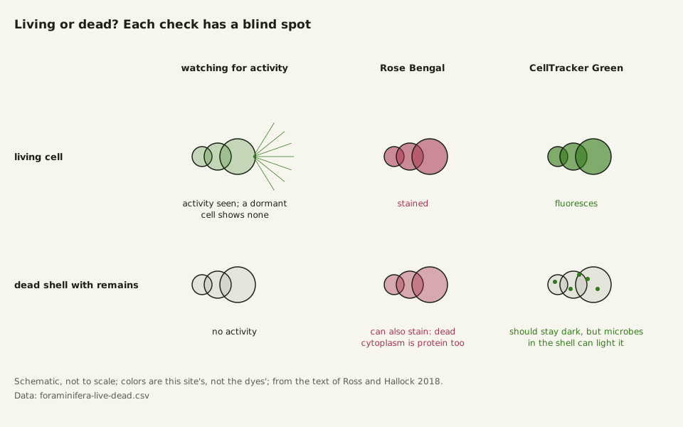
Not always by eye. In a 2018 paper, Ross and Hallock describe Amphistegina gibbosa slipping into a dormant state in which living individuals looked just like dead ones: no extended pseudopods, no attachment, no visible waste.
Stains help, and the color they add is borrowed color, not the cell's own. Rose Bengal stains proteins and can color dead cytoplasm as well as live, Ross and Hallock note. CellTracker Green becomes fluorescent only after esterases, enzymes common to living cells, cut it, so a cell must be alive to make it glow.
Slide the diagram sideways to read its labels, or open it full size.
Telling a living foraminifer from a dead one, in shells normally under 2 mm in the smaller kinds [10−3 m]: Rose Bengal can stain protein in both; CellTracker Green needs a living cell's enzymes, but microbes in a dead shell can set it off.
Schematic, not to scale; colors are this site's, not the dyes'. Data: foraminifera-live-dead.csv (sources, version and license, JSON).
"Living or dead? What each check can and cannot show in a foraminifer" by Ants, Cells & Atoms, https://antscellsandatoms.com/microscopic-life/foraminifera/ — CC BY 4.0 (https://creativecommons.org/licenses/by/4.0/)
Data: Ross BJ, Hallock P 2018, PeerJ 6:e5304, CC BY 4.0, https://doi.org/10.7717/peerj.5304, retrieved 2026-10-25; diagram computed 2026-10-25 for this page
Data: Ross BJ, Hallock P 2018, PeerJ 6:e5304, CC BY 4.0, https://doi.org/10.7717/peerj.5304, retrieved 2026-10-25
That check can be fooled too. In their experiments, 48% of specimens killed by heat and kept 48 hours in non-sterile seawater showed some fluorescence, which the authors tie to microbes growing in dead shells. As of that 2018 paper, the fluorescence readings did not settle whether a still Amphistegina was dormant or dead.
Sources
- Holzmann M, Nguyen NL, Angeles IB, Pawlowski J. "BFR2: a curated ribosomal reference dataset for benthic foraminifera." Scientific Data 11:1292 (2024). https://doi.org/10.1038/s41597-024-04137-8 Read: full text. Text license: CC BY 4.0 (https://creativecommons.org/licenses/by/4.0/).
- Rillo MC, Miller CG, Kucera M, Ezard THG. "Intraspecific size variation in planktonic foraminifera cannot be consistently predicted by the environment." Ecology and Evolution 10(20):11579–11590 (2020). https://doi.org/10.1002/ece3.6792 Read: full text. Text license: CC BY 4.0 (https://creativecommons.org/licenses/by/4.0/).
- Hohenegger J, Kinoshita S, Briguglio A, Eder W, Wöger J. "Lunar cycles and rainy seasons drive growth and reproduction in nummulitid foraminifera, important producers of carbonate buildups." Scientific Reports 9:8286 (2019). https://doi.org/10.1038/s41598-019-44646-w Read: full text. Text license: CC BY 4.0 (https://creativecommons.org/licenses/by/4.0/).
- Strabo. The Geography of Strabo, vol. 3, translated by H. C. Hamilton and W. Falconer (Books 7 onward by Falconer). London: Henry G. Bohn, 1857. Book 17, chapter 1, section 34, and the translators' notes 858–859. Project Gutenberg eBook #44886. https://www.gutenberg.org/ebooks/44886 Read: full text of Book 17, 1.34 and its notes.
- GBIF Secretariat. GBIF Backbone Taxonomy. Checklist dataset https://doi.org/10.15468/39omei, accessed 2026-10-25; Foraminifera (phylum, kingdom Chromista), usage key 8376456; Globorotalia menardii (d'Orbigny, 1865), 4382036; Nummulites Lamarck, 1801, 5428655; Nummulites gizehensis (Forskål, 1775), 8375775; Nummulites venosus (Fichtel & Moll, 1798), 7845048; Neogloboquadrina incompta (Cifelli, 1961), 7775876; Neogloboquadrina pachyderma (Ehrenberg, 1861), 7576430; Amphistegina gibbosa d'Orbigny, 1839, 8426787; Globigerinoides ruber (d'Orbigny, 1839), 8107071; Dicopomorpha echmepterygis Mockford, 1997, 1367990. Read: dataset record (species match and species records). Text license: CC BY 4.0 (https://creativecommons.org/licenses/by/4.0/).
- Sremac J, Bošnjak M, Firi KF, Šimičević A, Aščić Š. "Marine microfossils: Tiny archives of ocean changes through deep time." AIMS Microbiology 10(3):644–673 (2024). https://doi.org/10.3934/microbiol.2024030 Read: full text. Text license: CC BY 4.0 (https://creativecommons.org/licenses/by/4.0/).
- Li C, Du Y, Chen H, Han X, Wu W, Kong X, Zhang C, Yuan X. "Transmission infrared micro-spectroscopic study of individual human hair." Journal of Biomedical Optics 28(11):116501 (2023). https://doi.org/10.1117/1.JBO.28.11.116501 Read: full text. Text license: CC BY 4.0 (https://creativecommons.org/licenses/by/4.0/).
- Piegari G, De Biase D, d'Aquino I, Prisco F, Fico R, Ilsami R, Pozzato N, Genovese A, Paciello O. "Diagnosis of Drowning and the Value of the Diatom Test in Veterinary Forensic Pathology." Frontiers in Veterinary Science 6:404 (2019). https://doi.org/10.3389/fvets.2019.00404 Read: full text. Text license: CC BY 4.0 (https://creativecommons.org/licenses/by/4.0/).
- VanDusen BM, Fegley SR, Peterson CH. "Prey Distribution, Physical Habitat Features, and Guild Traits Interact to Produce Contrasting Shorebird Assemblages among Foraging Patches." PLoS ONE 7(12):e52694 (2012). https://doi.org/10.1371/journal.pone.0052694 Read: full text. Text license: CC BY 4.0 (https://creativecommons.org/licenses/by/4.0/).
- Polilov AA. "How small is the smallest? New record and remeasuring of Scydosella musawasensis Hall, 1999 (Coleoptera, Ptiliidae), the smallest known free-living insect." ZooKeys 526:61–64 (2015). https://doi.org/10.3897/zookeys.526.6531 Read: full text. Text license: CC BY 4.0 (https://creativecommons.org/licenses/by/4.0/).
- Gooday AJ, Durden JM, Smith CR. "Giant, highly diverse protists in the abyssal Pacific: vulnerability to impacts from seabed mining and potential for recovery." Communicative & Integrative Biology 13(1):189–197 (2020). https://doi.org/10.1080/19420889.2020.1843818 Read: full text. Text license: CC BY 4.0 (https://creativecommons.org/licenses/by/4.0/).
- Greco M, Westgård A, Sykes FE, Ezat MM, Meilland J. "Uncovering hidden structures: previously undescribed pseudopodia and ectoplasmic structures in planktonic foraminifera." Journal of Plankton Research 45(4):652–660 (2023). https://doi.org/10.1093/plankt/fbad031 Read: full text. Text license: CC BY 4.0 (https://creativecommons.org/licenses/by/4.0/).
- Ross BJ, Hallock P. "Challenges in using CellTracker Green on foraminifers that host algal endosymbionts." PeerJ 6:e5304 (2018). https://doi.org/10.7717/peerj.5304 Read: full text. Text license: CC BY 4.0 (https://creativecommons.org/licenses/by/4.0/).
Cite this page
Ants, Cells & Atoms. “Foraminifera: Single Cells Known for Shells, Some Coin-Sized.” Published 2026-10-25. https://antscellsandatoms.com/microscopic-life/foraminifera/. Accessed (add the date you read it).
No DOI is issued for entries. Figures made here are CC BY 4.0; see each caption for the license of photographs.
Related entries
 Illustration
Diatoms: Why Most Glass-Walled Algae Shrink as They Multiply
Image: Illustration: Ants, Cells & Atoms, all rights reserved
Illustration: Ants, Cells & Atoms. Generated schematic illustration — not a photograph, micrograph or specimen record. © Ants, Cells & Atoms, all rights reserved.
Plankton: From Bacteria to Jellyfish, Sorted in Size Steps
Image: “Diatomea. - Schachtellinge” by Ernst Haeckel (artist) and Adolf Giltsch (lithographer) (1904) · Public domain
"Diatomea. - Schachtellinge" by Ernst Haeckel (artist) and Adolf Giltsch (lithographer) (1904) — https://commons.wikimedia.org/wiki/File:Diatomea._-_Schachtellinge_LCCN2015647735.jpg — Public domain (artist Ernst Haeckel died 1919 and lithographer Adolf Giltsch died 1911, so the plate is out of copyright in the EU (life plus 70 years) and in the US (published 1904, before 1930); the scan is from the Library of Congress Prints and Photographs division (lccn.loc.gov/2015647735), which lists no rights restriction)

Paramecium: How a Nerve-Like Signal Sends One Cell Backward
Image: “Paramecium 1” by Nbnidhi (2017) · CC BY 4.0
"Paramecium 1" by Nbnidhi — https://commons.wikimedia.org/wiki/File:Paramecium_1.jpg — CC BY 4.0 (https://creativecommons.org/licenses/by/4.0/)
