Skip to the main content
Ants, Cells & Atoms

Life and matter below the width of a hair.

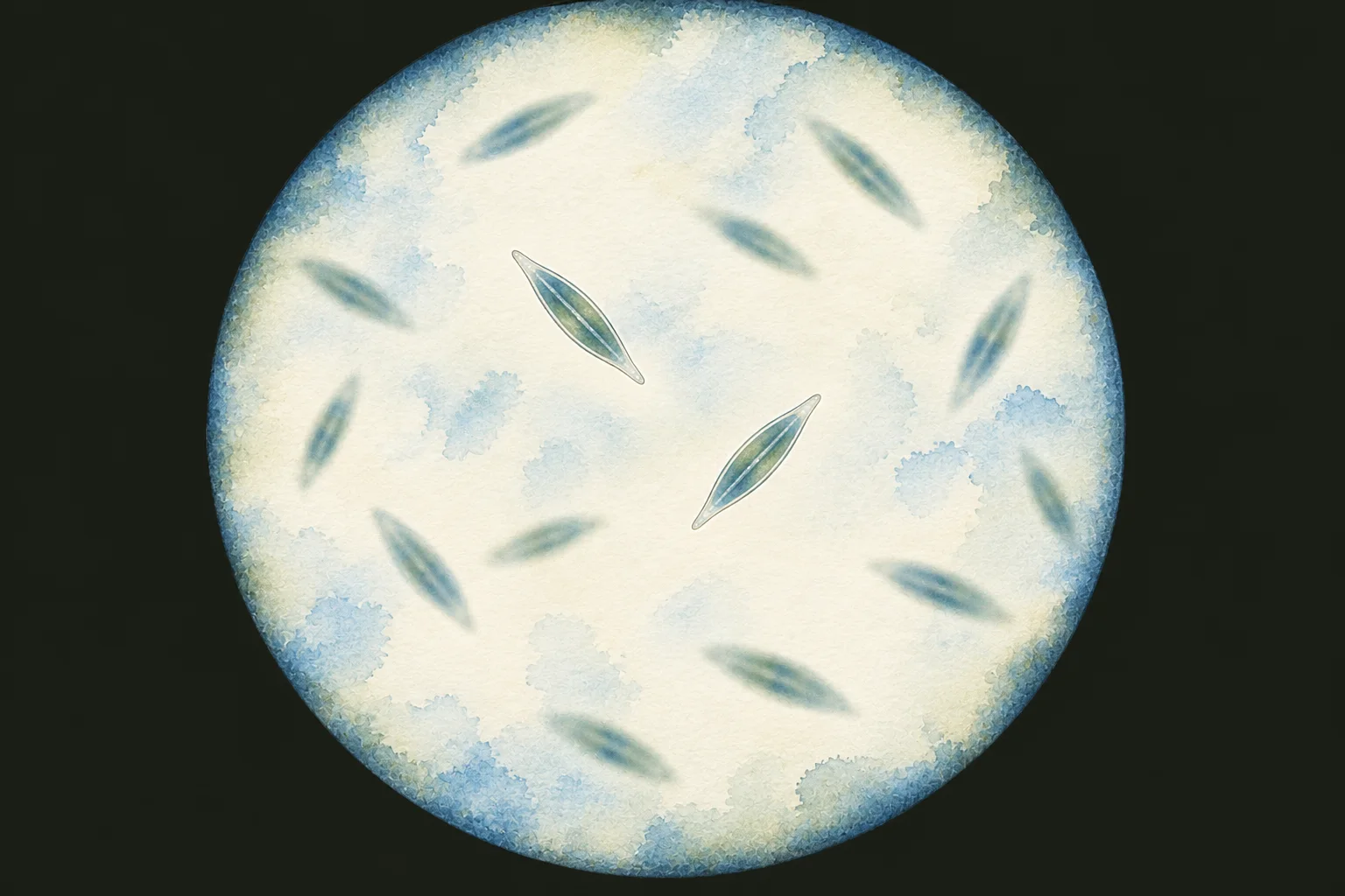
Illustration: spindle-shaped glassy cells drifting at different depths in a round field of view; two near the centre are in sharp focus. Illustration

Field-guide entry[10−5–10−4 m]

Diatoms: Why Most Glass-Walled Algae Shrink as They Multiply

Diatoms are single-celled algae in glass walls, 2–2,000 µm across species. Each new wall half forms inside an old one, so most lineages shrink as they divide.

Image: Illustration: Ants, Cells & Atoms, all rights reserved

Illustration: Ants, Cells & Atoms. Generated schematic illustration — not a photograph, micrograph or specimen record. © Ants, Cells & Atoms, all rights reserved.
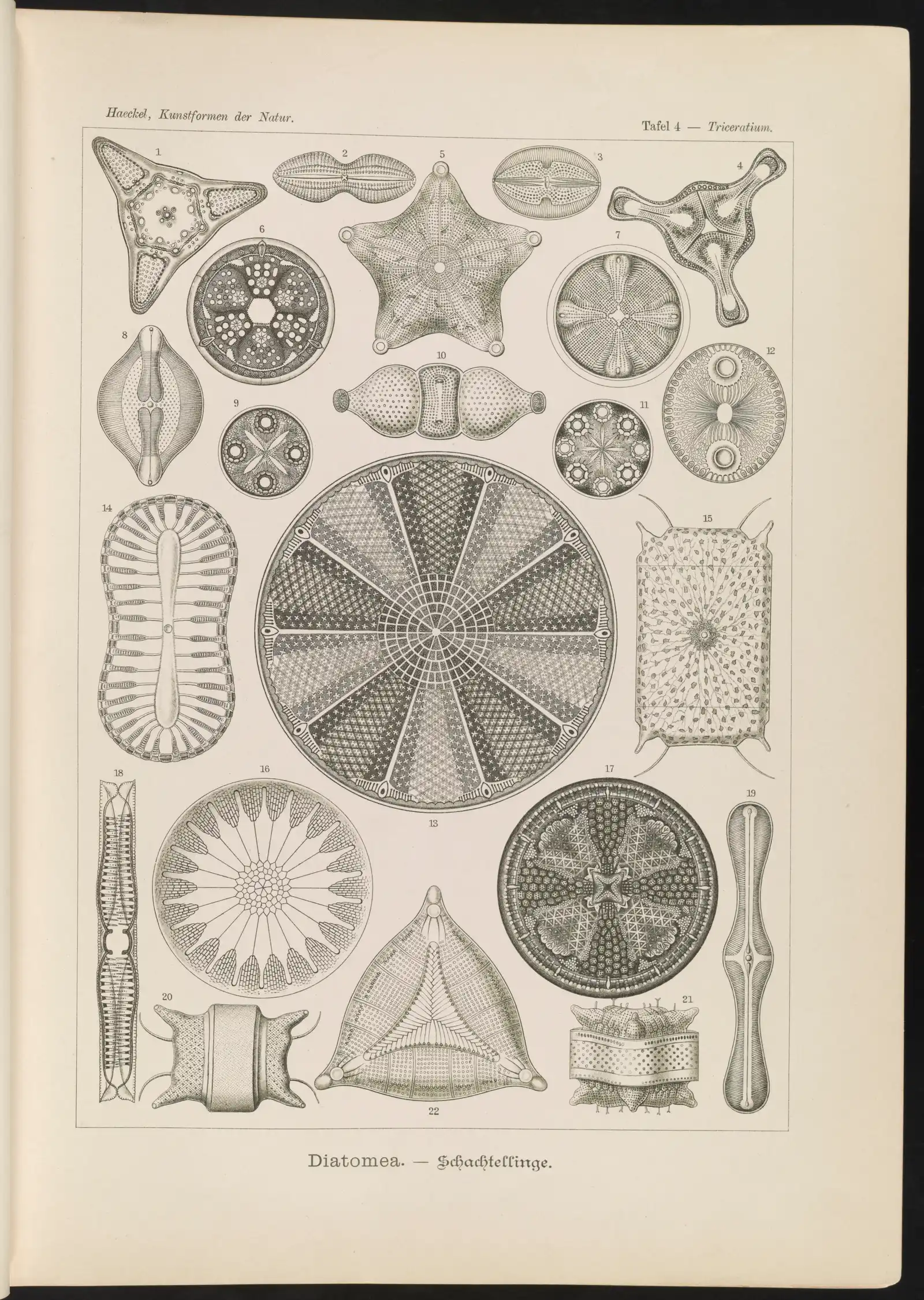
Diatoms drawn by Ernst Haeckel on a 1904 lithograph plate; the diatoms sized on this page span 7–138 µm; rows of symmetrical glass shells.

Field-guide entry[10−6–10−4 m]

Plankton: From Bacteria to Jellyfish, Sorted in Size Steps

Plankton are the living things that drift with the water, from bacteria under 1 µm to jellyfish over 2 m, sorted into size classes ten times apart.

Image: “Diatomea. - Schachtellinge” by Ernst Haeckel (artist) and Adolf Giltsch (lithographer) (1904) · Public domain

"Diatomea. - Schachtellinge" by Ernst Haeckel (artist) and Adolf Giltsch (lithographer) (1904) — https://commons.wikimedia.org/wiki/File:Diatomea._-_Schachtellinge_LCCN2015647735.jpg — Public domain (artist Ernst Haeckel died 1919 and lithographer Adolf Giltsch died 1911, so the plate is out of copyright in the EU (life plus 70 years) and in the US (published 1904, before 1930); the scan is from the Library of Congress Prints and Photographs division (lccn.loc.gov/2015647735), which lists no rights restriction)

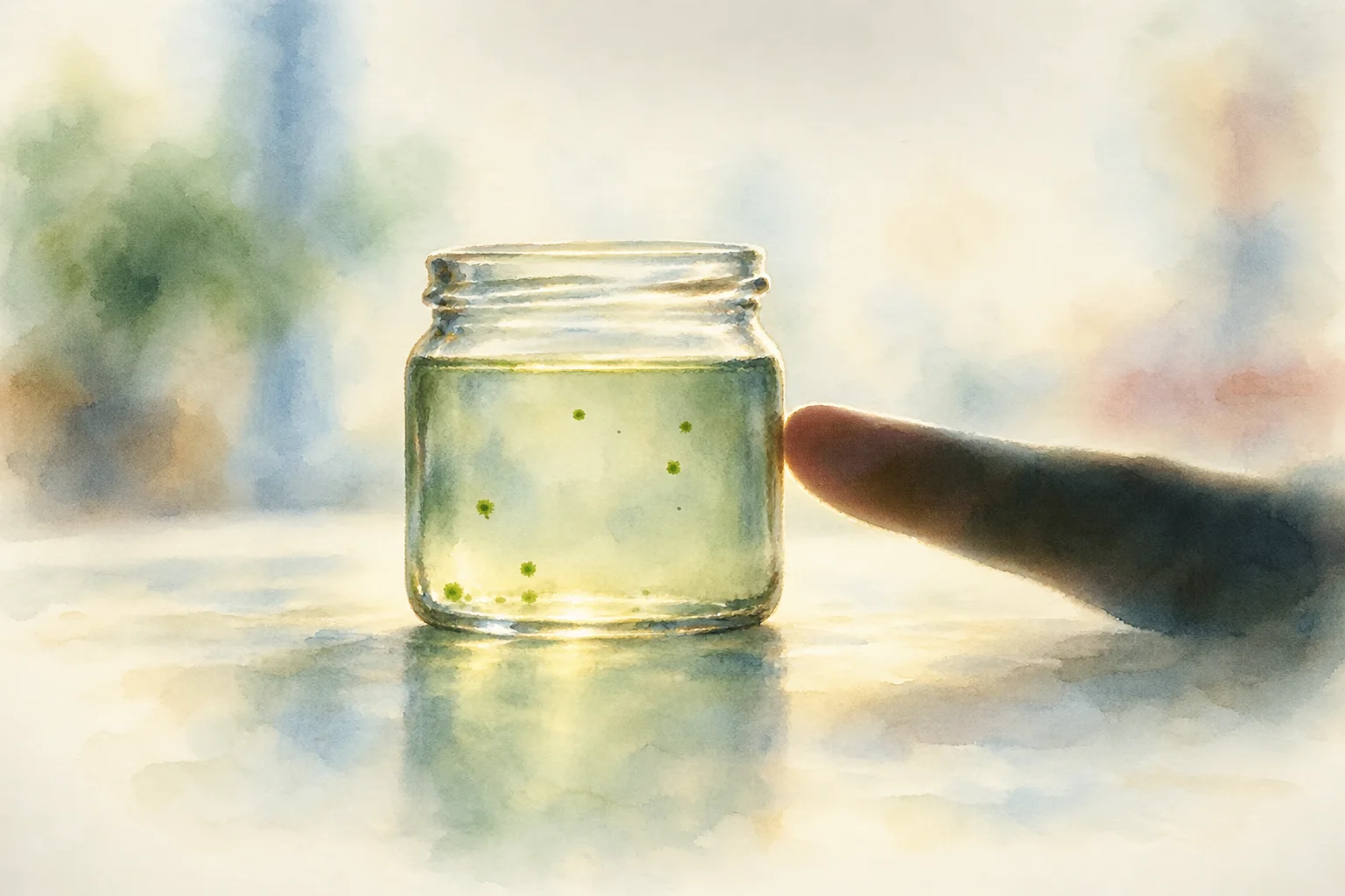
Illustration: a glass jar of green-tinged water with a fingertip touching its side, not to scale; a few green specks float at different heights. Illustration

Field-guide entry[10−4 m]

Volvox: Each Embryo Turns Inside Out Before It Can Swim

Volvox spheres reach 0.35–0.92 mm in seven species descriptions. Each starts as an embryo with flagellar ends facing in and turns inside out before it swims.

Image: Illustration: Ants, Cells & Atoms, all rights reserved

Illustration: Ants, Cells & Atoms. Generated schematic illustration — not a photograph, micrograph or specimen record. © Ants, Cells & Atoms, all rights reserved.
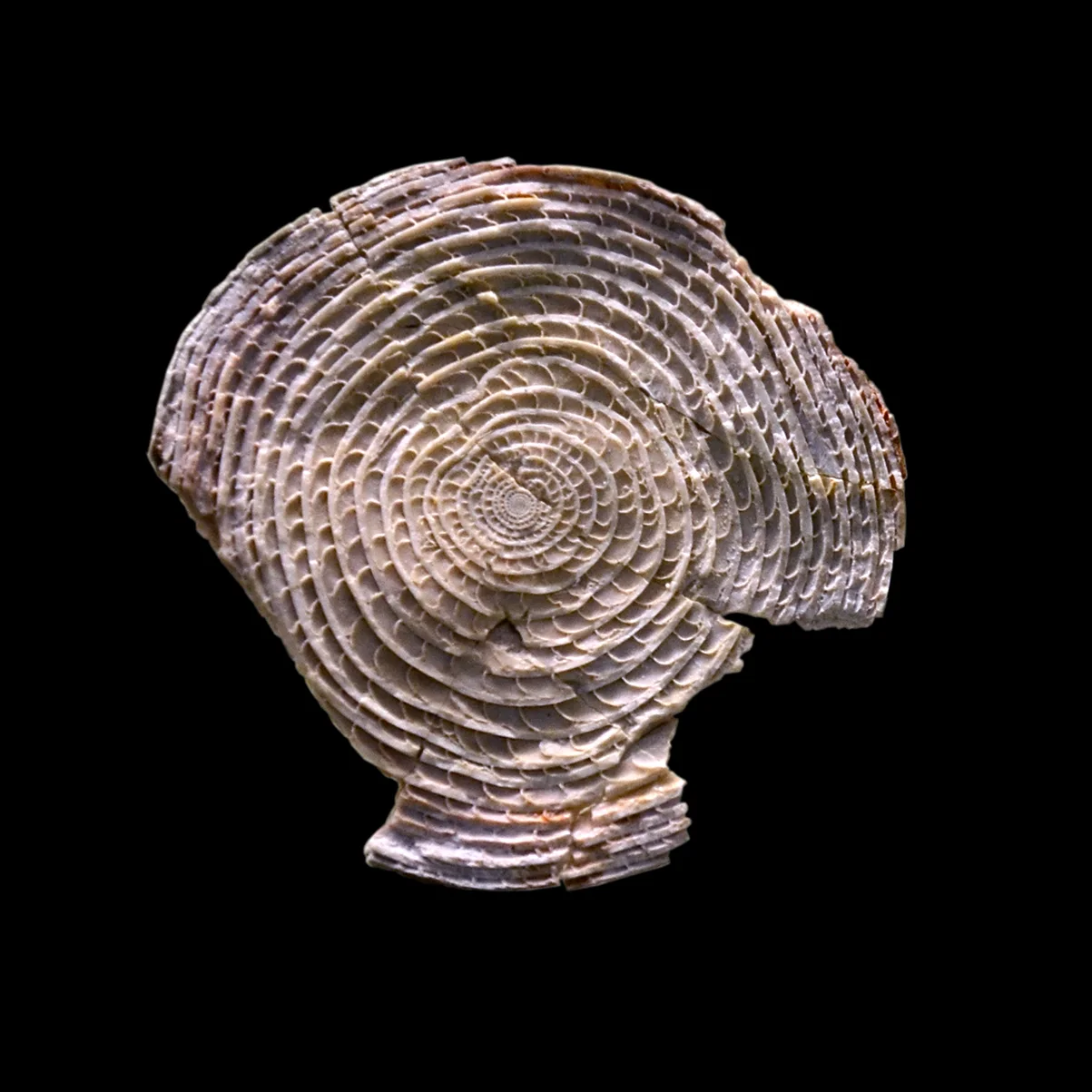
Fossil Nummulites puschi shell; the genus has 5 and 30 mm tests in one pyramid block; museum photo, natural color; a tight spiral of small chambers.

Field-guide entry[10−4–10−3 m]

Foraminifera: Single Cells Known for Shells, Some Coin-Sized

Foraminifera are single cells known for their shells. One drifting species has adult shells of 150–1,500 µm; fossil Nummulites in a pyramid block reach 30 mm.

Image: “Nummulite Naturhistorisches Museum Basel 27102013” by Vassil (2013) · CC0 1.0

"Nummulite Naturhistorisches Museum Basel 27102013" by Vassil — https://commons.wikimedia.org/wiki/File:Nummulite_Naturhistorisches_Museum_Basel_27102013.jpg — CC0 1.0 (https://creativecommons.org/publicdomain/zero/1.0/)

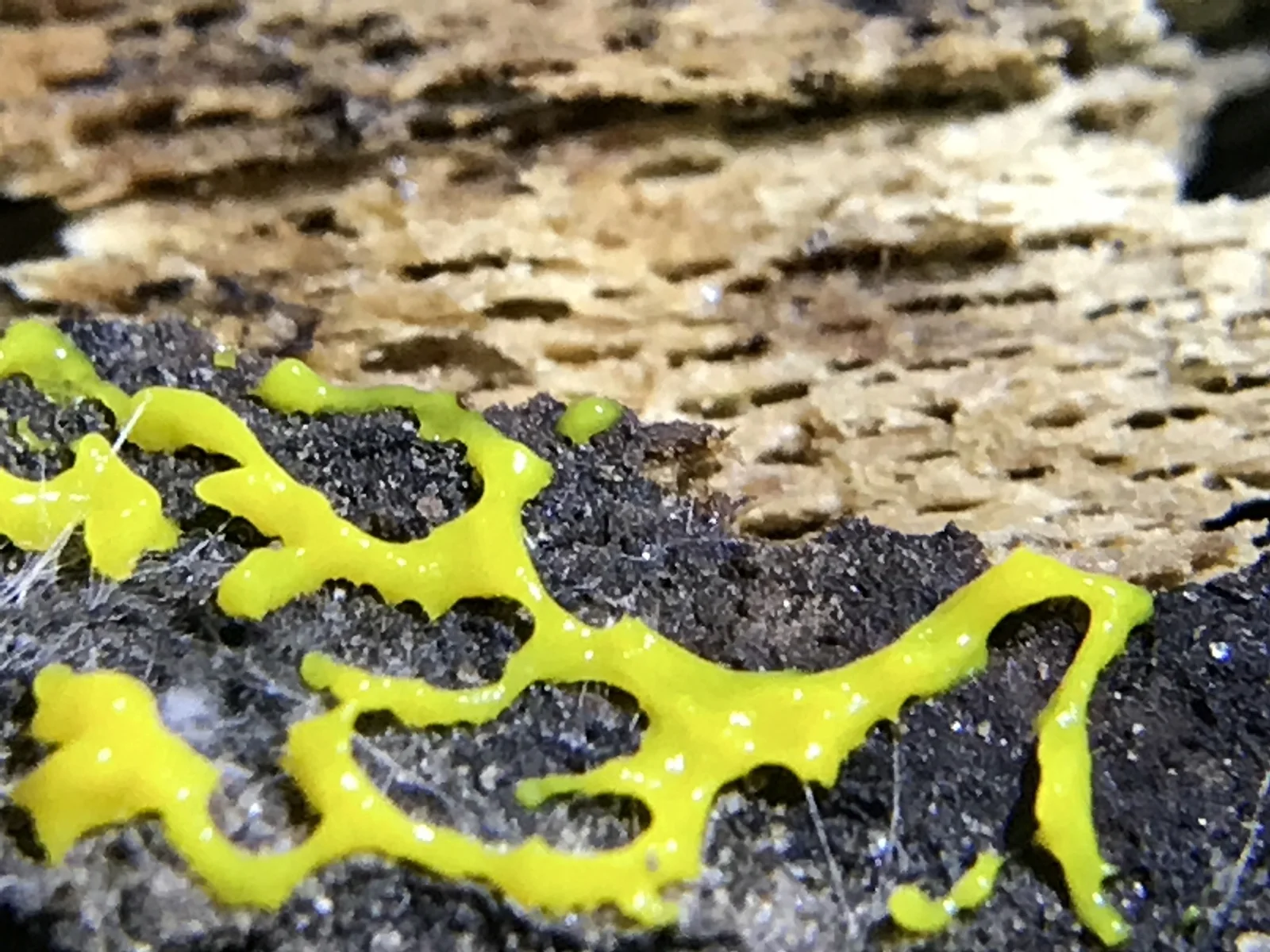
Physarum polycephalum plasmodium, one cell that can span tens of cm; close-up photo, native color; yellow branching strands on dark ground.

Field-guide entry[10−3–10−1 m]

Slime Mold: Not a Mold, and Its Body Can Be a Single Cell

Slime molds are not fungi. In Physarum polycephalum the feeding stage is one cell with up to millions of nuclei, its insides flowing back and forth in veins.

Image: “Physarum polycephalum 9986808” by jhubley (2017) · CC0 1.0

"Physarum polycephalum 9986808" by jhubley — https://commons.wikimedia.org/wiki/File:Physarum_polycephalum_9986808.jpg — CC0 1.0 (https://creativecommons.org/publicdomain/zero/1.0/)

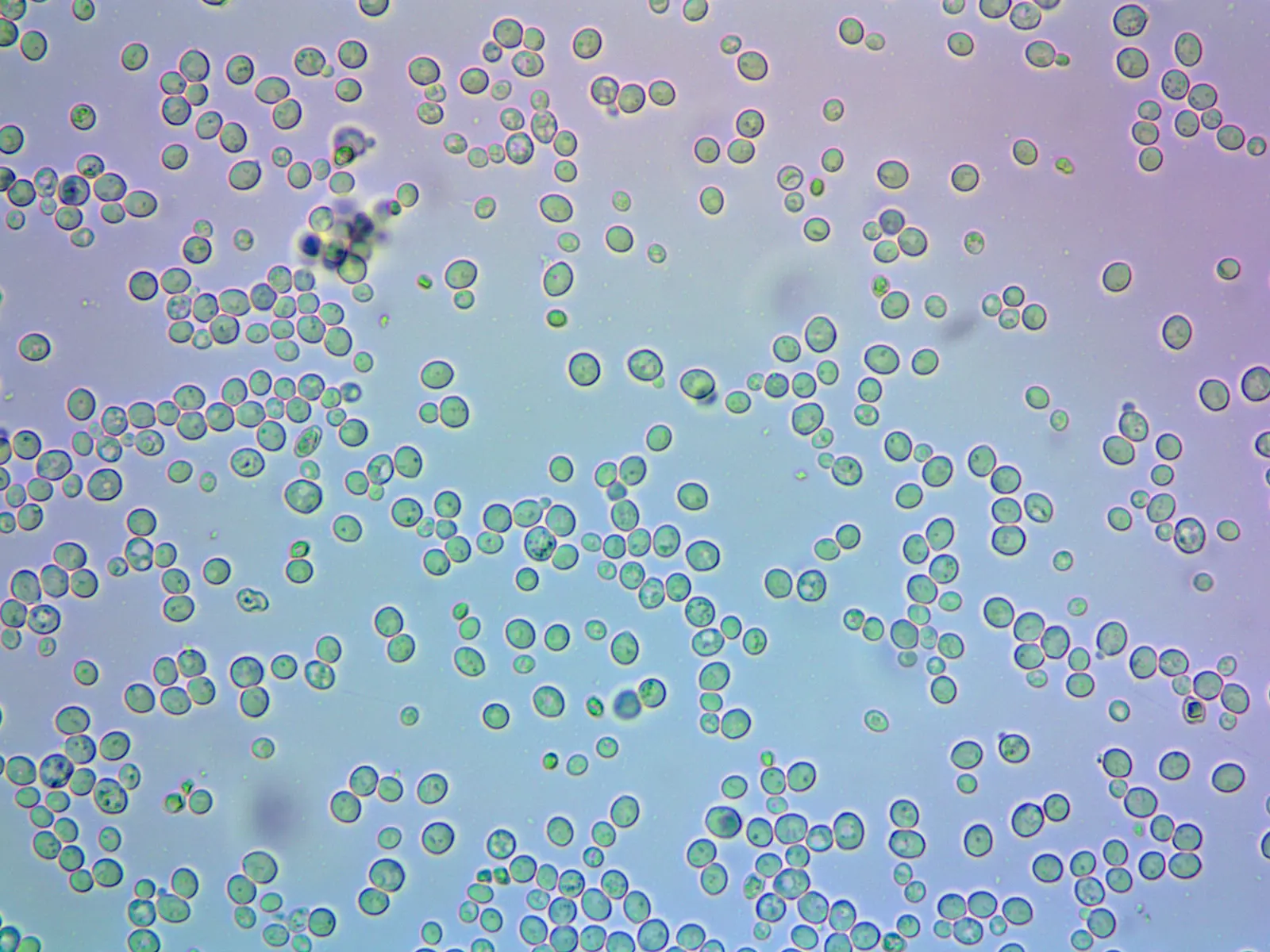
Compressed baker's yeast cells; cells of one lab strain run 3.6–11.0 µm; light micrograph, no stain stated; round cells, a few with small buds.

Field-guide entry[10−6–10−5 m]

Baker's Yeast: Each Bud Leaves a Scar on the Mother Cell

Baker's yeast cells run 3.6–11.0 µm across in one lab strain. Each bud leaves a ring scar of chitin on the mother, so the scars count its daughters.

Image: “Дрожжевые клетки без воздействия различных веществ (Yeast cells without exposure to various substances)” by Marina taikevich (2017) · CC BY 4.0

"Дрожжевые клетки без воздействия различных веществ (Yeast cells without exposure to various substances)" by Marina taikevich — https://commons.wikimedia.org/wiki/File:Дрожжевые_клетки_без_воздействия_различных_веществ.jpg — CC BY 4.0 (https://creativecommons.org/licenses/by/4.0/)

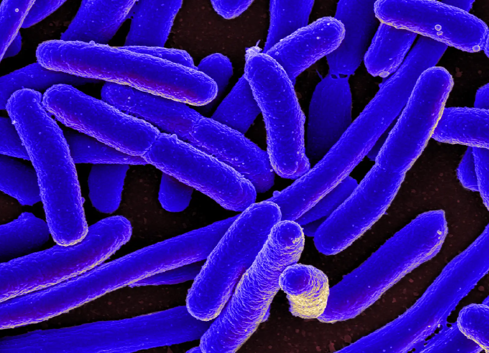
Escherichia coli, rods averaging 1.0–1.5 µm wide in one culture study, scanning electron micrograph colored afterwards; violet-blue on dark brown.

Mechanism explainer[10−5–10−4 m]

Borrowed Color: When a Micrograph's Hue Isn't the Cell's Own

Borrowed color is color in a micrograph that does not belong to the organism: a stain, a fluorescent marker, or false color added to a grey image.

Image: “E. coli Bacteria (16578744517)” by NIAID (2013) · CC BY 2.0

"E. coli Bacteria (16578744517)" by NIAID — https://commons.wikimedia.org/wiki/File:E._coli_Bacteria_(16578744517).jpg — CC BY 2.0 (https://creativecommons.org/licenses/by/2.0/)