Bacteriophage T4: About 57 µm of DNA in a Head 0.12 µm Long

Slide the ladder sideways to read its labels, or open it full size.
The head of bacteriophage T4: 86 nm wide and 120 nm long [10−8–10−7 m], with an E. coli cell's width, 1.0–1.5 µm, and T4's genome stretched out, about 57 µm, for scale.
Log scale from 10 cm to 0.1 nm, each rung ten times smaller; dashed lines: naked eye about 0.1 mm, light microscope about 0.2 µm (How to read the Zoom ladder). The DNA length is computed for this page. Data: bacteriophage-dimensions.csv (sources, version and license, JSON), bacteriophage-dna-packing.csv (sources, version and license, JSON).
"Zoom ladder: phage T4" by Ants, Cells & Atoms, https://antscellsandatoms.com/microscopic-life/bacteriophage/ — CC BY 4.0 (https://creativecommons.org/licenses/by/4.0/)
Data sources for this figure
Data: Head size: Rao VB, Black LW 2010, Virology Journal 7:356, CC BY 2.0, https://doi.org/10.1186/1743-422X-7-356 (retrieved 2026-10-25); Escherichia coli width 1.0–1.5 µm: Volkmer B, Heinemann M 2011, PLoS ONE 6(7):e23126, CC BY 4.0, https://doi.org/10.1371/journal.pone.0023126, via the shared cell-sizes.csv (retrieved 2026-10-25); Genome stretched out, about 57 µm: 171 kb (Rao and Black 2010) x 0.335 nm per base pair (RCSB PDB 1BNA, CC0, https://doi.org/10.2210/pdb1bna/pdb, as computed on the How Long Is DNA? page), computed 2026-10-25
Data sources for this figure
Data: Rao VB, Black LW 2010, Virology Journal 7:356, CC BY 2.0, https://doi.org/10.1186/1743-422X-7-356 (T4 head 120 nm long, 86 nm wide; retrieved 2026-10-25); Leiman PG, Arisaka F, van Raaij MJ, Kostyuchenko VA, Aksyuk AA, Kanamaru S, Rossmann MG 2010, Virology Journal 7:355, CC BY 2.0, https://doi.org/10.1186/1743-422X-7-355 (sheath, tube, baseplate and fibre sizes in Å, converted to nm; retrieved 2026-10-25); Yap ML, Rossmann MG 2014, Future Microbiology 9(12):1319–1327, https://doi.org/10.2217/fmb.14.91 (abstract: 925 Å tail; retrieved 2026-10-25); Smith DE et al. 2001, Nature 413:748–752, https://doi.org/10.1038/35099581 (abstract: phi29 capsid 42 x 54 nm; retrieved 2026-10-25); Lin YR, Lin CS 2012, BMC Genomics 13:224, CC BY 2.0, https://doi.org/10.1186/1471-2164-13-224 (phipp2 head and tail sizes; retrieved 2026-10-25); Valli J et al. 2021, Journal of Biological Chemistry 297(1):100791, CC BY 4.0, https://doi.org/10.1016/j.jbc.2021.100791 (light-microscope limit 170–250 nm, via the shared reference-objects.csv; retrieved 2026-10-25); Volkmer B, Heinemann M 2011, PLoS ONE 6(7):e23126, CC BY 4.0, https://doi.org/10.1371/journal.pone.0023126; Young KD in Marshall WF et al. 2012, BMC Biology 10:101, CC BY 2.0, https://doi.org/10.1186/1741-7007-10-101; Luef B et al. 2015, Nature Communications 6:6372, https://doi.org/10.1038/ncomms7372 (abstract) (bacterial sizes, via the shared cell-sizes.csv; retrieved 2026-10-25); unit conversions and comparison ratios computed 2026-10-25 for this page
Data sources for this figure
Data: Rao VB, Black LW 2010, Virology Journal 7:356, CC BY 2.0, https://doi.org/10.1186/1743-422X-7-356 (171 kb genome, full head about 1.02 genome lengths, about 500 mg/ml at 2.5 nm spacing, about 60 pN, up to about 2000 bp/s, about 5 minutes; retrieved 2026-10-25); Lin YR, Lin CS 2012, BMC Genomics 13:224, CC BY 2.0, https://doi.org/10.1186/1471-2164-13-224 (T4 genome 168,903 bp, GenBank NC_000866 as cited; retrieved 2026-10-25); Yap ML, Rossmann MG 2014, Future Microbiology 9(12):1319–1327, https://doi.org/10.2217/fmb.14.91 (abstract: 172 kbp; retrieved 2026-10-25); Smith DE et al. 2001, Nature 413:748–752, https://doi.org/10.1038/35099581 (abstract: phi29 DNA 6.6 µm in a 42 x 54 nm capsid, up to 57 pN, about 50 pN internal force; retrieved 2026-10-25); Evilevitch A et al. 2003, Proceedings of the National Academy of Sciences 100(16):9292–9295, https://doi.org/10.1073/pnas.1233721100 (abstract: 20 atmospheres stop lambda DNA ejection; retrieved 2026-10-25); RCSB PDB 1BNA, CC0, coordinates from Drew HR et al. 1981, https://doi.org/10.2210/pdb1bna/pdb (rise per base pair 0.335 nm, band 0.333–0.347 nm, computed on the How Long Is DNA? page; retrieved 2026-10-25); Nurk S et al. 2022, Science 376(6588):44–53, CC BY 4.0, https://doi.org/10.1126/science.abj6987, and Paulson JR et al. 2021, Seminars in Cell and Developmental Biology 117:7–29, CC BY 4.0, https://doi.org/10.1016/j.semcdb.2021.03.014 (human DNA per nucleated cell and a nucleus about 10 µm across, as on the How Long Is DNA? page; retrieved 2026-10-25); lengths and ratios computed 2026-10-25 for this page
Bench card
- Name
- bacteriophage (phage), a virus that infects bacteria; featured here: phage T4, species Tequatrovirus T4 (formerly Escherichia virus T4)Source: ICTV Master Species List 2025 (MSL41), https://doi.org/10.5281/zenodo.19154110; GBIF Backbone Taxonomy 12252006 and 9977478, https://doi.org/10.15468/39omei
- Actual size
- phage T4: head 86 nm wide and 120 nm long [10−8–10−7 m]; tail sheath 24 nm wide and 92.5 nm long before it contractsSource: Rao and Black 2010, https://doi.org/10.1186/1743-422X-7-356; Leiman et al. 2010, https://doi.org/10.1186/1743-422X-7-355; bacteriophage-dimensions.csv on this page
- Size in this image
- 100 nm scale bar printed on the opening micrograph, which shows phage ϕpp2, a T4-like phage of Vibrio, not T4; its authors measured its head at about 90–95 nm by 150–160 nmSource: Lin and Lin 2012, https://doi.org/10.1186/1471-2164-13-224
- Lives in
- seawater and other natural waters, alongside the bacteria it infectsSource: Mann 2005, https://doi.org/10.1371/journal.pbio.0030182; Bergh et al. 1989 (abstract), https://doi.org/10.1038/340467a0
- Seen with
- electron microscope for shape and size; in seawater stained with a fluorescent dye, a standard light microscope shows virus particles only as points of lightSource: Mann 2005, https://doi.org/10.1371/journal.pbio.0030182; Valli et al. 2021, https://doi.org/10.1016/j.jbc.2021.100791; figure captions on this page
- Color in images
- native in the electron micrographs (no color); fluorescence (SYBR Green) in the seawater imageSource: figure captions on this page
bacteriophage-bench.csv (CSV) · where each value comes from, and its license (JSON)
Data credits for this card
Data: International Committee on Taxonomy of Viruses, ICTV Master Species List 2025 (MSL41), version 1, CC BY 4.0, https://doi.org/10.5281/zenodo.19154110 (Tequatrovirus T4; retrieved 2026-10-25); GBIF Secretariat 2023, GBIF Backbone Taxonomy, Caudoviricetes (10794457), Tequatrovirus T4 (12252006), Escherichia virus T4 (9977478), checklist dataset https://doi.org/10.15468/39omei, CC BY 4.0, retrieved 2026-10-25; Rao VB, Black LW 2010, Virology Journal 7:356, CC BY 2.0, https://doi.org/10.1186/1743-422X-7-356; Leiman PG et al. 2010, Virology Journal 7:355, CC BY 2.0, https://doi.org/10.1186/1743-422X-7-355 (T4 head and sheath; retrieved 2026-10-25); Lin YR, Lin CS 2012, BMC Genomics 13:224, CC BY 2.0, https://doi.org/10.1186/1471-2164-13-224 (phipp2 head size; retrieved 2026-10-25); Mann NH 2005, PLoS Biology 3(5):e182, CC BY 4.0, https://doi.org/10.1371/journal.pbio.0030182; Bergh Ø et al. 1989, Nature 340:467–468, https://doi.org/10.1038/340467a0 (abstract); Valli J et al. 2021, Journal of Biological Chemistry 297(1):100791, CC BY 4.0, https://doi.org/10.1016/j.jbc.2021.100791 (habitat and seeing; retrieved 2026-10-25)
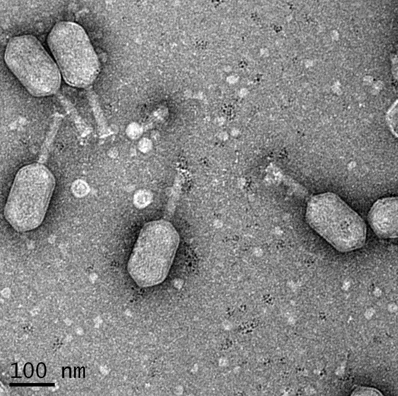
Phage ϕpp2, a T4-like phage that infects Vibrio bacteria, not T4 itself: heads about 90–95 nm wide and 150–160 nm long [10−8–10−7 m], beside a printed 100 nm bar.
Color: native (electron micrograph, no color)
"Phage.phiPP2" by Lin YR, Lin CS — https://commons.wikimedia.org/wiki/File:Phage.phiPP2.jpeg — CC BY 2.0 (https://creativecommons.org/licenses/by/2.0/)

How big is a bacteriophage?
Sizes vary from one phage to another, so this page follows one of them, phage T4. Rao and Black's 2010 review describes the phage's head as an elongated, or prolate, icosahedron, a many-faced shell drawn out along one axis, 120 nm long and 86 nm wide. The tail parts below come from Leiman and colleagues' 2010 review. Neither review gives the neck's length, so no total length appears here.
| Part of phage T4 | Size | Source |
|---|---|---|
| head, an elongated icosahedron | 120 nm long, 86 nm wide | Rao and Black 2010 |
| collar around the neck | 30 nm across, 4 nm thick | Leiman et al. 2010 |
| tail sheath, before contraction | 92.5 nm long, 24 nm wide, 23 rings | Leiman et al. 2010 |
| tail tube, inside the sheath | 9 nm across, 4 nm channel | Leiman et al. 2010 |
| baseplate, before contraction | about 52 nm across, about 27 nm high | Leiman et al. 2010 |
| one long tail fibre | about 144 nm, kinked | Leiman et al. 2010 |
| neck | length not given | — |
The micrograph above shows not T4 but ϕpp2, a T4-like phage that infects Vibrio bacteria. One of its hosts in Lin and Lin's 2012 study is a Vibrio species widely distributed in coastal waters, and they measured the phage's heads at 150–160 nm long, longer than T4's.
Names have shifted too. In a 2022 update reported by Turner and colleagues, the International Committee on Taxonomy of Viruses renamed T4's species Tequatrovirus T4 (formerly Escherichia virus T4), the name in its 2025 list, and dropped families based on tail shape, such as Myoviridae. Tailed phages with double-stranded DNA now sit in the class Caudoviricetes. As of its 2023 records, GBIF's backbone still files both names under Myoviridae.
Next to its host, T4 is small. Cultured Escherichia coli cells measure 1.0–1.5 µm wide across 22 growth conditions (Volkmer and Heinemann 2011), about 8–13 T4 head lengths, computed for this page. Even the smallest cells on the How Big Is a Cell? page are larger: the marine SAR11 bacteria, which Young puts at 0.2 µm, and ultra-small groundwater bacteria, 0.24–0.28 µm as spheres of equal volume, computed there from Luef and colleagues' 2015 volumes (abstract). Those are about 1.7 and 2.0–2.3 head lengths, computed for this page.
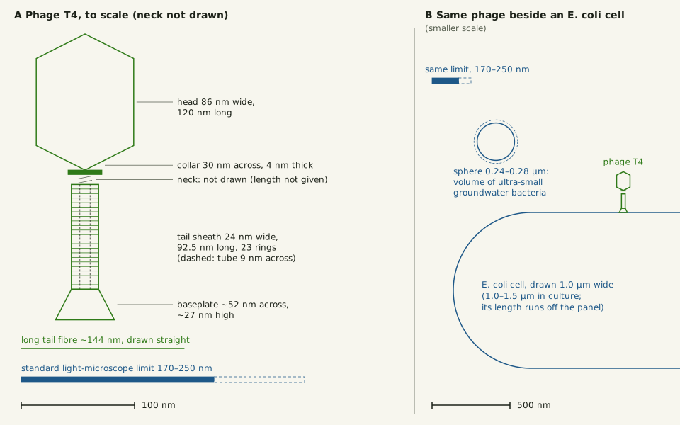
Slide the diagram sideways to read its labels, or open it full size.
Phage T4 drawn to scale: head 86 × 120 nm and tail sheath 24 × 92.5 nm [10−8–10−7 m], both below the 170–250 nm limit of a standard light microscope.
Panel B shows the same phage at a smaller scale beside an E. coli cell 1.0–1.5 µm wide. The neck is not drawn, because its length is not given in the sources used here. Data: bacteriophage-dimensions.csv (sources, version and license, JSON).
"Phage T4 to scale, beside an E. coli cell and the light-microscope limit" by Ants, Cells & Atoms, https://antscellsandatoms.com/microscopic-life/bacteriophage/ — CC BY 4.0 (https://creativecommons.org/licenses/by/4.0/)
Data sources for this figure
Data: Head: Rao VB, Black LW 2010, Virology Journal 7:356, CC BY 2.0, https://doi.org/10.1186/1743-422X-7-356 (retrieved 2026-10-25); Collar, tail sheath, tube, baseplate and long tail fibre: Leiman PG, Arisaka F, van Raaij MJ, Kostyuchenko VA, Aksyuk AA, Kanamaru S, Rossmann MG 2010, Virology Journal 7:355, CC BY 2.0, https://doi.org/10.1186/1743-422X-7-355 (retrieved 2026-10-25); Light-microscope limit 170–250 nm: Valli J et al. 2021, Journal of Biological Chemistry 297(1):100791, CC BY 4.0, https://doi.org/10.1016/j.jbc.2021.100791, via the shared reference-objects.csv (retrieved 2026-10-25); Escherichia coli width: Volkmer B, Heinemann M 2011, PLoS ONE 6(7):e23126, CC BY 4.0, https://doi.org/10.1371/journal.pone.0023126; ultra-small groundwater bacteria, equivalent-sphere diameter 0.24–0.28 µm computed on the How Big Is a Cell? page from Luef B et al. 2015, Nature Communications 6:6372, https://doi.org/10.1038/ncomms7372 (abstract); both via the shared cell-sizes.csv (retrieved 2026-10-25)
Data sources for this figure
Data: Rao VB, Black LW 2010, Virology Journal 7:356, CC BY 2.0, https://doi.org/10.1186/1743-422X-7-356 (T4 head 120 nm long, 86 nm wide; retrieved 2026-10-25); Leiman PG, Arisaka F, van Raaij MJ, Kostyuchenko VA, Aksyuk AA, Kanamaru S, Rossmann MG 2010, Virology Journal 7:355, CC BY 2.0, https://doi.org/10.1186/1743-422X-7-355 (sheath, tube, baseplate and fibre sizes in Å, converted to nm; retrieved 2026-10-25); Yap ML, Rossmann MG 2014, Future Microbiology 9(12):1319–1327, https://doi.org/10.2217/fmb.14.91 (abstract: 925 Å tail; retrieved 2026-10-25); Smith DE et al. 2001, Nature 413:748–752, https://doi.org/10.1038/35099581 (abstract: phi29 capsid 42 x 54 nm; retrieved 2026-10-25); Lin YR, Lin CS 2012, BMC Genomics 13:224, CC BY 2.0, https://doi.org/10.1186/1471-2164-13-224 (phipp2 head and tail sizes; retrieved 2026-10-25); Valli J et al. 2021, Journal of Biological Chemistry 297(1):100791, CC BY 4.0, https://doi.org/10.1016/j.jbc.2021.100791 (light-microscope limit 170–250 nm, via the shared reference-objects.csv; retrieved 2026-10-25); Volkmer B, Heinemann M 2011, PLoS ONE 6(7):e23126, CC BY 4.0, https://doi.org/10.1371/journal.pone.0023126; Young KD in Marshall WF et al. 2012, BMC Biology 10:101, CC BY 2.0, https://doi.org/10.1186/1741-7007-10-101; Luef B et al. 2015, Nature Communications 6:6372, https://doi.org/10.1038/ncomms7372 (abstract) (bacterial sizes, via the shared cell-sizes.csv; retrieved 2026-10-25); unit conversions and comparison ratios computed 2026-10-25 for this page
Can a light microscope see a phage?
Not its shape, at least through standard optics. The wave nature of light limits the finest detail a microscope can separate, and Abbe showed that the limit grows with the wavelength. For a standard fluorescence microscope detecting light of 470–700 nm, Valli and colleagues' 2021 review puts it at around 170–250 nm; two points closer than that blur into one spot. That is 2.0–2.9 times the width of T4's 86 nm head, computed for this page, and longer than the head's 120 nm length.
Super-resolution light microscopes do get below it, the same review notes, from about 120 nm with pixel-reassignment methods to a reported 1 nm with a technique called MINFLUX.
A standard microscope can still show that phages are there. In seawater stained with the dye SYBR Green, an epifluorescence microscope shows bacterial cells as larger dots and virus particles as smaller ones, in an image by Jed Fuhrman printed in Mann's review. That green is borrowed color, added by the dye rather than coming from the particles. Nor do the dots give sizes: light from even a single glowing molecule still spreads to the diffraction limit, Valli and colleagues explain.
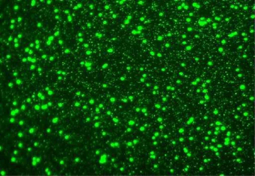
Seawater stained with SYBR Green under an epifluorescence microscope: larger dots are bacterial cells, smaller points virus particles; a standard light microscope blurs detail finer than 170–250 nm [10−7 m].
Color: fluorescence (SYBR Green)
"Seawater small life" by Jed Fuhrman — https://commons.wikimedia.org/wiki/File:Seawater_small_life.png — CC BY 2.5 (https://creativecommons.org/licenses/by/2.5/)

Shapes came with the electron microscope. The first phage electron micrographs were published in 1940 in Germany, Ackermann recounts in a 2011 history: H. Ruska showed phages mixed with bacteria at 14,000 times, and Pfankuch and Kausche, of the same Siemens & Halske laboratory, saw round particles 60 nm across at 22–25,000 times. Negative staining followed in 1959, introduced by Brenner and Horne: particles show pale against a darker stain, uranyl acetate in both electron micrographs here. By Ackermann's 2007 count, at least 5,568 phages had been examined by electron microscope since then, 96% of them with tails (abstract).
How does 57 µm of DNA fit in a 120 nm head?
Rao and Black give T4's genome as 171 kb, about 171,000 base pairs. At 0.335 nm per base pair, the spacing the How Long Is DNA? page computes from a B-DNA crystal (Protein Data Bank entry 1BNA), that is about 57.3 µm, computed for this page.
Other counts land close: the sequenced genome, 168,903 bp in GenBank as Lin and Lin cite it, gives 56.6 µm, and the 172 kbp in Yap and Rossmann's 2014 abstract gives 57.6 µm. All three use the same spacing. So they differ only in base pairs. A full head holds about 1.02 genome lengths, Rao and Black write: about 58 µm, computed for this page.
That genome runs about 480 head lengths, computed for this page, a ratio of lengths rather than a packing density. Phage φ29 packs 6.6 µm of DNA into a capsid, or head shell, 42 × 54 nm, Smith and colleagues reported in 2001 (abstract). That is about 120 times the capsid's longer side, computed for this page. By the How Long Is DNA? page's figures, a nucleated human cell keeps about 2 m in a nucleus about 10 µm across, some 200,000 times its width.
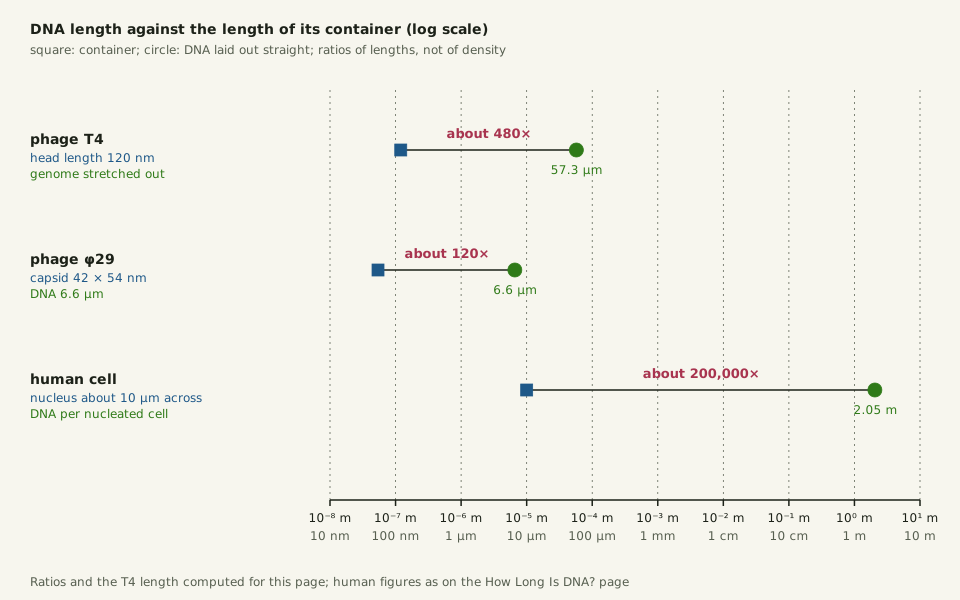
Slide the diagram sideways to read its labels, or open it full size.
Phage T4's genome, about 57 µm [10−5 m], against its 120 nm head, beside phage φ29 and a human nucleus; ratios of lengths, not of packing density.
Log scale. The T4 length and all three ratios are computed for this page; the human figures are those of the How Long Is DNA? page. Data: bacteriophage-dna-packing.csv (sources, version and license, JSON).
"DNA length against the length of its container" by Ants, Cells & Atoms, https://antscellsandatoms.com/microscopic-life/bacteriophage/ — CC BY 4.0 (https://creativecommons.org/licenses/by/4.0/)
Data sources for this figure
Data: Phage T4 genome and head: Rao VB, Black LW 2010, Virology Journal 7:356, CC BY 2.0, https://doi.org/10.1186/1743-422X-7-356 (retrieved 2026-10-25); rise per base pair from RCSB PDB 1BNA, CC0, https://doi.org/10.2210/pdb1bna/pdb, as computed on the How Long Is DNA? page; Phage phi29: Smith DE, Tans SJ, Smith SB, Grimes S, Anderson DL, Bustamante C 2001, Nature 413:748–752, https://doi.org/10.1038/35099581 (abstract; retrieved 2026-10-25); Human DNA per nucleated cell and a nucleus about 10 µm across: Nurk S et al. 2022, Science 376(6588):44–53, CC BY 4.0, https://doi.org/10.1126/science.abj6987, and Paulson JR et al. 2021, Seminars in Cell and Developmental Biology 117:7–29, CC BY 4.0, https://doi.org/10.1016/j.semcdb.2021.03.014, as on the How Long Is DNA? page (retrieved 2026-10-25); T4 length and the three ratios computed 2026-10-25 for this page
Data sources for this figure
Data: Rao VB, Black LW 2010, Virology Journal 7:356, CC BY 2.0, https://doi.org/10.1186/1743-422X-7-356 (171 kb genome, full head about 1.02 genome lengths, about 500 mg/ml at 2.5 nm spacing, about 60 pN, up to about 2000 bp/s, about 5 minutes; retrieved 2026-10-25); Lin YR, Lin CS 2012, BMC Genomics 13:224, CC BY 2.0, https://doi.org/10.1186/1471-2164-13-224 (T4 genome 168,903 bp, GenBank NC_000866 as cited; retrieved 2026-10-25); Yap ML, Rossmann MG 2014, Future Microbiology 9(12):1319–1327, https://doi.org/10.2217/fmb.14.91 (abstract: 172 kbp; retrieved 2026-10-25); Smith DE et al. 2001, Nature 413:748–752, https://doi.org/10.1038/35099581 (abstract: phi29 DNA 6.6 µm in a 42 x 54 nm capsid, up to 57 pN, about 50 pN internal force; retrieved 2026-10-25); Evilevitch A et al. 2003, Proceedings of the National Academy of Sciences 100(16):9292–9295, https://doi.org/10.1073/pnas.1233721100 (abstract: 20 atmospheres stop lambda DNA ejection; retrieved 2026-10-25); RCSB PDB 1BNA, CC0, coordinates from Drew HR et al. 1981, https://doi.org/10.2210/pdb1bna/pdb (rise per base pair 0.335 nm, band 0.333–0.347 nm, computed on the How Long Is DNA? page; retrieved 2026-10-25); Nurk S et al. 2022, Science 376(6588):44–53, CC BY 4.0, https://doi.org/10.1126/science.abj6987, and Paulson JR et al. 2021, Seminars in Cell and Developmental Biology 117:7–29, CC BY 4.0, https://doi.org/10.1016/j.semcdb.2021.03.014 (human DNA per nucleated cell and a nucleus about 10 µm across, as on the How Long Is DNA? page; retrieved 2026-10-25); lengths and ratios computed 2026-10-25 for this page
Inside T4 and other tailed phages with double-stranded DNA, the strands lie 2.5 nm apart at about 500 mg/ml, Rao and Black write. Packing DNA to near-crystalline density means overcoming its entropy, electrostatic repulsion and stiffness against bending, Smith and colleagues wrote of φ29 (abstract). In a 2026 review, Mateu notes that DNA's persistence length, a measure of that stiffness, is comparable to a capsid's diameter.
T4's motor, powered by ATP, pushes with up to about 60 pN (piconewtons), close to the loads of up to 57 pN on average that φ29's motor worked against (abstract). It feeds in up to about 2,000 base pairs a second, the fastest packaging motor reported as of 2010. Rao and Black put the whole genome at about 5 minutes, an average of about 570 base pairs a second, computed for this page.
How does a phage get its DNA into a bacterium?
Packed that tightly, the particle is under pressure. When double-stranded DNA is forced into a ready-made shell this way, the pressure inside may reach tens of atmospheres, Mateu's review says. The review adds that experiments show the pressure greatly helps inject the genome into the bacterial cell: lowering phage λ's pressure by shortening its DNA made release take more energy. In λ capsids opened in a test tube, Evilevitch and colleagues found that an osmotic pressure like that in the host's cytoplasm halved DNA ejection, and 20 atmospheres stopped it (abstract).
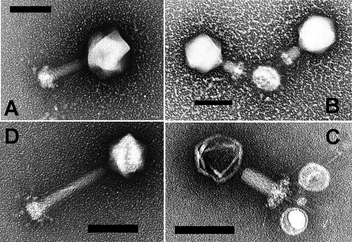
Phages P-SSM2 and P-SSM4 of the marine cyanobacterium Prochlorococcus, T4-like, though their heads are isometric rather than elongated: tails extended (A, D) and contracted (B, C), each panel with its own 100 nm bar [10−7 m].
Color: native (electron micrograph, no color)
"Cyanophages" by Sullivan MB, Coleman ML, Weigele P, Rohwer F, Chisholm SW — https://commons.wikimedia.org/wiki/File:Cyanophages.png — CC BY 2.5 (https://creativecommons.org/licenses/by/2.5/)

The bacteria these double-stranded DNA phages infect have a thick wall that is hard to get through, Mateu notes. By the same account, the phages inject their genome without entering the cell. Leiman and colleagues describe T4's long tail fibres as extended in solution, probably jiggling with thermal motion; once three or more bind receptors, they hold the phage perpendicular to the surface and, acting as levers, move the baseplate up to 100 nm toward it. Imaging T4 on E. coli minicells in 2015, Hu and colleagues instead found most long fibres folded back along the sheath until the phage attached for good (abstract).
Then the tail contracts, irreversibly, starting at the baseplate, which turns from a hexagon about 52 nm across into a star 61 nm across. The sheath shortens from 92.5 to 42 nm, to about 45% of its length (computed for this page), and widens from 24 to 33 nm. That drives the tube through the cell envelope, rotating almost a full turn, so in Leiman's account it drills rather than punctures the outer membrane. An enzyme in the tip protein gp5 digests the wall's peptidoglycan.
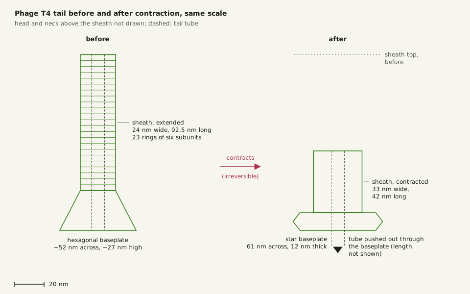
Slide the diagram sideways to read its labels, or open it full size.
Phage T4's tail before and after contraction, at one scale: sheath 24 × 92.5 nm, then 33 × 42 nm [10−8 m].
Head and neck not drawn; the tube's length after contraction is not shown. Data: bacteriophage-dimensions.csv (sources, version and license, JSON).
"Phage T4 tail before and after contraction" by Ants, Cells & Atoms, https://antscellsandatoms.com/microscopic-life/bacteriophage/ — CC BY 4.0 (https://creativecommons.org/licenses/by/4.0/)
Data: Sheath, tube and baseplate before and after contraction: Leiman PG, Arisaka F, van Raaij MJ, Kostyuchenko VA, Aksyuk AA, Kanamaru S, Rossmann MG 2010, Virology Journal 7:355, CC BY 2.0, https://doi.org/10.1186/1743-422X-7-355 (retrieved 2026-10-25); Shortening ratio computed 2026-10-25 for this page
Data sources for this figure
Data: Rao VB, Black LW 2010, Virology Journal 7:356, CC BY 2.0, https://doi.org/10.1186/1743-422X-7-356 (T4 head 120 nm long, 86 nm wide; retrieved 2026-10-25); Leiman PG, Arisaka F, van Raaij MJ, Kostyuchenko VA, Aksyuk AA, Kanamaru S, Rossmann MG 2010, Virology Journal 7:355, CC BY 2.0, https://doi.org/10.1186/1743-422X-7-355 (sheath, tube, baseplate and fibre sizes in Å, converted to nm; retrieved 2026-10-25); Yap ML, Rossmann MG 2014, Future Microbiology 9(12):1319–1327, https://doi.org/10.2217/fmb.14.91 (abstract: 925 Å tail; retrieved 2026-10-25); Smith DE et al. 2001, Nature 413:748–752, https://doi.org/10.1038/35099581 (abstract: phi29 capsid 42 x 54 nm; retrieved 2026-10-25); Lin YR, Lin CS 2012, BMC Genomics 13:224, CC BY 2.0, https://doi.org/10.1186/1471-2164-13-224 (phipp2 head and tail sizes; retrieved 2026-10-25); Valli J et al. 2021, Journal of Biological Chemistry 297(1):100791, CC BY 4.0, https://doi.org/10.1016/j.jbc.2021.100791 (light-microscope limit 170–250 nm, via the shared reference-objects.csv; retrieved 2026-10-25); Volkmer B, Heinemann M 2011, PLoS ONE 6(7):e23126, CC BY 4.0, https://doi.org/10.1371/journal.pone.0023126; Young KD in Marshall WF et al. 2012, BMC Biology 10:101, CC BY 2.0, https://doi.org/10.1186/1741-7007-10-101; Luef B et al. 2015, Nature Communications 6:6372, https://doi.org/10.1038/ncomms7372 (abstract) (bacterial sizes, via the shared cell-sizes.csv; retrieved 2026-10-25); unit conversions and comparison ratios computed 2026-10-25 for this page
Next, Hu and colleagues report, the tube pauses in the periplasm, between the outer and inner membranes, while it breaks down the peptidoglycan layer, and the inner membrane bulges outward and fuses with the tail tip (abstract). The DNA then passes through the tube into the cell.
How many phages are there?
Back on the beach, the hosts drift with the plankton. In the ocean's surface layers, bacteria may number 106–107 cells per millilitre, and phages are typically ten times as many, Mann wrote. Bergh and colleagues found up to 2.5 × 10⁸ virus particles per millilitre in natural waters in 1989 (abstract). Upper estimates for the oceans exceed 108 (Wigington and colleagues 2016, abstract).
| Count | Scope | Figure | Source |
|---|---|---|---|
| viruses per millilitre | seawater, typical | on the order of 107 | Mann 2005 |
| bacterial cells per millilitre | ocean surface layers | 106–107 | Mann 2005 |
| virus particles per millilitre | natural waters, highest in one 1989 study | up to 2.5 × 10⁸ | Bergh et al. 1989 (abstract) |
| virus particles per millilitre | oceans, upper estimates | more than 108 | Wigington et al. 2016 (abstract) |
| viruses | ocean | about 1030, estimate | Suttle 2007 (abstract) |
| phages | biosphere | 1030 or more, estimate | Mann 2005 |
| virus particles | Earth | close to 1031, best estimate | Mushegian 2020 (abstract) |
That tenfold ratio does not hold everywhere. Across 5,671 estimates from 25 marine surveys, Wigington and colleagues found that a 10:1 model has limited or no explanatory power: virus counts typically climb more slowly than cell counts, so the ratio falls where cells are dense (abstract). A 2017 correction removed 163 records from three freshwater datasets, leaving 5,508. The key results were qualitatively unchanged.
On average about 20% of marine heterotrophic bacteria are infected by viruses, and viruses break open 10–20% of the bacterial community each day, Suttle estimated in 1994, noting considerable variation around both figures (abstract).
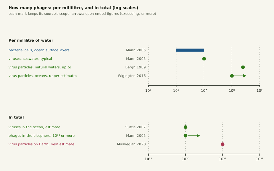
Slide the diagram sideways to read its labels, or open it full size.
Virus counts per millilitre and in total, each with its own scope and source; marine viruses are dominated by tailed phages, the group that includes T4 with its 86–120 nm head [10−8–10−7 m].
Log scales; arrows mark open-ended figures (exceeding, or more). Data: bacteriophage-counts.csv (sources, version and license, JSON).
"How many phages: counts per millilitre and totals" by Ants, Cells & Atoms, https://antscellsandatoms.com/microscopic-life/bacteriophage/ — CC BY 4.0 (https://creativecommons.org/licenses/by/4.0/)
Data sources for this figure
Data: Mann NH 2005, PLoS Biology 3(5):e182, CC BY 4.0, https://doi.org/10.1371/journal.pbio.0030182 (about 10^7 viruses per millilitre of seawater; 10^6–10^7 bacterioplankton cells; 10^30 or more phages in the biosphere; retrieved 2026-10-25); Bergh Ø, Børsheim KY, Bratbak G, Heldal M 1989, Nature 340:467–468, https://doi.org/10.1038/340467a0 (abstract: up to 2.5 x 10^8 per millilitre; retrieved 2026-10-25); Wigington CH et al. 2016, Nature Microbiology 1(3):15024, https://doi.org/10.1038/nmicrobiol.2015.24 (abstract: upper estimates exceeding 10^8 per millilitre; retrieved 2026-10-25); Suttle CA 2007, Nature Reviews Microbiology 5(10):801–812, https://doi.org/10.1038/nrmicro1750 (abstract: 10^30 viruses in the ocean; retrieved 2026-10-25); Mushegian AR 2020, Journal of Bacteriology 202(9):e00052-20, https://doi.org/10.1128/JB.00052-20 (abstract: best estimate close to 10^31 virus particles on Earth; retrieved 2026-10-25)
Data sources for this figure
Data: Mann NH 2005, PLoS Biology 3(5):e182, CC BY 4.0, https://doi.org/10.1371/journal.pbio.0030182 (about 10^7 viruses per millilitre of seawater; 10^6–10^7 bacterioplankton cells; 10^30 or more phages in the biosphere; retrieved 2026-10-25); Bergh Ø, Børsheim KY, Bratbak G, Heldal M 1989, Nature 340:467–468, https://doi.org/10.1038/340467a0 (abstract: up to 2.5 x 10^8 per millilitre; retrieved 2026-10-25); Wigington CH et al. 2016, Nature Microbiology 1(3):15024, https://doi.org/10.1038/nmicrobiol.2015.24, and Author Correction 2017, Nature Microbiology 2(11):1571, https://doi.org/10.1038/s41564-017-0042-1 (abstracts; retrieved 2026-10-25); Suttle CA 1994, Microbial Ecology 28(2):237–243, https://doi.org/10.1007/BF00166813 (abstract: about 20% infected, 10–20% lysed daily; retrieved 2026-10-25); Suttle CA 2007, Nature Reviews Microbiology 5(10):801–812, https://doi.org/10.1038/nrmicro1750 (abstract: 10^30 viruses in the ocean, about 10^23 infections per second; retrieved 2026-10-25); Mushegian AR 2020, Journal of Bacteriology 202(9):e00052-20, https://doi.org/10.1128/JB.00052-20 (abstract: about 10^31 virus particles on Earth, traced to Hendrix RW et al. 1999, https://doi.org/10.1073/pnas.96.5.2192; retrieved 2026-10-25); Ackermann HW 2007, Archives of Virology 152(2):227–243, https://doi.org/10.1007/s00705-006-0849-1 (abstract: at least 5568 phages examined, 96% tailed; retrieved 2026-10-25); one ratio computed 2026-10-25 for this page
Totals depend on what is counted. Suttle put the ocean's viruses at about 1030 in 2007, with about 1023 infections there every second (abstract); Mann put phages in the whole biosphere at 1030 or more. The 1031 frequently quoted for virus particles on Earth often comes without a source, Mushegian wrote in 2020. He traces it to Hendrix and colleagues in 1999 and concludes this best estimate remains close to 1031, unlikely to be much lower or higher (abstract).
Suttle offered a picture for the ocean's share: stretched end to end, those 1030 viruses would reach farther than the nearest 60 galaxies (abstract).
Sources
- Mann NH. "The Third Age of Phage." PLoS Biology 3(5):e182 (2005). https://doi.org/10.1371/journal.pbio.0030182 Read: full text. Text license: CC BY 4.0 (https://creativecommons.org/licenses/by/4.0/).
- Rao VB, Black LW. "Structure and assembly of bacteriophage T4 head." Virology Journal 7:356 (2010). https://doi.org/10.1186/1743-422X-7-356 Read: full text. Text license: CC BY 2.0 (https://creativecommons.org/licenses/by/2.0/).
- Valli J, Garcia-Burgos A, Rooney LM, Vale de Melo e Oliveira B, Duncan RR, Rickman C. "Seeing beyond the limit: A guide to choosing the right super-resolution microscopy technique." Journal of Biological Chemistry 297(1):100791 (2021). https://doi.org/10.1016/j.jbc.2021.100791 Read: full text. Text license: CC BY 4.0 (https://creativecommons.org/licenses/by/4.0/).
- Lin Y-R, Lin C-S. "Genome-wide characterization of vibrio phage ϕpp2 with unique arrangements of the mob-like genes." BMC Genomics 13:224 (2012). https://doi.org/10.1186/1471-2164-13-224 Read: full text. Text license: CC BY 2.0 (https://creativecommons.org/licenses/by/2.0/).
- Leiman PG, Arisaka F, van Raaij MJ, Kostyuchenko VA, Aksyuk AA, Kanamaru S, Rossmann MG. "Morphogenesis of the T4 tail and tail fibers." Virology Journal 7:355 (2010). https://doi.org/10.1186/1743-422X-7-355 Read: full text. Text license: CC BY 2.0 (https://creativecommons.org/licenses/by/2.0/).
- Turner D, Shkoporov AN, Lood C, Millard AD, Dutilh BE, et al. "Abolishment of morphology-based taxa and change to binomial species names: 2022 taxonomy update of the ICTV bacterial viruses subcommittee." Archives of Virology 168(2):74 (2023). https://doi.org/10.1007/s00705-022-05694-2 Read: full text. Text license: CC BY 4.0 (https://creativecommons.org/licenses/by/4.0/).
- International Committee on Taxonomy of Viruses (ICTV). ICTV Master Species List 2025 (MSL41), version 1 (2026). https://doi.org/10.5281/zenodo.19154110 Read: dataset record (the Tequatrovirus T4 row). Text license: CC BY 4.0 (https://creativecommons.org/licenses/by/4.0/).
- GBIF Secretariat. GBIF Backbone Taxonomy. Checklist dataset https://doi.org/10.15468/39omei, accessed 2026-10-25; Caudoviricetes, usage key 10794457; Tequatrovirus T4, usage key 12252006; Escherichia virus T4, usage key 9977478. Read: dataset record (name matches and species records). Text license: CC BY 4.0 (https://creativecommons.org/licenses/by/4.0/).
- Volkmer B, Heinemann M. "Condition-Dependent Cell Volume and Concentration of Escherichia coli to Facilitate Data Conversion for Systems Biology Modeling." PLoS ONE 6(7):e23126 (2011). https://doi.org/10.1371/journal.pone.0023126 Read: full text. Text license: CC BY 4.0 (https://creativecommons.org/licenses/by/4.0/).
- Marshall WF, Young KD, Swaffer M, Wood E, Nurse P, Kimura A, Frankel J, Wallingford J, Walbot V, Qu X, Roeder AHK. "What determines cell size?" BMC Biology 10:101 (2012). https://doi.org/10.1186/1741-7007-10-101 Read: full text. Text license: CC BY 2.0 (https://creativecommons.org/licenses/by/2.0/).
- Luef B, Frischkorn KR, Wrighton KC, Holman H-YN, Birarda G, Thomas BC, Singh A, Williams KH, Siegerist CE, Tringe SG, Downing KH, Comolli LR, Banfield JF. "Diverse uncultivated ultra-small bacterial cells in groundwater." Nature Communications 6:6372 (2015). https://doi.org/10.1038/ncomms7372 Read: abstract.
- Ackermann H-W. "The first phage electron micrographs." Bacteriophage 1(4):225–227 (2011). https://doi.org/10.4161/bact.1.4.17280 Read: full text. Text license: CC BY-NC 3.0 (https://creativecommons.org/licenses/by-nc/3.0/).
- Ackermann H-W. "5500 Phages examined in the electron microscope." Archives of Virology 152(2):227–243 (2007). https://doi.org/10.1007/s00705-006-0849-1 Read: abstract.
- Drew HR, Wing RM, Takano T, Broka C, Tanaka S, Itakura K, Dickerson RE. "Structure of a B-DNA dodecamer: conformation and dynamics." RCSB Protein Data Bank entry 1BNA (deposited 1981). https://doi.org/10.2210/pdb1bna/pdb Read: dataset record (atomic coordinates). Text license: CC0 1.0 (https://creativecommons.org/publicdomain/zero/1.0/).
- Yap ML, Rossmann MG. "Structure and function of bacteriophage T4." Future Microbiology 9(12):1319–1327 (2014). https://doi.org/10.2217/fmb.14.91 Read: abstract.
- Smith DE, Tans SJ, Smith SB, Grimes S, Anderson DL, Bustamante C. "The bacteriophage φ29 portal motor can package DNA against a large internal force." Nature 413:748–752 (2001). https://doi.org/10.1038/35099581 Read: abstract.
- Nurk S, Koren S, Rhie A, Rautiainen M, et al. "The complete sequence of a human genome." Science 376(6588):44–53 (2022). https://doi.org/10.1126/science.abj6987 Read: full text. Text license: CC BY 4.0 (https://creativecommons.org/licenses/by/4.0/).
- Paulson JR, Hudson DF, Cisneros-Soberanis F, Earnshaw WC. "Mitotic chromosomes." Seminars in Cell & Developmental Biology 117:7–29 (2021). https://doi.org/10.1016/j.semcdb.2021.03.014 Read: full text. Text license: CC BY 4.0 (https://creativecommons.org/licenses/by/4.0/).
- Mateu MG. "Viruses under mechanical force: Implications and applications." Virus Research 370:199767 (2026). https://doi.org/10.1016/j.virusres.2026.199767 Read: full text. Text license: CC BY 4.0 (https://creativecommons.org/licenses/by/4.0/).
- Evilevitch A, Lavelle L, Knobler CM, Raspaud E, Gelbart WM. "Osmotic pressure inhibition of DNA ejection from phage." Proceedings of the National Academy of Sciences of the USA 100(16):9292–9295 (2003). https://doi.org/10.1073/pnas.1233721100 Read: abstract.
- Sullivan MB, Coleman ML, Weigele P, Rohwer F, Chisholm SW. "Three Prochlorococcus Cyanophage Genomes: Signature Features and Ecological Interpretations." PLoS Biology 3(5):e144 (2005). https://doi.org/10.1371/journal.pbio.0030144 Read: full text. Text license: CC BY 4.0 (https://creativecommons.org/licenses/by/4.0/).
- Hu B, Margolin W, Molineux IJ, Liu J. "Structural remodeling of bacteriophage T4 and host membranes during infection initiation." Proceedings of the National Academy of Sciences of the USA 112(35):E4919–E4928 (2015). https://doi.org/10.1073/pnas.1501064112 Read: abstract.
- Bergh Ø, Børsheim KY, Bratbak G, Heldal M. "High abundance of viruses found in aquatic environments." Nature 340:467–468 (1989). https://doi.org/10.1038/340467a0 Read: abstract.
- Wigington CH, Sonderegger D, Brussaard CPD, Buchan A, Finke JF, Fuhrman JA, Lennon JT, Middelboe M, Suttle CA, Stock C, Wilson WH, Wommack KE, Wilhelm SW, Weitz JS. "Re-examination of the relationship between marine virus and microbial cell abundances." Nature Microbiology 1(3):15024 (2016); Author Correction, Nature Microbiology 2(11):1571 (2017). https://doi.org/10.1038/nmicrobiol.2015.24 Read: abstract (article and correction).
- Suttle CA. "The significance of viruses to mortality in aquatic microbial communities." Microbial Ecology 28(2):237–243 (1994). https://doi.org/10.1007/BF00166813 Read: abstract.
- Suttle CA. "Marine viruses — major players in the global ecosystem." Nature Reviews Microbiology 5(10):801–812 (2007). https://doi.org/10.1038/nrmicro1750 Read: abstract.
- Mushegian AR. "Are There 10³¹ Virus Particles on Earth, or More, or Fewer?" Journal of Bacteriology 202(9):e00052-20 (2020). https://doi.org/10.1128/JB.00052-20 Read: abstract.
- Hendrix RW, Smith MCM, Burns RN, Ford ME, Hatfull GF. "Evolutionary relationships among diverse bacteriophages and prophages: All the world's a phage." Proceedings of the National Academy of Sciences of the USA 96(5):2192–2197 (1999). https://doi.org/10.1073/pnas.96.5.2192 Read: abstract.
- Partensky F, Hess WR, Vaulot D. "Prochlorococcus, a Marine Photosynthetic Prokaryote of Global Significance." Microbiology and Molecular Biology Reviews 63(1):106–127 (1999). https://doi.org/10.1128/MMBR.63.1.106-127.1999 Read: abstract.
Cite this page
Ants, Cells & Atoms. “Bacteriophage T4: About 57 µm of DNA in a Head 0.12 µm Long.” Published 2026-10-25. https://antscellsandatoms.com/microscopic-life/bacteriophage/. Accessed (add the date you read it).
No DOI is issued for entries. Figures made here are CC BY 4.0; see each caption for the license of photographs.
Related entries
 Illustration
Diatoms: Why Most Glass-Walled Algae Shrink as They Multiply
Image: Illustration: Ants, Cells & Atoms, all rights reserved
Illustration: Ants, Cells & Atoms. Generated schematic illustration — not a photograph, micrograph or specimen record. © Ants, Cells & Atoms, all rights reserved.
Plankton: From Bacteria to Jellyfish, Sorted in Size Steps
Image: “Diatomea. - Schachtellinge” by Ernst Haeckel (artist) and Adolf Giltsch (lithographer) (1904) · Public domain
"Diatomea. - Schachtellinge" by Ernst Haeckel (artist) and Adolf Giltsch (lithographer) (1904) — https://commons.wikimedia.org/wiki/File:Diatomea._-_Schachtellinge_LCCN2015647735.jpg — Public domain (artist Ernst Haeckel died 1919 and lithographer Adolf Giltsch died 1911, so the plate is out of copyright in the EU (life plus 70 years) and in the US (published 1904, before 1930); the scan is from the Library of Congress Prints and Photographs division (lccn.loc.gov/2015647735), which lists no rights restriction)

Paramecium: How a Nerve-Like Signal Sends One Cell Backward
Image: “Paramecium 1” by Nbnidhi (2017) · CC BY 4.0
"Paramecium 1" by Nbnidhi — https://commons.wikimedia.org/wiki/File:Paramecium_1.jpg — CC BY 4.0 (https://creativecommons.org/licenses/by/4.0/)
