Plankton: From Bacteria to Jellyfish, Sorted in Size Steps

Slide the ladder sideways to read its labels, or open it full size.
Plankton: size classes from 0.2 µm to 10 cm, with drifters beyond them to over 2 m; the band this page centres on is the nano- and microplankton at 2–200 µm [10−6–10−4 m].
Log scale from 10 cm to 0.1 nm, each rung ten times smaller; dashed lines: naked eye about 0.1 mm, light microscope about 0.2 µm (How to read the Zoom ladder). Pink band: the syrup line, Re = 1 in water at 20 °C at 1–10 body lengths per second. Data: plankton-size-classes.csv (sources, version and license, JSON).
"Zoom ladder: plankton" by Ants, Cells & Atoms, https://antscellsandatoms.com/microscopic-life/plankton/ — CC BY 4.0 (https://creativecommons.org/licenses/by/4.0/)
Data sources for this figure
Data: Class limits: Hirata T et al. 2011, Biogeosciences 8:311, CC BY 3.0, https://doi.org/10.5194/bg-8-311-2011 (restating Sieburth JM, Smetacek V, Lenz J 1978, Limnology and Oceanography 23:1256); Hatton IA et al. 2021, Science Advances 7:eabh3732, CC BY 4.0, https://doi.org/10.1126/sciadv.abh3732, retrieved 2026-10-24; Largest example: Villanueva AA, Priya S 2014, PLoS ONE 9:e115220, CC BY 4.0, https://doi.org/10.1371/journal.pone.0115220, retrieved 2026-10-24; Syrup line band: Re = 1 for 1–10 body lengths per second in water at 20 °C, viscosity from IAPWS R12-08 (attribution: International Association for the Properties of Water and Steam), computed 2026-10-24
Data sources for this figure
Data: Class limits: Hirata T et al. 2011, Biogeosciences 8:311, CC BY 3.0, https://doi.org/10.5194/bg-8-311-2011 (restating Sieburth JM, Smetacek V, Lenz J 1978, Limnology and Oceanography 23:1256, https://doi.org/10.4319/lo.1978.23.6.1256); Hatton IA et al. 2021, Science Advances 7:eabh3732, CC BY 4.0, https://doi.org/10.1126/sciadv.abh3732, retrieved 2026-10-24; Examples: Partensky F, Hess WR, Vaulot D 1999, Microbiology and Molecular Biology Reviews 63:106 (abstract), https://doi.org/10.1128/MMBR.63.1.106-127.1999; Moore ER et al. 2017, PLoS ONE 12:e0181098, CC BY 4.0, https://doi.org/10.1371/journal.pone.0181098; Gastineau R et al. 2021, Biology 10:328, CC BY 4.0, https://doi.org/10.3390/biology10040328; Kiørboe T et al. 2010, Journal of the Royal Society Interface 7:1591 (abstract), https://doi.org/10.1098/rsif.2010.0176; Perry FA et al. 2019, PLoS ONE 14:e0219325, CC BY 4.0, https://doi.org/10.1371/journal.pone.0219325; Villanueva AA, Priya S 2014, PLoS ONE 9:e115220, CC BY 4.0, https://doi.org/10.1371/journal.pone.0115220, retrieved 2026-10-24; GBIF Secretariat 2023, GBIF Backbone Taxonomy, checklist dataset https://doi.org/10.15468/39omei, CC BY 4.0, accessed via api.gbif.org on 2026-10-24
Bench card
- Name
- Plankton (an ecological group, not a taxon): the organisms that drift with the waterSource: NOAA National Ocean Service, "What are plankton?"
- Group
- Bacteria, protists (diatoms, dinoflagellates, ciliates and others), crustaceans such as copepods and krill, larvae of many animals, jellyfish; representative taxa are listed with their GBIF keys in plankton-size-classes.csvSource: GBIF Backbone Taxonomy, https://doi.org/10.15468/39omei, accessed 2026-10-24
- Actual size
- 0.2 µm to over 2 m across the size classes; this page centres on the nano- and microplankton, 2–200 µm [10−6–10−4 m]Source: Hirata et al. 2011 (class limits after Sieburth et al. 1978); Villanueva & Priya 2014 (bell over 2 m); plankton-size-classes.csv
- Size in this image
- not measurable from this image
- Lives in
- oceans, lakes and ponds, carried by tides and currentsSource: NOAA National Ocean Service, "What are plankton?"
- Seen with
- naked eye for the largest classes; hand lens for mesoplankton; light microscope for the 2–200 µm band and, near its 0.2 µm limit, the smallest bacteria; electron microscope for their internal structureSource: plankton-size-classes.csv; Zoom ladder reference lines
- Color in images
- native in the lithograph (the plate's single printed ink) and the copepod photograph (no stain stated by its source); stain in the Ceratium light micrograph (magenta, not named by the photographer)Source: figure captions on this page
plankton-bench.csv (CSV) · where each value comes from, and its license (JSON)
Data credits for this card
Data: Definition and habitats: National Ocean Service (NOAA), "What are plankton?", https://oceanservice.noaa.gov/facts/plankton.html, US government work, retrieved 2026-10-24; Size range: Hirata T et al. 2011, Biogeosciences 8:311, CC BY 3.0, https://doi.org/10.5194/bg-8-311-2011; Villanueva AA, Priya S 2014, PLoS ONE 9:e115220, CC BY 4.0, https://doi.org/10.1371/journal.pone.0115220, retrieved 2026-10-24; GBIF Secretariat 2023, GBIF Backbone Taxonomy, checklist dataset https://doi.org/10.15468/39omei, CC BY 4.0, accessed via api.gbif.org on 2026-10-24
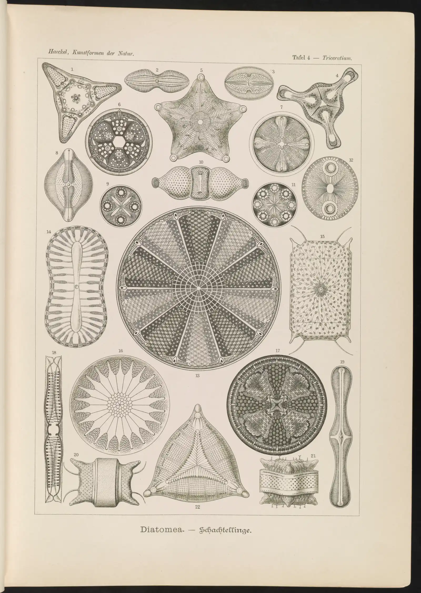
Diatomea, plate 4 of Ernst Haeckel's Kunstformen der Natur (1904): diatoms, single-celled drifters; the two species sized on this page measure 7–12 µm and 20.2–138.0 µm [10−6–10−4 m].
Color: native (the plate's single printed ink; a lithograph, not a micrograph)
“Diatomea. - Schachtellinge” by Ernst Haeckel (artist) and Adolf Giltsch (lithographer) (1904) · Public domain
Full credit
"Diatomea. - Schachtellinge" by Ernst Haeckel (artist) and Adolf Giltsch (lithographer) (1904) — https://commons.wikimedia.org/wiki/File:Diatomea._-_Schachtellinge_LCCN2015647735.jpg — Public domain (artist Ernst Haeckel died 1919 and lithographer Adolf Giltsch died 1911, so the plate is out of copyright in the EU (life plus 70 years) and in the US (published 1904, before 1930); the scan is from the Library of Congress Prints and Photographs division (lccn.loc.gov/2015647735), which lists no rights restriction)

The word comes from the Greek for drifter or wanderer, as the US National Ocean Service puts it. Some organisms drift for their whole lives. Others count as plankton only while young, then grow large enough to swim against the currents. The group has two basic halves: phytoplankton, which photosynthesise near the sunlit surface, and zooplankton, which eat them or one another.
What makes something plankton?
The test is behaviour in the water, not size and not ancestry. A copepod a millimetre long, a diatom a tenth of that and a jellyfish with a bell over 2 m across all count, because none of them can hold position against a current. The Ocean Service lists animals such as krill and sea snails, the young of larger invertebrates and fish, and weak swimmers like jellyfish among the zooplankton. It names copepods, planktonic crustaceans distantly related to shrimp and crabs, as one of the most common kinds in almost every ocean, sea and freshwater habitat.
Phytoplankton and zooplankton split by what they eat. Phytoplankton photosynthesise, so they stay where the light is; most zooplankton eat phytoplankton, and most are in turn eaten by larger animals or by each other. By day many zooplankton drift in deeper water, and at night they rise to feed near the surface, a daily movement the Ocean Service calls the largest migration on Earth. Drifting is a statement about the horizontal: a copepod can rise toward the surface each night and still be carried wherever the current goes.
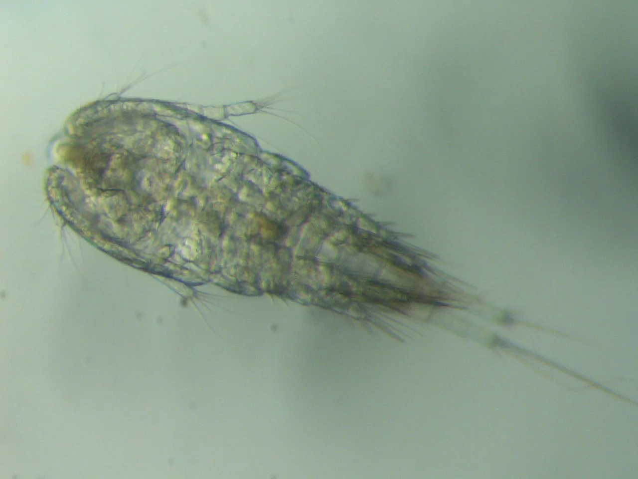
Diacyclops thomasi, a freshwater copepod from the Great Lakes, 2010; the three planktonic copepod species in Kiørboe et al. 2010 (abstract) were 0.3–3.0 mm long [10−4–10−3 m].
Color: native (no stain stated by the source)
"Cyclops thomasi (8741967598)" by NOAA Great Lakes Environmental Research Laboratory — https://commons.wikimedia.org/wiki/File:Cyclops_thomasi_(8741967598).jpg — CC BY-SA 2.0 (https://creativecommons.org/licenses/by-sa/2.0/)

Any colour in a picture like this that the animal did not make itself, from a dye or a false-colour overlay, is borrowed color. Every micrograph and photograph caption on this site declares which kind it shows.
Size classes ten times apart
Hirata and colleagues trace the scheme to Sieburth, Smetacek and Lenz in 1978. Only that paper's bibliographic record could be consulted for this page, so the class limits below come from later papers that restate or adapt it.
As Hirata and colleagues restate it in 2011, picoplankton are 0.2–2 µm, nanoplankton 2–20 µm and microplankton larger than 20 µm. Each limit is ten times the last, which is why the sequence fits a logarithmic axis and nothing else. On a ruler where 1 cm stands for 1 µm, a 2 m jellyfish would need a ruler 20 km long, computed for this page.
Above the microplankton the names continue, but the limits are conventions and differ between studies. A 2021 global survey by Hatton and colleagues used 200 µm to 2 cm for mesozooplankton and 0.2 to 10 cm for macrozooplankton. The two ranges overlap.
Romagnan and colleagues (2015) described a long-term series at Villefranche. It counted picoplankton below 2 µm by flow cytometry, imaged nanoplankton at 3–30 µm, and caught mesozooplankton with a 200 µm net and macroplankton with a 680 µm net. The table keeps each limit as its source states it.
| Size class | Limits | Limits as stated by | One measured example |
|---|---|---|---|
| picoplankton | 0.2–2 µm | Hirata et al. 2011, after Sieburth et al. 1978 | Prochlorococcus, 0.5–0.7 µm across (Partensky et al. 1999, abstract) |
| nanoplankton | 2–20 µm | Hirata et al. 2011, after Sieburth et al. 1978 | Thalassiosira pseudonana initial cells, 7–12 µm (Moore et al. 2017) |
| microplankton | 20–200 µm | lower limit Hirata et al. 2011; upper limit Hatton et al. 2021 | Haslea silbo, 20.2–138.0 µm long (Gastineau et al. 2021) |
| mesoplankton | 200 µm–2 cm | Hatton et al. 2021 | three planktonic copepod species, 0.3–3.0 mm (Kiørboe et al. 2010, abstract) |
| macroplankton | 0.2–10 cm | Hatton et al. 2021 | adult Antarctic krill, over 30 mm (Perry et al. 2019) |
| beyond the macroplankton limit | over 10 cm | no readable source gives an upper limit | lion's mane jellyfish, bell over 2 m (Villanueva and Priya 2014) |
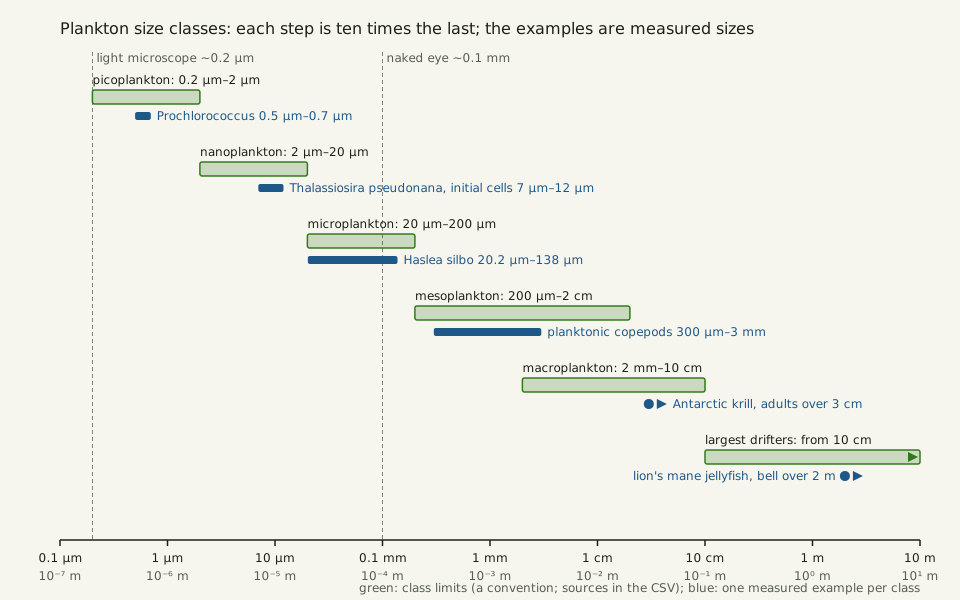
Slide the diagram sideways to read its labels, or open it full size.
Plankton size classes from 0.2 µm to 10 cm and the largest drifters beyond, one measured example in each [10−7–100 m].
Green bars are class limits as each source states them (a convention; the two larger classes overlap); blue marks are measured sizes. Data: plankton-size-classes.csv (sources, version and license, JSON).
"Plankton size classes, ten times apart" by Ants, Cells & Atoms, https://antscellsandatoms.com/microscopic-life/plankton/ — CC BY 4.0 (https://creativecommons.org/licenses/by/4.0/)
Data sources for this figure
Data: Class limits: Hirata T et al. 2011, Biogeosciences 8:311, CC BY 3.0, https://doi.org/10.5194/bg-8-311-2011 (restating Sieburth JM, Smetacek V, Lenz J 1978, Limnology and Oceanography 23:1256, https://doi.org/10.4319/lo.1978.23.6.1256); Hatton IA et al. 2021, Science Advances 7:eabh3732, CC BY 4.0, https://doi.org/10.1126/sciadv.abh3732, retrieved 2026-10-24; Examples: Partensky F, Hess WR, Vaulot D 1999, Microbiology and Molecular Biology Reviews 63:106 (abstract), https://doi.org/10.1128/MMBR.63.1.106-127.1999; Moore ER et al. 2017, PLoS ONE 12:e0181098, CC BY 4.0, https://doi.org/10.1371/journal.pone.0181098; Gastineau R et al. 2021, Biology 10:328, CC BY 4.0, https://doi.org/10.3390/biology10040328; Kiørboe T et al. 2010, Journal of the Royal Society Interface 7:1591 (abstract), https://doi.org/10.1098/rsif.2010.0176; Perry FA et al. 2019, PLoS ONE 14:e0219325, CC BY 4.0, https://doi.org/10.1371/journal.pone.0219325; Villanueva AA, Priya S 2014, PLoS ONE 9:e115220, CC BY 4.0, https://doi.org/10.1371/journal.pone.0115220, retrieved 2026-10-24
Data sources for this figure
Data: Class limits: Hirata T et al. 2011, Biogeosciences 8:311, CC BY 3.0, https://doi.org/10.5194/bg-8-311-2011 (restating Sieburth JM, Smetacek V, Lenz J 1978, Limnology and Oceanography 23:1256, https://doi.org/10.4319/lo.1978.23.6.1256); Hatton IA et al. 2021, Science Advances 7:eabh3732, CC BY 4.0, https://doi.org/10.1126/sciadv.abh3732, retrieved 2026-10-24; Examples: Partensky F, Hess WR, Vaulot D 1999, Microbiology and Molecular Biology Reviews 63:106 (abstract), https://doi.org/10.1128/MMBR.63.1.106-127.1999; Moore ER et al. 2017, PLoS ONE 12:e0181098, CC BY 4.0, https://doi.org/10.1371/journal.pone.0181098; Gastineau R et al. 2021, Biology 10:328, CC BY 4.0, https://doi.org/10.3390/biology10040328; Kiørboe T et al. 2010, Journal of the Royal Society Interface 7:1591 (abstract), https://doi.org/10.1098/rsif.2010.0176; Perry FA et al. 2019, PLoS ONE 14:e0219325, CC BY 4.0, https://doi.org/10.1371/journal.pone.0219325; Villanueva AA, Priya S 2014, PLoS ONE 9:e115220, CC BY 4.0, https://doi.org/10.1371/journal.pone.0115220, retrieved 2026-10-24; GBIF Secretariat 2023, GBIF Backbone Taxonomy, checklist dataset https://doi.org/10.15468/39omei, CC BY 4.0, accessed via api.gbif.org on 2026-10-24
Can you see plankton without a microscope? The naked-eye line on the ladder sits near 0.1 mm, inside the microplankton class, so the larger diatoms, most copepods, krill and the jellyfish are visible unaided. Picoplankton and nanoplankton need a light microscope, and the smallest bacteria push against its 0.2 µm limit. Where a 2 µm cell and a 200 µm one sit among cells in general is the subject of How Big Is a Cell?
Who drifts in each class?
Start at the bottom. Prochlorococcus, a marine cyanobacterium 0.5–0.7 µm in diameter, is the smallest known photosynthetic organism, by the account of Partensky, Hess and Vaulot in 1999 (abstract). They also call it presumably the most abundant photosynthetic organism on Earth. Smaller than any cell here are the viruses of seawater, typically on the order of ten million per millilitre and dominated by tailed bacteriophages, which infect bacteria, by Mann's 2005 account.
The bacterium Pelagibacter ubique, a Candidatus name in Zhao's paper, belongs to the SAR11 group that Rappé and colleagues first grew in culture in 2002 (abstract). Its average cell volume is 0.037 µm3 in frozen-hydrated cells, as Zhao and colleagues reported in 2017 (abstract); a sphere of that volume is about 0.4 µm across, computed for this page. Bacteria of about this size are counted on another page, How Many Cells in the Human Body?
One step up, nanoplankton include small diatoms. Newly formed initial cells of Thalassiosira pseudonana measured 7–12 µm, the largest size reported for the species, in Moore and colleagues' 2017 cultures.
In the microplankton, the blue diatom Haslea silbo ranges from 138.0 µm in the largest initial cells down to 20.2 µm in final-stage cultures (Gastineau et al. 2021). That shrinking by division is the story told under Diatoms. Dinoflagellates such as Ceratium furca (now Tripos furca in the GBIF Backbone), with a typical size of 122.5 µm in Lisicki and colleagues' 2019 compilation, share the class and swim with flagella.
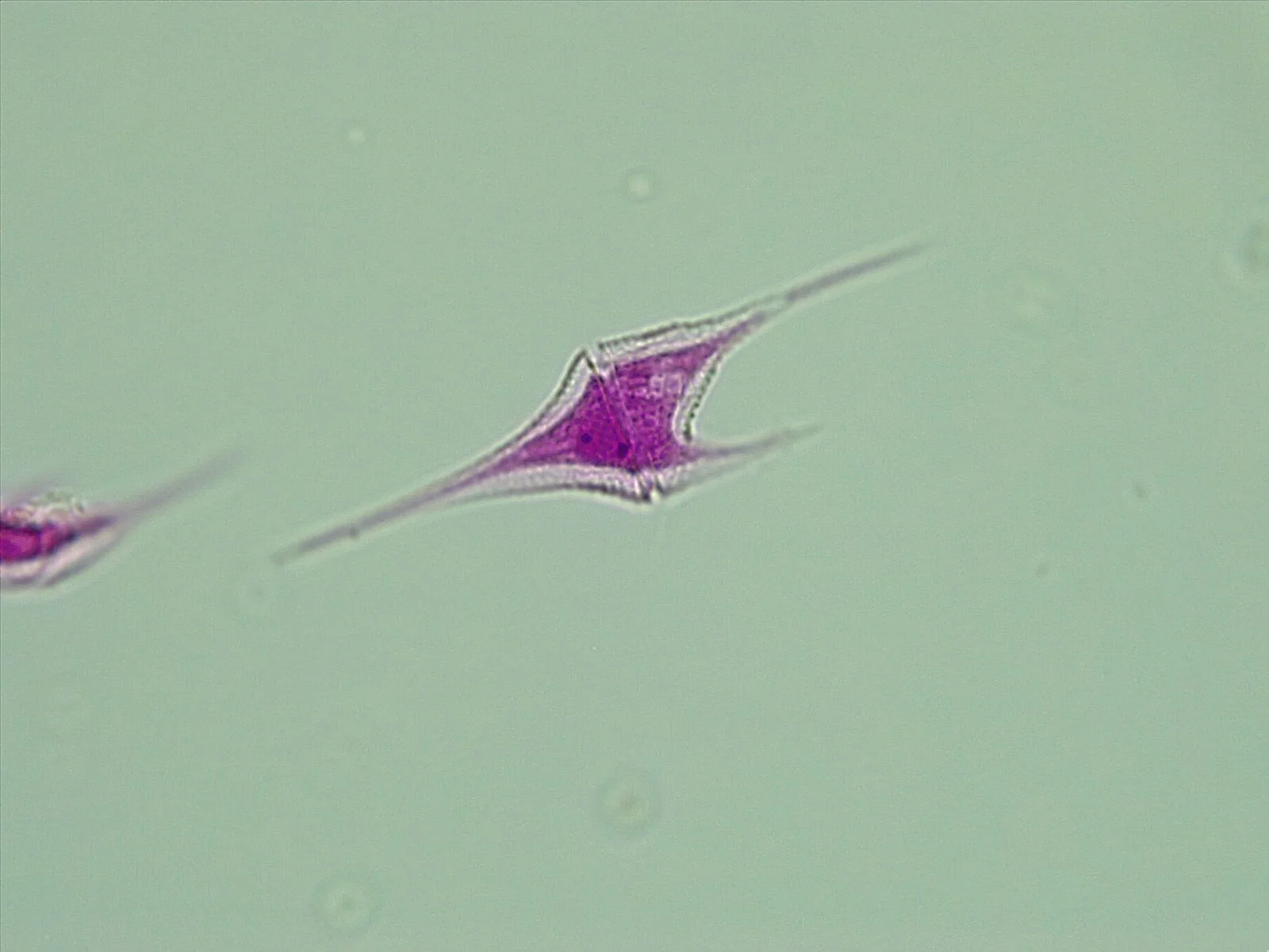
Ceratium, a dinoflagellate genus whose species have typical sizes of about 50 to 400 µm [10−5–10−4 m] in the Lisicki et al. 2019 compilation; light micrograph.
Color: stain (magenta, not named by the photographer)
"Ceratium 1" by Bwiltz — https://commons.wikimedia.org/wiki/File:Ceratium_1.jpg — CC BY-SA 4.0 (https://creativecommons.org/licenses/by-sa/4.0/)

Mesoplankton is copepod country. Kiørboe and colleagues studied escape jumps in three species 0.3–3.0 mm long (2010, abstract). Hatton and colleagues list copepods, larvaceans, amphipods and giant rhizaria, the group to which foraminifera belong, among the mesozooplankton. Their macrozooplankton includes arrow worms, krill, tunicates, fish larvae, comb jellies and jellyfish. Adult Antarctic krill are defined as longer than 30 mm in the survey databases Perry and colleagues used in 2019, with juveniles at 15–30 mm.
At the top, the lion's mane jellyfish Cyanea capillata can exceed 2 m in bell diameter, with tentacles 36 m long, according to Villanueva and Priya (2014). That puts it beyond the 10 cm macrozooplankton limit and off the 10 cm end of the Zoom ladder. Across the whole span, Hatton and colleagues found the biomass within most order-of-magnitude size classes near 1 gigatonne wet weight, with bacteria above that line and large mammals below it.
Can plankton swim at all?
Most of them can, just not against a current. Lisicki, Velho Rodrigues, Goldstein and Lauga compiled swimming speeds for 189 single-celled eukaryotes in 2019. Flagellates average 31 µm in size with a median speed of 127 µm/s, ciliates 132 µm and 784 µm/s, and both groups keep a Reynolds number below 1. A ciliate such as Paramecium caudatum, listed in their appendix at 225.5 µm and 2,489 µm/s, is the fastest single cell on this page and still sits on the viscous side; see Paramecium.
Among green algae, the same authors note, the smaller flagellated species and the largest multicellular ones differ in whether stirring or diffusion matters more for what reaches the cell. Volvox is the multicellular end of that range on this site. The chart below puts every swimmer with a published size and speed on one pair of logarithmic axes, with the Reynolds number computed for this page.
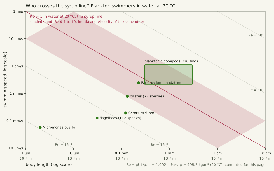
Slide the diagram sideways to read its labels, or open it full size.
Plankton swimmers from a 2 µm flagellate to 3 mm copepods: body length against speed, with the Re = 1 line in water at 20 °C [10−6–10−3 m].
Points and the box are published sizes and speeds; Re is computed for this page with the viscosity of water from the IAPWS 2008 formulation. Data: plankton-swimmers-reynolds.csv (sources, version and license, JSON).
"Who crosses the syrup line? Plankton swimmers in water at 20 °C" by Ants, Cells & Atoms, https://antscellsandatoms.com/microscopic-life/plankton/ — CC BY 4.0 (https://creativecommons.org/licenses/by/4.0/)
Data sources for this figure
Data: Sizes and speeds: Lisicki M, Velho Rodrigues MF, Goldstein RE, Lauga E 2019, eLife 8:e44907, CC BY 4.0, https://doi.org/10.7554/eLife.44907; Seuront L 2011, PLoS ONE 6:e26283, CC BY 4.0, https://doi.org/10.1371/journal.pone.0026283; Kiørboe T et al. 2010, Journal of the Royal Society Interface 7:1591 (abstract), https://doi.org/10.1098/rsif.2010.0176, retrieved 2026-10-24; Viscosity of water: International Association for the Properties of Water and Steam, IAPWS R12-08 (2008), https://iapws.org/technical-guidance/release/viscosity; Reynolds numbers computed 2026-10-24
Data sources for this figure
Data: Sizes and speeds: Lisicki M, Velho Rodrigues MF, Goldstein RE, Lauga E 2019, eLife 8:e44907, CC BY 4.0, https://doi.org/10.7554/eLife.44907; Seuront L 2011, PLoS ONE 6:e26283, CC BY 4.0, https://doi.org/10.1371/journal.pone.0026283; Kiørboe T et al. 2010, Journal of the Royal Society Interface 7:1591 (abstract), https://doi.org/10.1098/rsif.2010.0176, retrieved 2026-10-24; Viscosity of water: International Association for the Properties of Water and Steam, IAPWS R12-08 (2008), https://iapws.org/technical-guidance/release/viscosity; Reynolds numbers computed 2026-10-24; GBIF Secretariat 2023, GBIF Backbone Taxonomy, checklist dataset https://doi.org/10.15468/39omei, CC BY 4.0, accessed via api.gbif.org on 2026-10-24
| Swimmer | Size | Speed | Re in water at 20 °C (computed for this page) |
|---|---|---|---|
| Micromonas pusilla, a green flagellate | 2 µm | 58.5 µm/s | 0.0001 |
| flagellates, 112 species | 31 µm (average) | 127 µm/s (median) | 0.004 |
| Ceratium furca, a dinoflagellate | 122.5 µm | 194 µm/s | 0.02 |
| ciliates, 77 species | 132 µm (average) | 784 µm/s (median) | 0.1 |
| Paramecium caudatum | 225.5 µm | 2,489 µm/s | 0.6 |
| planktonic copepods, cruising (lengths: three species, Kiørboe et al. 2010, abstract; speeds: Temora longicornis, Seuront 2011) | 0.3–3.0 mm | 2.1–11.3 mm/s | 0.6–34 |
Where is the syrup line in water?
That Reynolds number — a ratio of inertia to stickiness — sets the syrup line: the body length where Re reaches 1 for a stated fluid, temperature and speed. In water at 20 °C, a swimmer cruising at 1–10 body lengths per second crosses it at 0.32–1.0 mm, computed for this page. Only well below it, at Re ≪ 1, can inertia be ignored. Change the speed or fluid and the line moves.
Planktonic copepods straddle it. Temora longicornis adults cruised at 2.1–11.3 mm/s at 18 °C in Seuront's 2011 experiments. For 0.3–3.0 mm bodies, that gives Re of about 0.6–34, computed for this page: inertia and viscosity of the same order at the low end, inertia more than ten times viscosity at the high end. Copepods, Kiørboe and colleagues note, use their feeding appendages for steady swimming and their swimming legs for short, intense escape jumps (abstract).
Jellyfish are further across. Pitt and colleagues (2013) describe jellyfish swimming as inertia-dominated, while a tentacle 0.01–0.1 mm thick moved at 1 cm/s falls to Re 0.1–1, where both forces count. The water itself jostles the smallest drifters; see Brownian Motion.
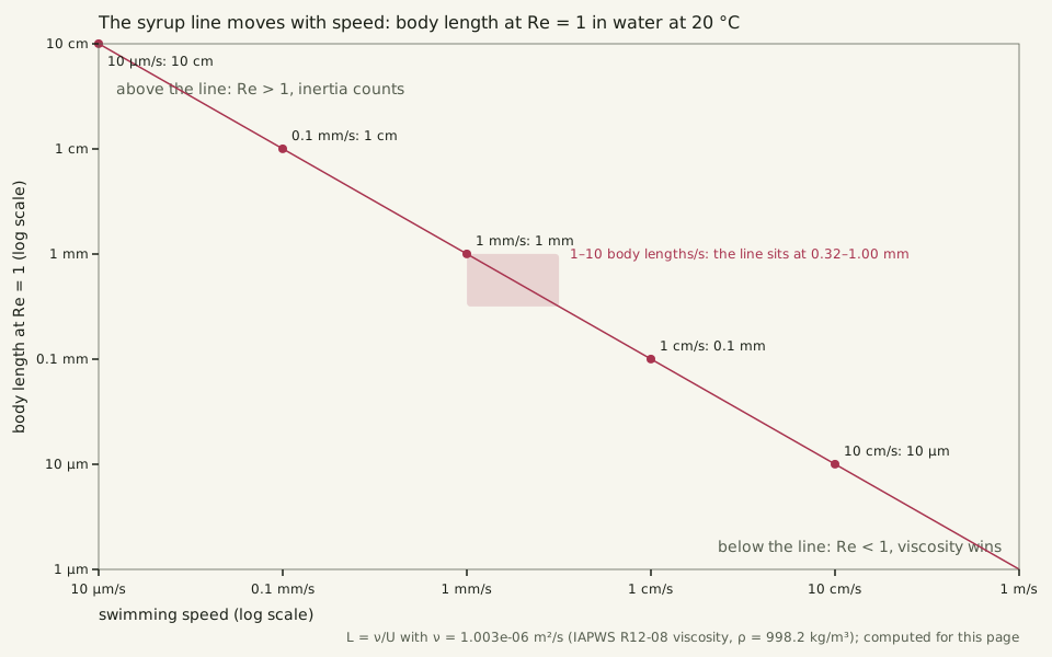
Slide the diagram sideways to read its labels, or open it full size.
The syrup line moves with speed: in water at 20 °C the body length at Re = 1 runs from 10 cm at 10 µm/s to 10 µm at 10 cm/s [10−5–10−1 m].
The line is L = ν/U, computed for this page with the viscosity of water from the IAPWS 2008 formulation; the pink box is the 1–10 body lengths per second band drawn on the Zoom ladder.
"The syrup line moves with speed: body length at Re = 1 in water at 20 °C" by Ants, Cells & Atoms, https://antscellsandatoms.com/microscopic-life/plankton/ — CC BY 4.0 (https://creativecommons.org/licenses/by/4.0/)
Data: Viscosity of water: International Association for the Properties of Water and Steam, IAPWS R12-08 (2008), https://iapws.org/technical-guidance/release/viscosity; body lengths at Re = 1 computed 2026-10-24
Instruments change every few rungs: flow cytometry below 2 µm, microscopes up to a few hundred micrometres, 200 and 680 µm mesh nets above that. Hatton and colleagues note that surveys of the largest animals cover whole oceans. Their size spectrum, stitched from such methods, spans 23 orders of magnitude in body mass, and the ladder is least certain at the seams. Under a microscope, a drop from the jar shows you one rung; a research ship's net shows another.
Sources
- National Ocean Service, National Oceanic and Atmospheric Administration. "What are plankton?" Ocean Facts. https://oceanservice.noaa.gov/facts/plankton.html Read: full text.
- Hirata T, Hardman-Mountford NJ, Brewin RJW, Aiken J, Barlow R, Suzuki K, Isada T, Howell E, Hashioka T, Noguchi-Aita M, Yamanaka Y. "Synoptic relationships between surface Chlorophyll-a and diagnostic pigments specific to phytoplankton functional types." Biogeosciences 8(2):311–327 (2011). https://doi.org/10.5194/bg-8-311-2011 Read: full text. Text license: CC BY 3.0 (https://creativecommons.org/licenses/by/3.0/).
- Sieburth JM, Smetacek V, Lenz J. "Pelagic ecosystem structure: Heterotrophic compartments of the plankton and their relationship to plankton size fractions." Limnology and Oceanography 23(6):1256–1263 (1978). https://doi.org/10.4319/lo.1978.23.6.1256 Read: bibliographic record only.
- Hatton IA, Heneghan RF, Bar-On YM, Galbraith ED. "The global ocean size spectrum from bacteria to whales." Science Advances 7(46):eabh3732 (2021). https://doi.org/10.1126/sciadv.abh3732 Read: full text.
- Romagnan J-B, Legendre L, Guidi L, Jamet J-L, Jamet D, Mousseau L, Pedrotti M-L, Picheral M, Gorsky G, Sardet C, Stemmann L. "Comprehensive Model of Annual Plankton Succession Based on the Whole-Plankton Time Series Approach." PLoS ONE 10(3):e0119219 (2015). https://doi.org/10.1371/journal.pone.0119219 Read: full text.
- Partensky F, Hess WR, Vaulot D. "Prochlorococcus, a Marine Photosynthetic Prokaryote of Global Significance." Microbiology and Molecular Biology Reviews 63(1):106–127 (1999). https://doi.org/10.1128/MMBR.63.1.106-127.1999 Read: abstract.
- Rappé MS, Connon SA, Vergin KL, Giovannoni SJ. "Cultivation of the ubiquitous SAR11 marine bacterioplankton clade." Nature 418(6898):630–633 (2002). https://doi.org/10.1038/nature00917 Read: abstract.
- Zhao X, Schwartz CL, Pierson J, Giovannoni SJ, McIntosh JR, Nicastro D. "Three-Dimensional Structure of the Ultraoligotrophic Marine Bacterium "Candidatus Pelagibacter ubique"." Applied and Environmental Microbiology 83(3):e02807-16 (2017). https://doi.org/10.1128/AEM.02807-16 Read: abstract.
- Moore ER, Bullington BS, Weisberg AJ, Jiang Y, Chang J, Halsey KH. "Morphological and transcriptomic evidence for ammonium induction of sexual reproduction in Thalassiosira pseudonana and other centric diatoms." PLoS ONE 12(7):e0181098 (2017). https://doi.org/10.1371/journal.pone.0181098 Read: full text.
- Gastineau R, Hansen G, Poulin M, Lemieux C, Turmel M, Bardeau J-F, Leignel V, Hardivillier Y, Morançais M, Fleurence J, Gaudin P, Méléder V, Cox EJ, Davidovich NA, Davidovich OI, Witkowski A, Kociolek JP, Mouget J-L. "Haslea silbo, A Novel Cosmopolitan Species of Blue Diatoms." Biology 10(4):328 (2021). https://doi.org/10.3390/biology10040328 Read: full text.
- Lisicki M, Velho Rodrigues MF, Goldstein RE, Lauga E. "Swimming eukaryotic microorganisms exhibit a universal speed distribution." eLife 8:e44907 (2019). https://doi.org/10.7554/eLife.44907 Read: full text, including Appendix 1 (flagellates) and Appendix 2 (ciliates).
- Kiørboe T, Andersen A, Langlois VJ, Jakobsen HH. "Unsteady motion: escape jumps in planktonic copepods, their kinematics and energetics." Journal of the Royal Society Interface 7(52):1591–1602 (2010). https://doi.org/10.1098/rsif.2010.0176 Read: abstract.
- Perry FA, Atkinson A, Sailley SF, Tarling GA, Hill SL, Lucas CH, Mayor DJ. "Habitat partitioning in Antarctic krill: Spawning hotspots and nursery areas." PLoS ONE 14(7):e0219325 (2019). https://doi.org/10.1371/journal.pone.0219325 Read: full text.
- Villanueva AA, Priya S. "Cyanea capillata Bell Kinematics Analysis through Corrected In Situ Imaging and Modeling Using Strategic Nodes." PLoS ONE 9(12):e115220 (2014). https://doi.org/10.1371/journal.pone.0115220 Read: full text.
- Pitt KA, Duarte CM, Lucas CH, Sutherland KR, Condon RH, Mianzan H, Purcell JE, Robinson KL, Uye S-I. "Jellyfish Body Plans Provide Allometric Advantages beyond Low Carbon Content." PLoS ONE 8(8):e72683 (2013). https://doi.org/10.1371/journal.pone.0072683 Read: full text.
- Seuront L. "Hydrocarbon Contamination Decreases Mating Success in a Marine Planktonic Copepod." PLoS ONE 6(10):e26283 (2011). https://doi.org/10.1371/journal.pone.0026283 Read: full text.
- International Association for the Properties of Water and Steam. "Release on the IAPWS Formulation 2008 for the Viscosity of Ordinary Water Substance." IAPWS R12-08 (2008). https://iapws.org/technical-guidance/release/viscosity Read: full text (PDF): equation (10), Tables 1, 2 and 4.
- GBIF Secretariat. GBIF Backbone Taxonomy. Checklist dataset https://doi.org/10.15468/39omei, accessed 2026-10-24; usage keys for the taxa named on this page are listed in plankton-size-classes.csv and plankton-swimmers-reynolds.csv. Read: dataset record (species match and species records for the taxa named). Text license: CC BY 4.0 (https://creativecommons.org/licenses/by/4.0/).
- WoRMS (2026). Foraminifera. World Register of Marine Species. Accessed at https://www.marinespecies.org/aphia.php?p=taxdetails&id=1410 on 2026-10-24. https://doi.org/10.14284/170 Read: dataset record (classification of the phylum Foraminifera under the infrakingdom Rhizaria). Text license: CC BY 4.0 (https://creativecommons.org/licenses/by/4.0/).
- Mann NH. "The Third Age of Phage." PLoS Biology 3(5):e182 (2005). https://doi.org/10.1371/journal.pbio.0030182 Read: full text. Text license: CC BY 4.0 (https://creativecommons.org/licenses/by/4.0/).
Cite this page
Ants, Cells & Atoms. “Plankton: From Bacteria to Jellyfish, Sorted in Size Steps.” Published 2026-10-25. https://antscellsandatoms.com/microscopic-life/plankton/. Accessed (add the date you read it).
No DOI is issued for entries. Figures made here are CC BY 4.0; see each caption for the license of photographs.
Related entries
 Illustration
Diatoms: Why Most Glass-Walled Algae Shrink as They Multiply
Image: Illustration: Ants, Cells & Atoms, all rights reserved
Illustration: Ants, Cells & Atoms. Generated schematic illustration — not a photograph, micrograph or specimen record. © Ants, Cells & Atoms, all rights reserved.
Paramecium: How a Nerve-Like Signal Sends One Cell Backward
Image: “Paramecium 1” by Nbnidhi (2017) · CC BY 4.0
"Paramecium 1" by Nbnidhi — https://commons.wikimedia.org/wiki/File:Paramecium_1.jpg — CC BY 4.0 (https://creativecommons.org/licenses/by/4.0/)

 Illustration
Volvox: Each Embryo Turns Inside Out Before It Can Swim
Image: Illustration: Ants, Cells & Atoms, all rights reserved
Illustration: Ants, Cells & Atoms. Generated schematic illustration — not a photograph, micrograph or specimen record. © Ants, Cells & Atoms, all rights reserved.