Volvox: Each Embryo Turns Inside Out Before It Can Swim

Slide the ladder sideways to read its labels, or open it full size.
Volvox: the largest spheres 0.35–0.92 mm long in seven species descriptions [10−4 m], with a V. globator embryo about to turn inside out (70–90 µm) and one V. carteri cell (up to 10 µm) for scale.
Log scale from 10 cm to 0.1 nm, each rung ten times smaller; dashed lines: naked eye about 0.1 mm, light microscope about 0.2 µm (How to read the Zoom ladder). Pink band: the syrup line, Re = 1 in water at 20 °C at 1–10 body lengths per second. Data: volvox-sizes.csv (sources, version and license, JSON).
"Zoom ladder: Volvox" by Ants, Cells & Atoms, https://antscellsandatoms.com/microscopic-life/volvox/ — CC BY 4.0 (https://creativecommons.org/licenses/by/4.0/)
Data sources for this figure
Data: Largest spheroid lengths: Nozaki H et al. 2015, PLOS ONE 10(11):e0142632, CC BY 4.0, https://doi.org/10.1371/journal.pone.0142632; Nozaki H et al. 2018, Botanical Studies 59:10, CC BY 4.0, https://doi.org/10.1186/s40529-018-0227-9; retrieved 2026-10-25; Embryo diameter: Höhn S, Hallmann A 2011, BMC Biology 9:89, CC BY 2.0, https://doi.org/10.1186/1741-7007-9-89, retrieved 2026-10-25; Syrup line band: body length at Re = 1 for 1–10 body lengths per second in water at 20 °C, density and viscosity from IAPWS R6-95(2018) and IAPWS R12-08 (attribution: International Association for the Properties of Water and Steam), as listed in this site's the-syrup-line-bands.csv, computed 2026-10-24
Data sources for this figure
Data: Höhn S, Hallmann A 2011, BMC Biology 9:89, CC BY 2.0, https://doi.org/10.1186/1741-7007-9-89, retrieved 2026-10-25; Nozaki H et al. 2015, PLOS ONE 10(11):e0142632, CC BY 4.0, https://doi.org/10.1371/journal.pone.0142632, retrieved 2026-10-25; Nozaki H et al. 2016, PLOS ONE 11(11):e0167148, CC BY 4.0, https://doi.org/10.1371/journal.pone.0167148, retrieved 2026-10-25; Nozaki H et al. 2018, Botanical Studies 59:10, CC BY 4.0, https://doi.org/10.1186/s40529-018-0227-9, retrieved 2026-10-25; Nozaki H et al. 2024, PLOS ONE 19(9):e0310549, CC BY 4.0, https://doi.org/10.1371/journal.pone.0310549, retrieved 2026-10-25; Ueki N, Matsunaga S, Inouye I, Hallmann A 2010, BMC Biology 8:103, CC BY 2.0, https://doi.org/10.1186/1741-7007-8-103, retrieved 2026-10-25; Polilov AA 2015, ZooKeys 526:61-64, CC BY 4.0, https://doi.org/10.3897/zookeys.526.6531, retrieved 2026-10-25; Photo measurement: Jon Houseman and Matthew Ford, Volvox asexual 2, CC BY-SA 4.0, https://commons.wikimedia.org/wiki/File:Volvox_asexual_2.jpg; sizes computed 2026-10-25; Photo measurement: sgharvey, Volvox 1, CC BY 2.0, https://commons.wikimedia.org/wiki/File:Volvox_1.jpg; lengths computed 2026-10-25


Bench card
- Name
- Volvox Linnaeus, 1758, a genus of spherical green algae; the best-studied species is Volvox carteriSource: https://www.gbif.org/species/2639376; Umen 2020 (best-studied species), https://doi.org/10.1186/s13227-020-00158-7
- Actual size
- largest spheroids 0.35–0.92 mm long in seven species descriptions, varying by species [10−4 m]; about 800 to 11,800 cells per spheroid in the same descriptionsSource: Nozaki et al. 2015 (up to 350–400 µm), https://doi.org/10.1371/journal.pone.0142632; Nozaki et al. 2018 (up to 920 µm), https://doi.org/10.1186/s40529-018-0227-9; Kimbara et al. 2019 (11,800 cells), https://doi.org/10.1371/journal.pone.0221632
- Size in this image
- not measurable from this image
- Lives in
- fresh water; Volvox carteri in ponds, rice paddies, lakes and reservoirsSource: Umen 2020, https://doi.org/10.1186/s13227-020-00158-7
- Seen with
- naked eye, just barely, as a speck; light microscope for its cells and embryosSource: Umen 2020, https://doi.org/10.1186/s13227-020-00158-7
- Color in images
- stain in the opening photo (magenta, not named by the photographer); native, color-balanced by the photographer, in the photo with the 0.1 mm bar; native green, grayscale and false color in the inversion figureSource: figure captions on this page
volvox-bench.csv (CSV) · where each value comes from, and its license (JSON)
Data credits for this card
Data: GBIF Secretariat 2023, GBIF Backbone Taxonomy, Volvox Linnaeus, 1758 (taxon key 2639376), checklist dataset https://doi.org/10.15468/39omei, CC BY 4.0, retrieved 2026-10-25; Nozaki H et al. 2015, PLOS ONE 10(11):e0142632, CC BY 4.0, https://doi.org/10.1371/journal.pone.0142632, retrieved 2026-10-25; Nozaki H et al. 2018, Botanical Studies 59:10, CC BY 4.0, https://doi.org/10.1186/s40529-018-0227-9, retrieved 2026-10-25; Kimbara R et al. 2019, PLOS ONE 14(8):e0221632, CC BY 4.0, https://doi.org/10.1371/journal.pone.0221632, retrieved 2026-10-25; Umen JG 2020, EvoDevo 11:13, CC BY 4.0, https://doi.org/10.1186/s13227-020-00158-7, retrieved 2026-10-25; Höhn S, Hallmann A 2011, BMC Biology 9:89, CC BY 2.0, https://doi.org/10.1186/1741-7007-9-89, retrieved 2026-10-25
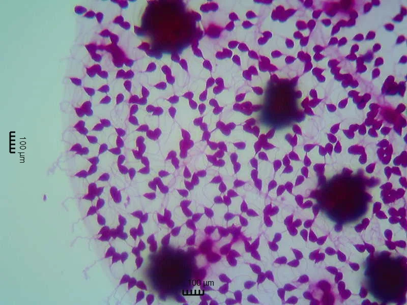
Part of a Volvox sphere, a genus whose largest spheres measure 0.35–0.92 mm long in seven species descriptions [10−4 m].
Color: stain (magenta, not named by the photographer)
"Volvox 1" by sgharvey — https://commons.wikimedia.org/wiki/File:Volvox_1.jpg — CC BY 2.0 (https://creativecommons.org/licenses/by/2.0/)
The even magenta of that photo looks like a dye, which its source does not name: borrowed color, not the alga's own green. The two 100 µm bars printed on it do not fit the cells either. By those bars a typical cell would be 39–54 µm long, computed for this page, while the species descriptions cited below give Volvox cells of 7 to 16 µm at most, so no size on this page is taken from that photo.
 Illustration
A jar of green-tinged water beside a fingertip, with a few green specks drifting in it.
Illustration — not to scale
Illustration: Ants, Cells & Atoms. Generated schematic illustration — not a photograph, micrograph or specimen record. © Ants, Cells & Atoms, all rights reserved.
How big is a Volvox sphere?
It depends on the species. In recent species descriptions the largest spheres, which biologists call spheroids, run from 0.35–0.40 mm long in V. africanus and V. reticuliferus (Nozaki and colleagues, 2015) to 0.92 mm in V. carteri from Taiwan (Nozaki and colleagues, 2018). Mature spheroids of cultured Japanese V. rousseletii strains measured 0.35–0.48 mm (Kimbara and colleagues, 2019). Cell counts vary as widely, from about 800 to 11,800 cells per spheroid in those descriptions, while V. barberi holds 10,000–50,000, the most recorded for any Volvox species as of 2008, by Solari and colleagues' account (abstract), and swims fast enough for daily vertical trips of 2–3 m an hour.
Umen's 2020 review describes single spheroids in a culture flask as just barely visible to the naked eye. That fits this site's Zoom ladder, where the naked-eye line sits near 0.1 mm. On the ladder a Volvox sphere shares the 10−4 m rung with Paramecium and diatoms, while each of its somatic cells, up to 7–10 µm in three of those descriptions, sits one or two rungs below among other cells. About three males of the smallest known parasitoid insect, each 139 µm long by Polilov's 2015 account, would span a 0.4 mm sphere, computed for this page; the smallest insects have a page of their own.
The photo below carries a bar that its source gives as 0.1 mm. Against that bar, the two whole spheres in the top row measure about 0.28–0.29 mm and 0.38–0.41 mm across, computed for this page from the bar's 140-pixel length. The photographer describes the five spheres as stages of asexual reproduction.
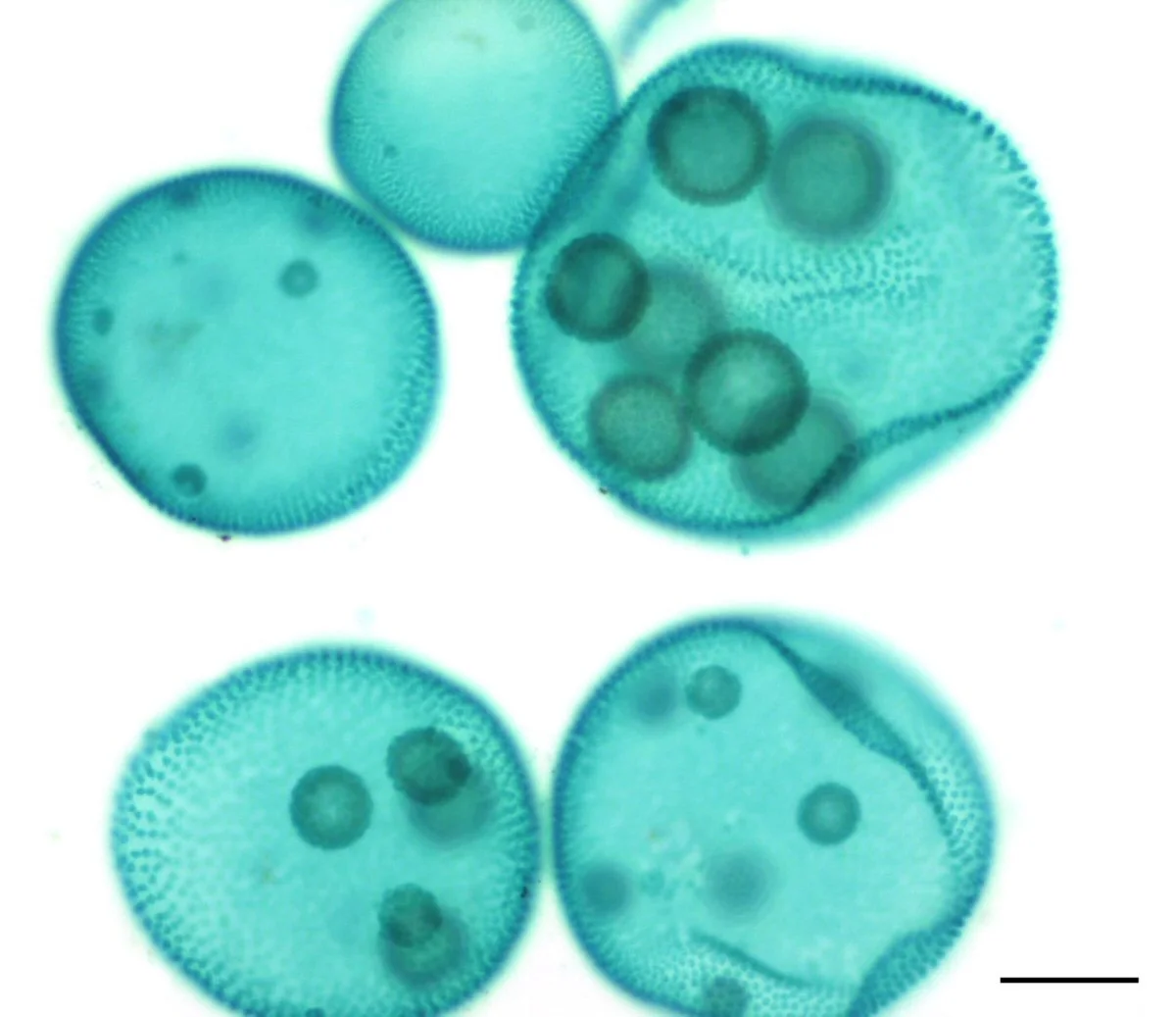
Volvox spheres at stages of asexual reproduction; the two whole ones in the top row are about 0.28 and 0.4 mm across by the printed 0.1 mm bar [10−4 m].
Color: native, color-balanced and background-adjusted by the photographer (no stain stated)
"Volvox asexual 2" by Jon Houseman and Matthew Ford — https://commons.wikimedia.org/wiki/File:Volvox_asexual_2.jpg — CC BY-SA 4.0 (https://creativecommons.org/licenses/by-sa/4.0/)
How does an embryo turn itself inside out?
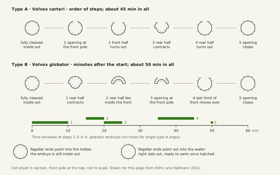
A new sphere starts inside its parent. In V. carteri, one of the 12–16 large reproductive cells, called gonidia, divides into about 2,000 small somatic cells and 12–16 new gonidia, laid out as a hollow ball one cell thick (Umen, 2020). At that stage every cell's flagellar end faces the hollow, not the water outside where flagella work for swimming, Höhn and Hallmann write, so the embryo must turn right side out. Its cells stay joined by cytoplasmic bridges, strands of cell material left when each division stops short of separating the daughters.
V. carteri does it in about 45 minutes, starting roughly an hour after the last division, by Höhn and Hallmann's account of earlier work. An opening called the phialopore appears at the front pole, its lips curl outward and back over the rear half, and the opening finally closes, leaving the flagellar ends outside and the gonidia inside. As the bend travels, the cells change shape and move along the bridges. A motor protein called InvA is thought to drive that movement.
V. globator runs the steps in another order, called type B. In Höhn and Hallmann's study of embryos with 2,000–6,000 cells, the rear half contracted and moved into the front half first, and only 20–25 minutes in did the phialopore open, letting the front half slide back over the inverted rear. The flip took about 50 minutes, and the embryo shrank from about 80 to 60 µm across.
Slide the diagram sideways to read its labels, or open it full size.
Two orders of the same flip in Volvox embryos about 50–90 µm across [10−5 m]: type A in V. carteri (about 45 minutes) and type B in V. globator (about 50 minutes).
Schematic, not to scale, drawn for this page from Höhn and Hallmann's 2011 text. Data: volvox-inversion-steps.csv (sources, version and license, JSON).
"Volvox inversion: two orders of the same flip" by Ants, Cells & Atoms, https://antscellsandatoms.com/microscopic-life/volvox/ — CC BY 4.0 (https://creativecommons.org/licenses/by/4.0/)
Data: Order and timing of inversion: Höhn S, Hallmann A 2011, BMC Biology 9:89, CC BY 2.0, https://doi.org/10.1186/1741-7007-9-89, retrieved 2026-10-25
Data: Höhn S, Hallmann A 2011, BMC Biology 9:89, CC BY 2.0, https://doi.org/10.1186/1741-7007-9-89, retrieved 2026-10-25
| Step | V. carteri (type A) | V. globator (type B), minutes after the start |
|---|---|---|
| 1 | Opening appears at the front pole | Rear half contracts and moves into the front half (0–10) |
| 2 | Front half turns out through it | Rear half lies inside the front half (15–20) |
| 3 | Rear half contracts | Opening appears at the front pole and widens (20–25) |
| 4 | Rear half turns out | Last third of the front half moves over the inverted rear half (35–45) |
| 5 | Opening closes, about 45 minutes in all | Opening closes (about 50) |
Genetic trees place Volvox species on more than one branch, Höhn and Hallmann note, and since the two types differ in almost every detail, they suggest the two flips arose separately. In 2015 Höhn and colleagues imaged inversion in living embryos in three dimensions and described it as an elastic shell whose cells change its local curvature (abstract). The figure below comes from that paper.
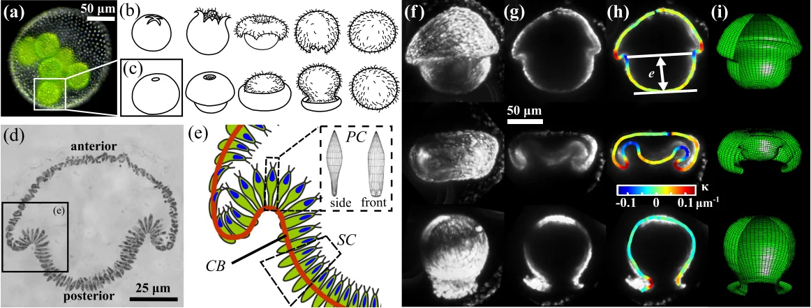
Embryonic inversion in Volvox, embryos 50–90 µm across [10−5 m]: a V. globator spheroid holding embryos (a), outlines of type A and type B (b, c), and sections and 3D views of V. globator embryos (d–i).
Color: native green in (a); grayscale in (d), (f) and (g); false color in the drawing (e), the curvature map (h) and the computed surfaces (i)
"Inversion Process for Volvox" by Höhn S, Honerkamp-Smith AR, Haas PA, Khuc Trong P, Goldstein RE — https://commons.wikimedia.org/wiki/File:Inversion_Process_for_Volvox.png — CC BY 3.0 (https://creativecommons.org/licenses/by/3.0/)

How does a sphere of cells swim and steer?
Once a young sphere hatches, its surface bristles with flagella, two per somatic cell, beating rearward in nearly parallel planes. The push is slightly off-axis, so the sphere rotates as it swims, once every 1 to 3 seconds in V. rousseletii (Ueki and colleagues, 2010). Linnaeus named the genus in 1758 for that spinning, by Pedley and colleagues' account.
Steering needs no known direct signals between cells. Front cells carry the largest eyespots, light-sensing spots about 3.6 µm wide in V. rousseletii, while rear eyespots are far smaller or missing; as rotation carries front cells into the light, they reverse their beat and the sphere turns toward it.
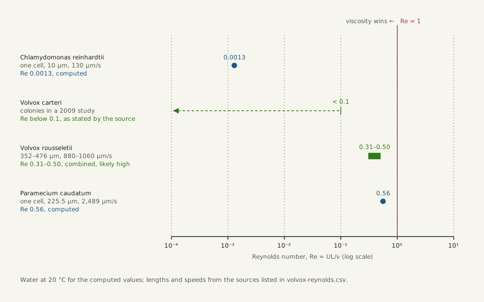
In Ueki's measurements, V. rousseletii hatchlings swam about 880 µm/s upward and 1,060 µm/s sideways. Paired with the 0.35–0.48 mm length of mature spheroids from other strains in the 2019 study, that gives a Reynolds number — a ratio of inertia to stickiness — of about 0.31–0.50 in water at 20 °C, a combined estimate computed for this page.
That figure is likely high: spheres keep growing after hatching, as Pedley and colleagues describe for V. carteri, so the hatchlings were probably shorter. Pedley's team puts even the largest V. carteri colonies in a 2009 data set below 0.1.
Slide the diagram sideways to read its labels, or open it full size.
Volvox spheres, 0.35–0.48 mm long in one species [10−4 m]: Reynolds number 0.31–0.50 (combined estimate, likely high) and below 0.1 (stated), beside two single cells.
Re = UL/ν; computed values use water at 20 °C from the IAPWS formulations; the single-cell values come from this site's swimmers table. Data: volvox-reynolds.csv (sources, version and license, JSON).
"Reynolds number: Volvox beside two single cells" by Ants, Cells & Atoms, https://antscellsandatoms.com/microscopic-life/volvox/ — CC BY 4.0 (https://creativecommons.org/licenses/by/4.0/)
Data sources for this figure
Data: Volvox rousseletii length: Kimbara R et al. 2019, PLOS ONE 14(8):e0221632, CC BY 4.0, https://doi.org/10.1371/journal.pone.0221632, retrieved 2026-10-25; Volvox rousseletii speed: Ueki N, Matsunaga S, Inouye I, Hallmann A 2010, BMC Biology 8:103, CC BY 2.0, https://doi.org/10.1186/1741-7007-8-103, retrieved 2026-10-25; Volvox carteri value: Pedley TJ, Brumley DR, Goldstein RE 2016, Journal of Fluid Mechanics 798:165-186, CC BY 4.0, https://doi.org/10.1017/jfm.2016.306, retrieved 2026-10-25; Chlamydomonas reinhardtii and Paramecium caudatum lengths and speeds: Lisicki M, Velho Rodrigues MF, Goldstein RE, Lauga E 2019, eLife 8:e44907, CC BY 4.0, https://doi.org/10.7554/eLife.44907, as listed in this site's swimmers-reynolds.csv, retrieved 2026-10-24; Water density and viscosity at 20 °C: International Association for the Properties of Water and Steam, IAPWS R6-95(2018) and IAPWS R12-08, as listed in this site's water-properties.csv; Reynolds numbers computed 2026-10-25
Data sources for this figure
Data: Kimbara R et al. 2019, PLOS ONE 14(8):e0221632, CC BY 4.0, https://doi.org/10.1371/journal.pone.0221632, retrieved 2026-10-25; Ueki N, Matsunaga S, Inouye I, Hallmann A 2010, BMC Biology 8:103, CC BY 2.0, https://doi.org/10.1186/1741-7007-8-103, retrieved 2026-10-25; Pedley TJ, Brumley DR, Goldstein RE 2016, Journal of Fluid Mechanics 798:165-186, CC BY 4.0, https://doi.org/10.1017/jfm.2016.306, retrieved 2026-10-25; Lisicki M, Velho Rodrigues MF, Goldstein RE, Lauga E 2019, eLife 8:e44907, CC BY 4.0, https://doi.org/10.7554/eLife.44907, as listed in this site's swimmers-reynolds.csv, retrieved 2026-10-24; Water density and viscosity at 20 °C: International Association for the Properties of Water and Steam, IAPWS R6-95(2018) and IAPWS R12-08, as listed in this site's water-properties.csv; Reynolds numbers computed 2026-10-25
At 0.31–0.50, inertia and viscosity are of the same order, with viscosity still winning; below 0.1, viscous forces are more than ten times larger. At 0.88 mm/s in water at 20 °C, the syrup line falls at a body length of about 1.1 mm, computed for this page, so these spheres stay under it. Volvox's closest single-celled relative, Chlamydomonas reinhardtii, sits near 0.0013, computed from Lisicki and colleagues' 10 µm and 130 µm/s.
Is Volvox one organism or a colony?
GBIF files Volvox under kingdom Plantae, phylum Chlorophyta: a green alga, and one of the freshwater plankton, taken from a lake's water column in the 2019 V. rousseletii study. Whether each sphere is one organism or a colony depends on whom you read: Pedley and colleagues call it a colony, while Ueki and colleagues rank Volvox among the multicellular genera and Eudorina among the colonial ones.
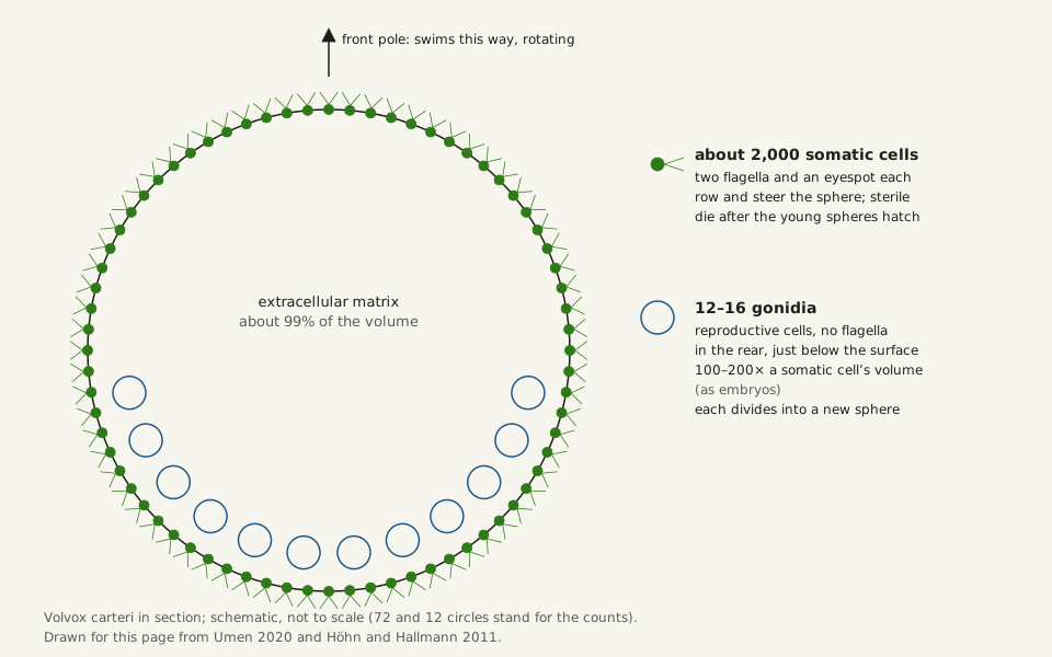
The case for one organism rests on a division of labour. In V. carteri about 2,000 small somatic cells, each similar in size to a Chlamydomonas cell, swim and are sterile, while only the 12–16 gonidia make new spheres; Umen's 2020 review calls that complete split a hallmark of complex multicellularity. After the young spheres hatch, the parent's somatic cells die.
Slide the diagram sideways to read its labels, or open it full size.
Volvox carteri in section, a sphere several hundred µm across [10−4 m]: about 2,000 sterile somatic cells at the surface, 12–16 gonidia below it, and a matrix filling about 99% of the volume.
Schematic, not to scale, drawn for this page from Umen's 2020 review and Höhn and Hallmann's 2011 study.
"Volvox carteri: two kinds of cell in one sphere" by Ants, Cells & Atoms, https://antscellsandatoms.com/microscopic-life/volvox/ — CC BY 4.0 (https://creativecommons.org/licenses/by/4.0/)
Data: Umen JG 2020, EvoDevo 11:13, CC BY 4.0, https://doi.org/10.1186/s13227-020-00158-7, retrieved 2026-10-25; Höhn S, Hallmann A 2011, BMC Biology 9:89, CC BY 2.0, https://doi.org/10.1186/1741-7007-9-89, retrieved 2026-10-25
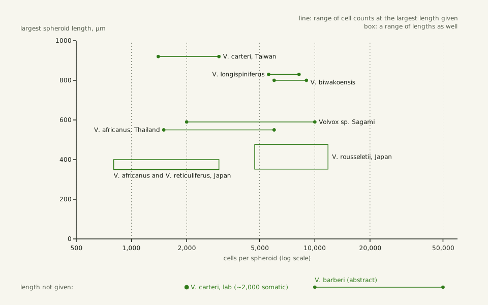
Most of a sphere is not cells at all. A clear extracellular matrix, the jelly the cells sit in, fills around 99% of a V. carteri spheroid's volume (Umen, 2020). In the species descriptions charted below, more cells do not always mean a bigger sphere. Taiwanese V. carteri reaches 0.92 mm with 1,400–3,000 somatic cells, while cultured V. rousseletii, with 4,700–11,800 cells, stayed under 0.48 mm.
Slide the diagram sideways to read its labels, or open it full size.
Volvox spheres, the largest 0.35–0.92 mm long by species [10−4 m], against their 800 to 11,800 cells; V. barberi holds 10,000–50,000 by one abstract, with no length given.
Lines: cell counts at the largest length each description gives; boxes: a range of lengths as well. Data: volvox-species.csv (sources, version and license, JSON).
"Volvox: cells per sphere against sphere length, by species" by Ants, Cells & Atoms, https://antscellsandatoms.com/microscopic-life/volvox/ — CC BY 4.0 (https://creativecommons.org/licenses/by/4.0/)
Data sources for this figure
Data: Nozaki H et al. 2015, PLOS ONE 10(11):e0142632, CC BY 4.0, https://doi.org/10.1371/journal.pone.0142632, retrieved 2026-10-25; Nozaki H et al. 2016, PLOS ONE 11(11):e0167148, CC BY 4.0, https://doi.org/10.1371/journal.pone.0167148, retrieved 2026-10-25; Nozaki H et al. 2018, Botanical Studies 59:10, CC BY 4.0, https://doi.org/10.1186/s40529-018-0227-9, retrieved 2026-10-25; Kimbara R et al. 2019, PLOS ONE 14(8):e0221632, CC BY 4.0, https://doi.org/10.1371/journal.pone.0221632, retrieved 2026-10-25; Nozaki H et al. 2020, PLOS ONE 15(7):e0235622, CC BY 4.0, https://doi.org/10.1371/journal.pone.0235622, retrieved 2026-10-25; Nozaki H et al. 2022, Botanical Studies 63:1, CC BY 4.0, https://doi.org/10.1186/s40529-022-00332-1, retrieved 2026-10-25; Nozaki H et al. 2024, PLOS ONE 19(9):e0310549, CC BY 4.0, https://doi.org/10.1371/journal.pone.0310549, retrieved 2026-10-25; Volvox barberi cell number (abstract): Solari CA, Michod RE, Goldstein RE 2008, Journal of Phycology 44:1395-1398, https://doi.org/10.1111/j.1529-8817.2008.00603.x, retrieved 2026-10-25; Volvox carteri laboratory strains: Umen JG 2020, EvoDevo 11:13, CC BY 4.0, https://doi.org/10.1186/s13227-020-00158-7, retrieved 2026-10-25
Data sources for this figure
Data: Umen JG 2020, EvoDevo 11:13, CC BY 4.0, https://doi.org/10.1186/s13227-020-00158-7, retrieved 2026-10-25; Nozaki H et al. 2015, PLOS ONE 10(11):e0142632, CC BY 4.0, https://doi.org/10.1371/journal.pone.0142632, retrieved 2026-10-25; Nozaki H et al. 2016, PLOS ONE 11(11):e0167148, CC BY 4.0, https://doi.org/10.1371/journal.pone.0167148, retrieved 2026-10-25; Nozaki H et al. 2018, Botanical Studies 59:10, CC BY 4.0, https://doi.org/10.1186/s40529-018-0227-9, retrieved 2026-10-25; Kimbara R et al. 2019, PLOS ONE 14(8):e0221632, CC BY 4.0, https://doi.org/10.1371/journal.pone.0221632, retrieved 2026-10-25; Nozaki H et al. 2020, PLOS ONE 15(7):e0235622, CC BY 4.0, https://doi.org/10.1371/journal.pone.0235622, retrieved 2026-10-25; Nozaki H et al. 2022, Botanical Studies 63:1, CC BY 4.0, https://doi.org/10.1186/s40529-022-00332-1, retrieved 2026-10-25; Nozaki H et al. 2024, PLOS ONE 19(9):e0310549, CC BY 4.0, https://doi.org/10.1371/journal.pone.0310549, retrieved 2026-10-25; Solari CA, Michod RE, Goldstein RE 2008, Journal of Phycology 44:1395-1398 (abstract), https://doi.org/10.1111/j.1529-8817.2008.00603.x, retrieved 2026-10-25; GBIF Secretariat 2023, GBIF Backbone Taxonomy, Volvox (2639376), Volvox carteri (2639377), Volvox longispiniferus (11439135), checklist dataset https://doi.org/10.15468/39omei, CC BY 4.0, retrieved 2026-10-25
Sexual spheres arise when a hormone called sex inducer turns V. carteri gonidia into males or females, Umen writes. When the group became multicellular is still being dated: about 200 million years ago by the estimate Höhn and Hallmann cite, or about 250 million years for the split from single-celled relatives in Umen's review, which notes that sparse green-algal fossils make such dates hard to calibrate.
Sources
- Höhn S, Hallmann A. "There is more than one way to turn a spherical cellular monolayer inside out: type B embryo inversion in Volvox globator." BMC Biology 9:89 (2011). https://doi.org/10.1186/1741-7007-9-89 Read: full text. Text license: CC BY 2.0 (https://creativecommons.org/licenses/by/2.0/).
- Nozaki H, Ueki N, Isaka N, Saigo T, Yamamoto K, Matsuzaki R, Takahashi F, Wakabayashi K-i, Kawachi M. "A New Morphological Type of Volvox from Japanese Large Lakes and Recent Divergence of this Type and V. ferrisii in Two Different Freshwater Habitats." PLOS ONE 11(11):e0167148 (2016). https://doi.org/10.1371/journal.pone.0167148 Read: full text. Text license: CC BY 4.0 (https://creativecommons.org/licenses/by/4.0/).
- Nozaki H, Matsuzaki R, Shimotori K, Ueki N, Heman W, Mahakham W, Yamaguchi H, Tanabe Y, Kawachi M. "Two species of the green algae Volvox sect. Volvox from the Japanese ancient lake, Lake Biwa." PLOS ONE 19(9):e0310549 (2024). https://doi.org/10.1371/journal.pone.0310549 Read: full text. Text license: CC BY 4.0 (https://creativecommons.org/licenses/by/4.0/).
- Nozaki H, Matsuzaki R, Yamamoto K, Kawachi M, Takahashi F. "Delineating a New Heterothallic Species of Volvox (Volvocaceae, Chlorophyceae) Using New Strains of 'Volvox africanus'." PLOS ONE 10(11):e0142632 (2015). https://doi.org/10.1371/journal.pone.0142632 Read: full text. Text license: CC BY 4.0 (https://creativecommons.org/licenses/by/4.0/).
- Nozaki H, Ueki N, Takusagawa M, Yamashita S, Misumi O, Matsuzaki R, Kawachi M, Chiang Y-R, Wu J-T. "Morphology, taxonomy and mating-type loci in natural populations of Volvox carteri in Taiwan." Botanical Studies 59:10 (2018). https://doi.org/10.1186/s40529-018-0227-9 Read: full text. Text license: CC BY 4.0 (https://creativecommons.org/licenses/by/4.0/).
- Kimbara R, Isaka N, Matsuzaki R, Kawai-Toyooka H, Kawachi M, Nozaki H. "Morphological and molecular identification of the dioecious 'African species' Volvox rousseletii (Chlorophyceae) in the water column of a Japanese lake based on field-collected and cultured materials." PLOS ONE 14(8):e0221632 (2019). https://doi.org/10.1371/journal.pone.0221632 Read: full text. Text license: CC BY 4.0 (https://creativecommons.org/licenses/by/4.0/).
- Solari CA, Michod RE, Goldstein RE. "Volvox barberi, the fastest swimmer of the Volvocales (Chlorophyceae)." Journal of Phycology 44(6):1395–1398 (2008). https://doi.org/10.1111/j.1529-8817.2008.00603.x Read: abstract.
- Umen JG. "Volvox and volvocine green algae." EvoDevo 11:13 (2020). https://doi.org/10.1186/s13227-020-00158-7 Read: full text. Text license: CC BY 4.0 (https://creativecommons.org/licenses/by/4.0/).
- Polilov AA. "How small is the smallest? New record and remeasuring of Scydosella musawasensis Hall, 1999 (Coleoptera, Ptiliidae), the smallest known free-living insect." ZooKeys 526:61–64 (2015). https://doi.org/10.3897/zookeys.526.6531 Read: full text. Text license: CC BY 4.0 (https://creativecommons.org/licenses/by/4.0/).
- Höhn S, Honerkamp-Smith AR, Haas PA, Khuc Trong P, Goldstein RE. "Dynamics of a Volvox Embryo Turning Itself Inside Out." Physical Review Letters 114(17):178101 (2015). https://doi.org/10.1103/PhysRevLett.114.178101 Read: abstract. Text license: CC BY 3.0 (https://creativecommons.org/licenses/by/3.0/).
- Ueki N, Matsunaga S, Inouye I, Hallmann A. "How 5000 independent rowers coordinate their strokes in order to row into the sunlight: Phototaxis in the multicellular green alga Volvox." BMC Biology 8:103 (2010). https://doi.org/10.1186/1741-7007-8-103 Read: full text. Text license: CC BY 2.0 (https://creativecommons.org/licenses/by/2.0/).
- Pedley TJ, Brumley DR, Goldstein RE. "Squirmers with swirl: a model for Volvox swimming." Journal of Fluid Mechanics 798:165–186 (2016). https://doi.org/10.1017/jfm.2016.306 Read: full text. Text license: CC BY 4.0 (https://creativecommons.org/licenses/by/4.0/).
- International Association for the Properties of Water and Steam. IAPWS R6-95(2018), Revised Release on the IAPWS Formulation 1995 for the Thermodynamic Properties of Ordinary Water Substance for General and Scientific Use (2018). https://iapws.org/technical-guidance/release/IAPWS-95 Read: values as computed from the release equations in this site's water-properties table. Text license: IAPWS: publication in whole or in part allowed with attribution (https://iapws.org/technical-guidance/release/IAPWS-95).
- International Association for the Properties of Water and Steam. IAPWS R12-08, Release on the IAPWS Formulation 2008 for the Viscosity of Ordinary Water Substance (2008). https://iapws.org/technical-guidance/release/viscosity Read: values as computed from the release equations in this site's water-properties table. Text license: IAPWS: publication in whole or in part allowed with attribution (https://iapws.org/technical-guidance/release/viscosity).
- Lisicki M, Velho Rodrigues MF, Goldstein RE, Lauga E. "Swimming eukaryotic microorganisms exhibit a universal speed distribution." eLife 8:e44907 (2019). https://doi.org/10.7554/eLife.44907 Read: full text. Text license: CC BY 4.0 (https://creativecommons.org/licenses/by/4.0/).
- GBIF Secretariat. GBIF Backbone Taxonomy. Checklist dataset https://doi.org/10.15468/39omei, accessed 2026-10-25; Volvox Linnaeus, 1758, usage key 2639376; Volvox carteri, usage key 2639377; Volvox globator L., usage key 2639390; Volvox longispiniferus Nozaki & Mahakham, 2020, usage key 11439135. Read: dataset record (species match and species records). Text license: CC BY 4.0 (https://creativecommons.org/licenses/by/4.0/).
- Nozaki H, Mahakham W, Heman W, Matsuzaki R, Kawachi M. "A new preferentially outcrossing monoicous species of Volvox sect. Volvox (Chlorophyta) from Thailand." PLOS ONE 15(7):e0235622 (2020). https://doi.org/10.1371/journal.pone.0235622 Read: full text. Text license: CC BY 4.0 (https://creativecommons.org/licenses/by/4.0/).
- Nozaki H, Mahakham W, Heman W, Matsuzaki R, Kawachi M. "Morphology, mating system and taxonomy of Volvox africanus (Volvocaceae, Chlorophyceae) from Thailand." Botanical Studies 63:1 (2022). https://doi.org/10.1186/s40529-022-00332-1 Read: full text. Text license: CC BY 4.0 (https://creativecommons.org/licenses/by/4.0/).
Cite this page
Ants, Cells & Atoms. “Volvox: Each Embryo Turns Inside Out Before It Can Swim.” Published 2026-10-25. https://antscellsandatoms.com/microscopic-life/volvox/. Accessed (add the date you read it).
No DOI is issued for entries. Figures made here are CC BY 4.0; see each caption for the license of photographs.
Related entries
 Illustration
Diatoms: Why Most Glass-Walled Algae Shrink as They Multiply
Image: Illustration: Ants, Cells & Atoms, all rights reserved
Illustration: Ants, Cells & Atoms. Generated schematic illustration — not a photograph, micrograph or specimen record. © Ants, Cells & Atoms, all rights reserved.
Plankton: From Bacteria to Jellyfish, Sorted in Size Steps
Image: “Diatomea. - Schachtellinge” by Ernst Haeckel (artist) and Adolf Giltsch (lithographer) (1904) · Public domain
"Diatomea. - Schachtellinge" by Ernst Haeckel (artist) and Adolf Giltsch (lithographer) (1904) — https://commons.wikimedia.org/wiki/File:Diatomea._-_Schachtellinge_LCCN2015647735.jpg — Public domain (artist Ernst Haeckel died 1919 and lithographer Adolf Giltsch died 1911, so the plate is out of copyright in the EU (life plus 70 years) and in the US (published 1904, before 1930); the scan is from the Library of Congress Prints and Photographs division (lccn.loc.gov/2015647735), which lists no rights restriction)

Paramecium: How a Nerve-Like Signal Sends One Cell Backward
Image: “Paramecium 1” by Nbnidhi (2017) · CC BY 4.0
"Paramecium 1" by Nbnidhi — https://commons.wikimedia.org/wiki/File:Paramecium_1.jpg — CC BY 4.0 (https://creativecommons.org/licenses/by/4.0/)
