Paramecium: How a Nerve-Like Signal Sends One Cell Backward

Slide the ladder sideways to read its labels, or open it full size.
Paramecium caudatum: cells 140–311 µm long in a 2021 database [10−4 m], with a Paramecium cilium, about 10 µm long and 0.2 µm thick, for scale.
Log scale from 10 cm to 0.1 nm, each rung ten times smaller; dashed lines: naked eye about 0.1 mm, light microscope about 0.2 µm (How to read the Zoom ladder). Pink band: the syrup line, Re = 1 in water at 20 °C at 1–10 body lengths per second. Data: paramecium-sizes.csv (sources, version and license, JSON).
"Zoom ladder: Paramecium caudatum" by Ants, Cells & Atoms, https://antscellsandatoms.com/microscopic-life/paramecium/ — CC BY 4.0 (https://creativecommons.org/licenses/by/4.0/)
Data sources for this figure
Data: Cell length: Velho Rodrigues MF, Lisicki M, Lauga E 2021, PLOS ONE 16(6):e0252291, CC BY 4.0, https://doi.org/10.1371/journal.pone.0252291, retrieved 2026-10-24; Cilium length and thickness: Brette R 2021, eNeuro 8(3):ENEURO.0018-21.2021, CC BY 4.0, https://doi.org/10.1523/ENEURO.0018-21.2021, retrieved 2026-10-24; Syrup line band: body length at Re = 1 for 1–10 body lengths per second in water at 20 °C, density and viscosity from IAPWS R6-95(2018) and IAPWS R12-08 (attribution: International Association for the Properties of Water and Steam), as listed in this site's the-syrup-line-bands.csv, computed 2026-10-24
Data sources for this figure
Data: Velho Rodrigues MF, Lisicki M, Lauga E 2021, PLOS ONE 16(6):e0252291, CC BY 4.0, https://doi.org/10.1371/journal.pone.0252291, retrieved 2026-10-24; Lisicki M, Velho Rodrigues MF, Goldstein RE, Lauga E 2019, eLife 8:e44907, CC BY 4.0, https://doi.org/10.7554/eLife.44907, retrieved 2026-10-24; Brette R 2021, eNeuro 8(3):ENEURO.0018-21.2021, CC BY 4.0, https://doi.org/10.1523/ENEURO.0018-21.2021, retrieved 2026-10-24; Long H et al. 2023, Annual Review of Genetics 57:391–410 (author manuscript), CC BY 4.0, https://doi.org/10.1146/annurev-genet-071819-104035, retrieved 2026-10-24; Scale-bar measurement: Josef Reischig, Paramecium caudatum (248 06) Native preparation, CC BY-SA 3.0, https://commons.wikimedia.org/wiki/File:Paramecium_caudatum_(248_06)_Native_preparation.jpg; length computed 2026-10-24; GBIF Secretariat 2023, GBIF Backbone Taxonomy, Paramecium caudatum (taxon key 8122199) and Ciliophora (7765738), checklist dataset https://doi.org/10.15468/39omei, CC BY 4.0, retrieved 2026-10-24

Bench card
- Actual size
- 140–311 µm long [10−4 m] and 35–70 µm wide across compiled studiesSource: Velho Rodrigues et al. 2021 (BOSO-Micro), https://doi.org/10.1371/journal.pone.0252291
- Size in this image
- not measurable from this image
- Lives in
- fresh water, in ponds and lakes around the worldSource: Brette 2021, https://doi.org/10.1523/ENEURO.0018-21.2021
- Seen with
- naked eye, just, as a speck; light microscope for its shape and ciliaSource: Long et al. 2023, https://doi.org/10.1146/annurev-genet-071819-104035
- Color in images
- stain in the opening photo (pink, not named by the photographer) and in the inside view (dark violet, the dye not named); native in the phase-contrast photoSource: figure captions on this page
paramecium-bench.csv (CSV) · where each value comes from, and its license (JSON)
Data credits for this card
Data: GBIF Secretariat 2023, GBIF Backbone Taxonomy, Paramecium caudatum (taxon key 8122199) and Ciliophora (7765738), checklist dataset https://doi.org/10.15468/39omei, CC BY 4.0, retrieved 2026-10-24; Velho Rodrigues MF, Lisicki M, Lauga E 2021, PLOS ONE 16(6):e0252291, CC BY 4.0, https://doi.org/10.1371/journal.pone.0252291, retrieved 2026-10-24; Brette R 2021, eNeuro 8(3):ENEURO.0018-21.2021, CC BY 4.0, https://doi.org/10.1523/ENEURO.0018-21.2021, retrieved 2026-10-24; Long H et al. 2023, Annual Review of Genetics 57:391–410 (author manuscript), CC BY 4.0, https://doi.org/10.1146/annurev-genet-071819-104035, retrieved 2026-10-24
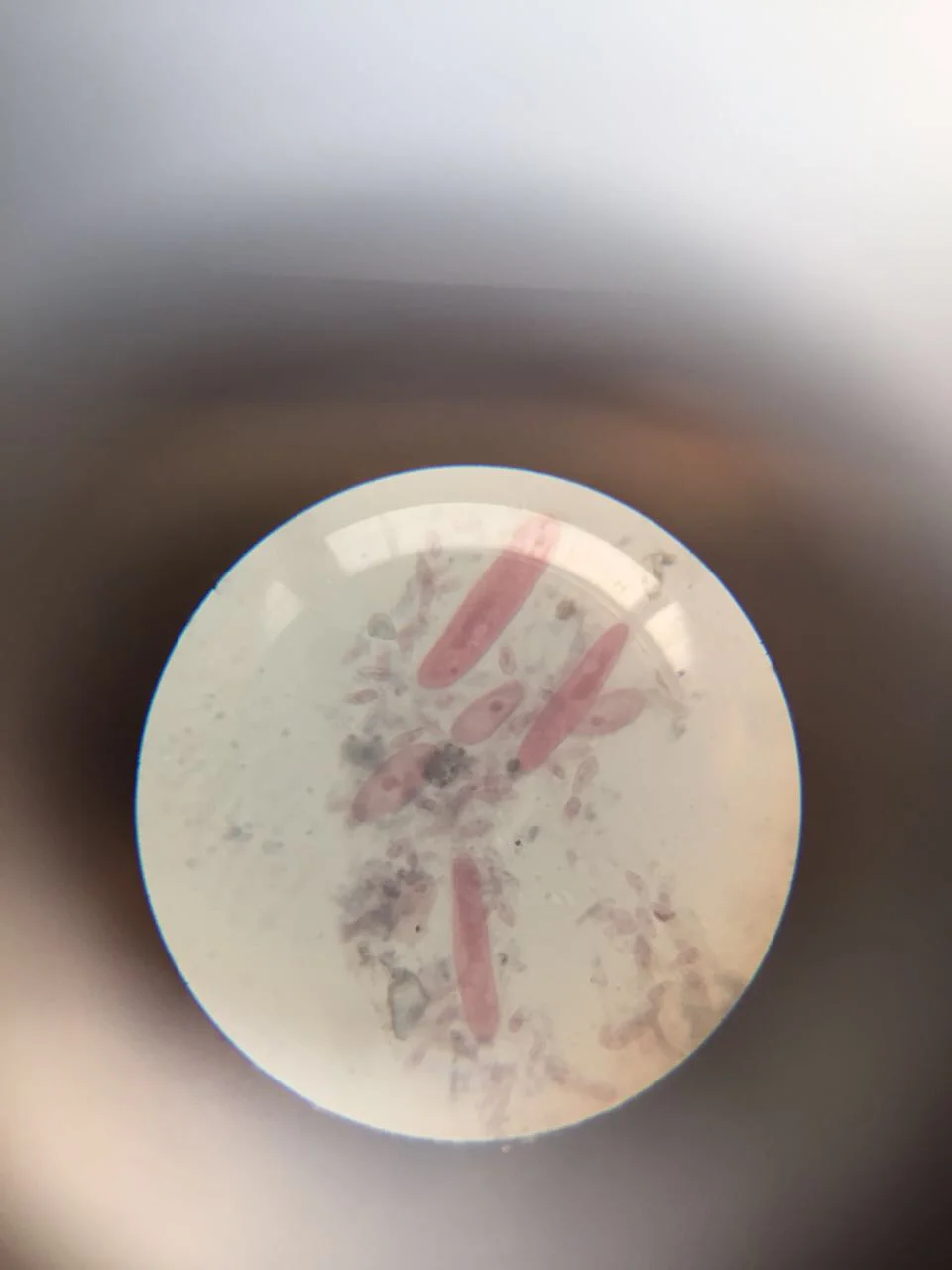
Paramecium cells photographed through a microscope eyepiece; most species in the genus are about 50–300 µm long, a 2023 review says [10−5–10−4 m].
Color: stain (pink, not named by the photographer)
"Paramecium 1" by Nbnidhi — https://commons.wikimedia.org/wiki/File:Paramecium_1.jpg — CC BY 4.0 (https://creativecommons.org/licenses/by/4.0/)

How big is a paramecium?
It depends on the species. Two recent reviews put most species at about 50–300 µm long (Long and colleagues, 2023) or at 100–300 µm, depending on species (Brette, 2021). A 2019 compilation by Lisicki and colleagues gives one typical size for each of ten entries, from 91 µm for P. polycaryum (P. polycarium in GBIF) to 251 µm for P. multimicronucleatum.
Brette describes P. caudatum as a large species, about 200 µm long. Across the studies gathered in the BOSO-Micro database (Velho Rodrigues, Lisicki and Lauga, 2021), its cells run 140–311 µm long and 35–70 µm wide.
Long and colleagues add that cells this size are just visible to the naked eye. This site's Zoom ladder puts that limit at about 0.1 mm, a guide that shifts with viewing distance, contrast and eyesight. P. caudatum sits just above it: a speck at best, with its shape left to a light microscope. Next to other single cells it is large: the flagellates in Lisicki's compilation average 31 µm, about a seventh of the 225.5 µm listed there for P. caudatum.
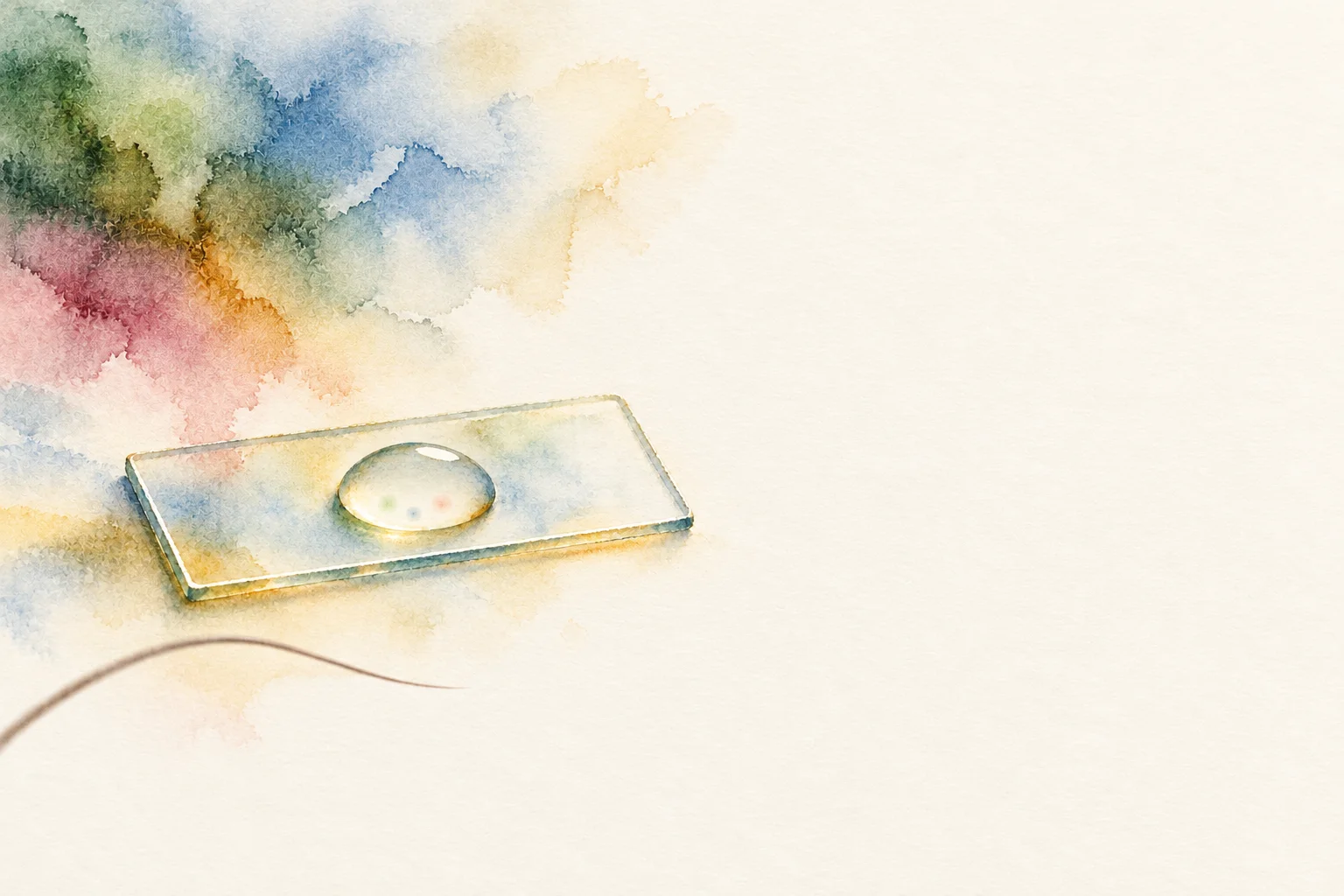 Illustration
A drop of water on a glass slide, a hair beside it; to the naked eye a paramecium is a speck at best.
Illustration — not to scale
Illustration: Ants, Cells & Atoms. Generated schematic illustration — not a photograph, micrograph or specimen record. © Ants, Cells & Atoms, all rights reserved.
In the photo below, the cell is about twice as long as the printed 0.1 mm scale bar: 197–200 µm, computed for this page from the bar's length in pixels. On the ladder, that is the rung this page shares with diatoms and Volvox.
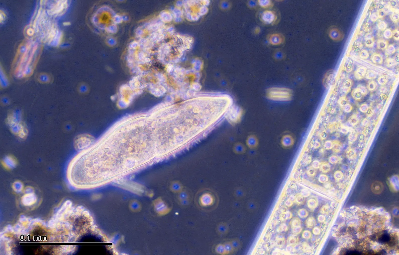
Paramecium caudatum in negative phase contrast, about twice the length of the 0.1 mm bar printed on the photo [10−4 m].
Color: native (unstained preparation, negative phase contrast)
"Paramecium caudatum (248 06) Native preparation" by Josef Reischig — https://commons.wikimedia.org/wiki/File:Paramecium_caudatum_(248_06)_Native_preparation.jpg — CC BY-SA 3.0 (https://creativecommons.org/licenses/by-sa/3.0/)
Size also sets how far anything dissolved in the water must travel to reach the middle of the cell. No point inside a P. caudatum cell, at most 70 µm wide in that database, lies more than 35 µm from its surface. Oxygen in water at 25 °C has a diffusion coefficient of 2.1×10⁻⁵ cm2/s, by Somerville and Proctor's 2013 figure, so it typically spreads 35 µm along one direction in about 0.3 s, computed for this page from Vestergaard and colleagues' 2012 one-dimensional diffusion time, x2/(2D) (paramecium-oxygen-diffusion.csv (sources, version and license, JSON) Data: Somerville GA, Proctor RA 2013, BMC Microbiology 13:9, CC BY 2.0, https://doi.org/10.1186/1471-2180-13-9, retrieved 2026-10-24; Velho Rodrigues MF, Lisicki M, Lauga E 2021, PLOS ONE 16(6):e0252291, CC BY 4.0, https://doi.org/10.1371/journal.pone.0252291, retrieved 2026-10-24; One-dimensional diffusion time t = x²/(2D): Vestergaard CL, Flyvbjerg H, Møller IM 2012, Frontiers in Plant Science 3:295, CC BY 3.0, https://doi.org/10.3389/fpls.2012.00295, retrieved 2026-10-24; Diffusion times computed 2026-10-24). Across 1 cm, the same spread takes about 6.6 hours; for that arithmetic at insect size, see insect breathing.
What is inside the cell, and how does it eat?
Food goes in at one place. Cilia lining the oral groove, a furrow along one side of the cell, sweep bacteria, yeast or algae toward the mouth, and these oral cilia behave differently from the ones that row the cell, Brette writes. In P. caudatum, a new food vacuole, a membrane sac that holds the meal, grows at the cytopharynx, the gullet; spent vacuoles are emptied at a site called the cytoproct, as Allen described in 1974 (abstract). A well-fed paramecium can divide every 6 hours, depending on temperature.
The other traffic is water. The cell's contents hold about five times fewer ions than an animal cell's, Brette notes, but fresh water typically holds fewer still, so water keeps seeping in by osmosis; contractile vacuoles expel it.
Just under the surface sit trichocysts, needle-like bodies the cell can fire. When a laser heats a paramecium by 5–10 °C, the cell can throw them and jump away within 5 ms at about 10 mm/s, in experiments Brette describes from a 2011 study by Hamel and colleagues.
Each cell also carries two kinds of nucleus, Long and colleagues explain: a germline micronucleus that holds the genome passed to offspring, and a somatic macronucleus with hundreds of copies of each chromosome, whose genes are in constant use. The dark violet in the photo below is borrowed color: the photographer's notes say the cells were stained but do not name the dye.
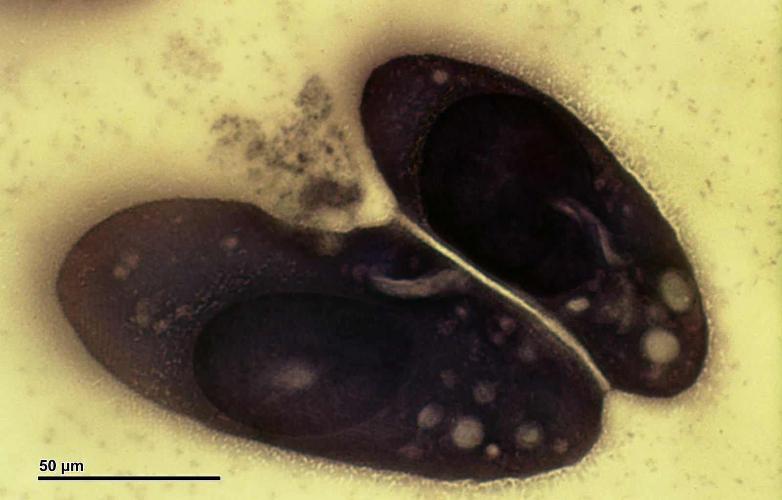
Two stained Paramecium caudatum cells with the 50 µm bar printed on the photo; the species measures 140–311 µm long in a 2021 database [10−4 m].
Color: stain (dark violet, not named by the photographer)
"Paramecium caudatum (250 20) Cultured" by Josef Reischig — https://commons.wikimedia.org/wiki/File:Paramecium_caudatum_(250_20)_Cultured.jpg — CC BY-SA 3.0 (https://creativecommons.org/licenses/by-sa/3.0/)

How do thousands of cilia move one cell?
Several thousand cilia cover a paramecium; Brette's review cites a count of about 4,000 for the smaller P. tetraurelia, each cilium about 10 µm long and 0.2 µm thick. That thickness sits right on the ladder's light-microscope line, about 0.2 µm. In forward swimming each cilium beats 10–20 times a second, its power stroke toward the rear and to the right, so the cell rolls about its long axis, about once a second; its path is a spiral.
Neighbouring cilia beat slightly out of step. That offset sends waves of beating over the surface at about 1 µm per millisecond. No control centre times these metachronal waves; Brette credits the flow of water between cilia and the mechanical coupling through the cell body.
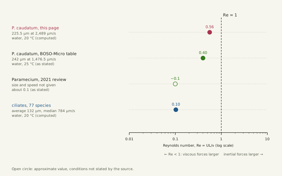
The Reynolds number — a ratio of inertia to stickiness — tells how water treats a swimmer of this size. With the 225.5 µm and 2,489 µm/s that Lisicki's compilation gives for P. caudatum, it comes to about 0.56 in water at 20 °C, computed for this page (Reynolds Number). BOSO-Micro's own table gives 0.40 in water at 25 °C from its averages of 242 µm and 1,476.5 µm/s, and Brette's review puts it at about 0.1. Measured speeds of P. caudatum run from 478.7 to 4,500 µm/s across the studies in that database.
All three sit below 1. At the two compiled figures for P. caudatum, 0.40–0.56, inertia and viscosity are of the same order, with viscosity still winning; at Brette's 0.1, viscous forces are about ten times larger. The syrup line moves with speed: at 2.49 mm/s in water at 20 °C, Re reaches 1 at a body length of about 0.40 mm, computed for this page, a little under twice the cell's 225.5 µm. The plankton page lays out where other drifting swimmers fall against the same line.
Slide the diagram sideways to read its labels, or open it full size.
Paramecium caudatum at 225.5 µm (a typical size) and 242 µm (an average length) in two compilations [10−4 m]: Reynolds number 0.40–0.56, beside a review's 0.1 for the genus and 0.10 for the average ciliate.
Re = UL/ν; values marked computed use the viscosity of water from the IAPWS 2008 formulation, the others are as stated by their sources. Data: paramecium-reynolds.csv (sources, version and license, JSON).
"Paramecium Reynolds number: four estimates below Re = 1" by Ants, Cells & Atoms, https://antscellsandatoms.com/microscopic-life/paramecium/ — CC BY 4.0 (https://creativecommons.org/licenses/by/4.0/)
Data sources for this figure
Data: Paramecium caudatum and ciliate sizes and speeds: Lisicki M, Velho Rodrigues MF, Goldstein RE, Lauga E 2019, eLife 8:e44907, CC BY 4.0, https://doi.org/10.7554/eLife.44907, retrieved 2026-10-24; Database Reynolds number: Velho Rodrigues MF, Lisicki M, Lauga E 2021, PLOS ONE 16(6):e0252291, CC BY 4.0, https://doi.org/10.1371/journal.pone.0252291, retrieved 2026-10-24; Review value: Brette R 2021, eNeuro 8(3):ENEURO.0018-21.2021, CC BY 4.0, https://doi.org/10.1523/ENEURO.0018-21.2021, retrieved 2026-10-24; Water density and viscosity at 20 °C: International Association for the Properties of Water and Steam, IAPWS R6-95(2018) and IAPWS R12-08, as listed in this site's water-properties.csv; Reynolds numbers computed 2026-10-24
Data sources for this figure
Data: Lisicki M, Velho Rodrigues MF, Goldstein RE, Lauga E 2019, eLife 8:e44907, CC BY 4.0, https://doi.org/10.7554/eLife.44907, retrieved 2026-10-24; Velho Rodrigues MF, Lisicki M, Lauga E 2021, PLOS ONE 16(6):e0252291, CC BY 4.0, https://doi.org/10.1371/journal.pone.0252291, retrieved 2026-10-24; Brette R 2021, eNeuro 8(3):ENEURO.0018-21.2021, CC BY 4.0, https://doi.org/10.1523/ENEURO.0018-21.2021, retrieved 2026-10-24; Water density and viscosity at 20 °C: International Association for the Properties of Water and Steam, IAPWS R6-95(2018) and IAPWS R12-08, as listed in this site's water-properties.csv; Reynolds numbers computed 2026-10-24
What happens when it bumps into something?
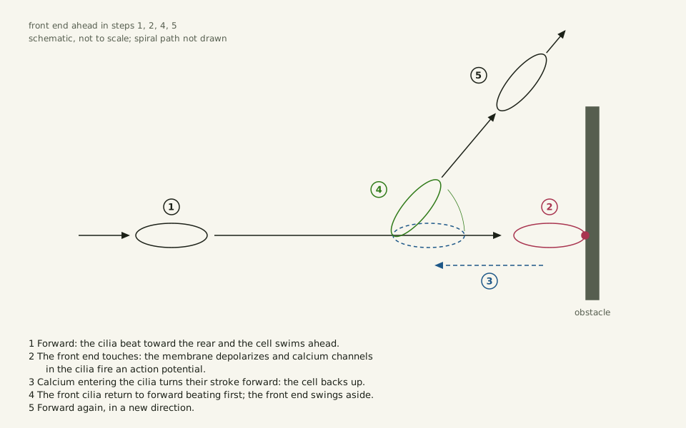
Touch the front end, and calcium ions flow in through channels that open under mechanical pressure, as Kunita and colleagues summarize. That shifts the voltage across the membrane. If the shift is large enough, calcium channels in the cilia fire an action potential — a brief electrical spike of the kind nerve cells use — a finding Brette's review credits to Eckert's 1972 work. Some authors, Brette notes, call the cell a "swimming neuron".
Calcium flowing into the cilia turns their power stroke toward the front, and the cell swims backward. Potassium channels then bring the voltage back, calcium is pumped out or bound up, and the cilia return to their usual stroke. The front cilia switch back first. While the rear ones still beat in reverse, the front end swings aside, and the cell sets off in a new direction.
Slide the diagram sideways to read its labels, or open it full size.
The avoiding reaction of Paramecium, a cell 100–300 µm long in one review, the length varying with species [10−4 m], in five steps: forward, touch, back up, swing the front end aside, forward in a new direction.
Schematic, not to scale, drawn for this page from Brette's 2021 description; the spiral path and the cell's rotation are left out.
"Paramecium avoiding reaction: back up, turn, go" by Ants, Cells & Atoms, https://antscellsandatoms.com/microscopic-life/paramecium/ — CC BY 4.0 (https://creativecommons.org/licenses/by/4.0/)
Data: Sequence of the avoiding reaction: Brette R 2021, eNeuro 8(3):ENEURO.0018-21.2021, CC BY 4.0, https://doi.org/10.1523/ENEURO.0018-21.2021, retrieved 2026-10-24; Kunita I, Kuroda S, Ohki K, Nakagaki T 2014, Frontiers in Microbiology 5:270, CC BY 3.0, https://doi.org/10.3389/fmicb.2014.00270, retrieved 2026-10-24
The reaction is graded. A weak stimulus may only swing the front end, a stronger one adds a stretch of backward swimming, and a very strong one can bring a long retreat and a full circle. Citing Machemer and Eckert's 1973 work, Brette writes that how long the cell swims backward tracks how far the stimulus shifted the voltage. A touch at the rear does the opposite: the voltage swings the other way and the cell speeds forward, its cilia beating up to twice as fast.
How far does it back up, and which way does it turn?
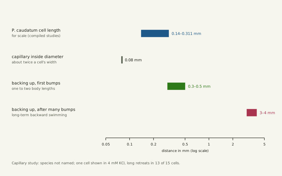
In glass capillaries 0.08 mm wide, about twice a cell's width and too narrow to turn in, paramecia of an unnamed species met a dead end in a 2014 study by Kunita and colleagues. After the first bumps they backed up 0.3–0.5 mm, one to two body lengths; after many tries the retreats grew to 3–4 mm, and 13 of the 15 cells tested under those conditions behaved this way.
Slide the diagram sideways to read its labels, or open it full size.
Backing-up distances of paramecia in a dead-end capillary, 0.3–0.5 mm and then 3–4 mm, beside Paramecium caudatum cells 0.14–0.311 mm long [10−4–10−3 m].
Log scale in millimetres; capillary values from Kunita and colleagues 2014, whose cells were of an unnamed species. Data: paramecium-backing-distances.csv (sources, version and license, JSON).
"Paramecium backing up in a dead-end capillary" by Ants, Cells & Atoms, https://antscellsandatoms.com/microscopic-life/paramecium/ — CC BY 4.0 (https://creativecommons.org/licenses/by/4.0/)
Data: Kunita I, Kuroda S, Ohki K, Nakagaki T 2014, Frontiers in Microbiology 5:270, CC BY 3.0, https://doi.org/10.3389/fmicb.2014.00270, retrieved 2026-10-24; Cell length for scale: Velho Rodrigues MF, Lisicki M, Lauga E 2021, PLOS ONE 16(6):e0252291, CC BY 4.0, https://doi.org/10.1371/journal.pone.0252291, retrieved 2026-10-24
Data: Kunita I, Kuroda S, Ohki K, Nakagaki T 2014, Frontiers in Microbiology 5:270, CC BY 3.0, https://doi.org/10.3389/fmicb.2014.00270, retrieved 2026-10-24; Velho Rodrigues MF, Lisicki M, Lauga E 2021, PLOS ONE 16(6):e0252291, CC BY 4.0, https://doi.org/10.1371/journal.pone.0252291, retrieved 2026-10-24
Which way the cell turns is less settled than it looks. By Brette's account, Jennings reported in 1899 that it always swings toward the side away from its oral groove, but as of the 2021 review systematic measurements were lacking, and the details of how the cell turns, likely through uneven beating by different groups of cilia, were still missing.
Sources
- Velho Rodrigues MF, Lisicki M, Lauga E. "The bank of swimming organisms at the micron scale (BOSO-Micro)." PLOS ONE 16(6):e0252291 (2021). https://doi.org/10.1371/journal.pone.0252291 Read: full text. Text license: CC BY 4.0 (https://creativecommons.org/licenses/by/4.0/).
- Brette R. "Integrative Neuroscience of Paramecium, a ‘Swimming Neuron’." eNeuro 8(3):ENEURO.0018-21.2021 (2021). https://doi.org/10.1523/ENEURO.0018-21.2021 Read: full text. Text license: CC BY 4.0 (https://creativecommons.org/licenses/by/4.0/).
- Long H, Johri P, Gout J-F, Ni J, Hao Y, Licknack T, Wang Y, Pan J, Jiménez-Marín B, Lynch M. "Paramecium Genetics, Genomics, and Evolution." Annual Review of Genetics 57:391–410 (2023). https://doi.org/10.1146/annurev-genet-071819-104035 Read: full text (author manuscript). Text license: CC BY 4.0 (https://creativecommons.org/licenses/by/4.0/).
- Lisicki M, Velho Rodrigues MF, Goldstein RE, Lauga E. "Swimming eukaryotic microorganisms exhibit a universal speed distribution." eLife 8:e44907 (2019). https://doi.org/10.7554/eLife.44907 Read: full text. Text license: CC BY 4.0 (https://creativecommons.org/licenses/by/4.0/).
- Somerville GA, Proctor RA. "Cultivation conditions and the diffusion of oxygen into culture media: The rationale for the flask-to-medium ratio in microbiology." BMC Microbiology 13:9 (2013). https://doi.org/10.1186/1471-2180-13-9 Read: full text. Text license: CC BY 2.0 (https://creativecommons.org/licenses/by/2.0/).
- Vestergaard CL, Flyvbjerg H, Møller IM. "Intracellular signaling by diffusion: can waves of hydrogen peroxide transmit intracellular information in plant cells?" Frontiers in Plant Science 3:295 (2012). https://doi.org/10.3389/fpls.2012.00295 Read: full text. Text license: CC BY 3.0 (https://creativecommons.org/licenses/by/3.0/).
- Allen RD. "Food vacuole membrane growth with microtubule-associated membrane transport in Paramecium." Journal of Cell Biology 63(3):904–922 (1974). https://doi.org/10.1083/jcb.63.3.904 Read: abstract. Text license: CC BY-NC-SA 4.0 (https://creativecommons.org/licenses/by-nc-sa/4.0/).
- International Association for the Properties of Water and Steam. IAPWS R6-95(2018), Revised Release on the IAPWS Formulation 1995 for the Thermodynamic Properties of Ordinary Water Substance for General and Scientific Use (2018). https://iapws.org/technical-guidance/release/IAPWS-95 Read: values as computed from the release equations in this site's water-properties table. Text license: IAPWS: publication in whole or in part allowed with attribution (https://iapws.org/technical-guidance/release/IAPWS-95).
- International Association for the Properties of Water and Steam. IAPWS R12-08, Release on the IAPWS Formulation 2008 for the Viscosity of Ordinary Water Substance (2008). https://iapws.org/technical-guidance/release/viscosity Read: values as computed from the release equations in this site's water-properties table. Text license: IAPWS: publication in whole or in part allowed with attribution (https://iapws.org/technical-guidance/release/viscosity).
- Kunita I, Kuroda S, Ohki K, Nakagaki T. "Attempts to retreat from a dead-ended long capillary by backward swimming in Paramecium." Frontiers in Microbiology 5:270 (2014). https://doi.org/10.3389/fmicb.2014.00270 Read: full text. Text license: CC BY 3.0 (https://creativecommons.org/licenses/by/3.0/).
- Sender R, Fuchs S, Milo R. "Revised Estimates for the Number of Human and Bacteria Cells in the Body." PLOS Biology 14(8):e1002533 (2016). https://doi.org/10.1371/journal.pbio.1002533 Read: full text. Text license: CC BY 4.0 (https://creativecommons.org/licenses/by/4.0/).
- Polilov AA. "How small is the smallest? New record and remeasuring of Scydosella musawasensis Hall, 1999 (Coleoptera, Ptiliidae), the smallest known free-living insect." ZooKeys 526:61–64 (2015). https://doi.org/10.3897/zookeys.526.6531 Read: full text. Text license: CC BY 4.0 (https://creativecommons.org/licenses/by/4.0/).
- GBIF Secretariat. GBIF Backbone Taxonomy. Checklist dataset https://doi.org/10.15468/39omei, accessed 2026-10-24; Paramecium caudatum Ehrenberg, 1834, usage key 8122199; Ciliophora, usage key 7765738; Paramecium polycarium Woodruff, 1923, usage key 8323167. Read: dataset record (species match and species records). Text license: CC BY 4.0 (https://creativecommons.org/licenses/by/4.0/).
Cite this page
Ants, Cells & Atoms. “Paramecium: How a Nerve-Like Signal Sends One Cell Backward.” Published 2026-10-25. https://antscellsandatoms.com/microscopic-life/paramecium/. Accessed (add the date you read it).
No DOI is issued for entries. Figures made here are CC BY 4.0; see each caption for the license of photographs.
Related entries
 Illustration
Diatoms: Why Most Glass-Walled Algae Shrink as They Multiply
Image: Illustration: Ants, Cells & Atoms, all rights reserved
Illustration: Ants, Cells & Atoms. Generated schematic illustration — not a photograph, micrograph or specimen record. © Ants, Cells & Atoms, all rights reserved.
Plankton: From Bacteria to Jellyfish, Sorted in Size Steps
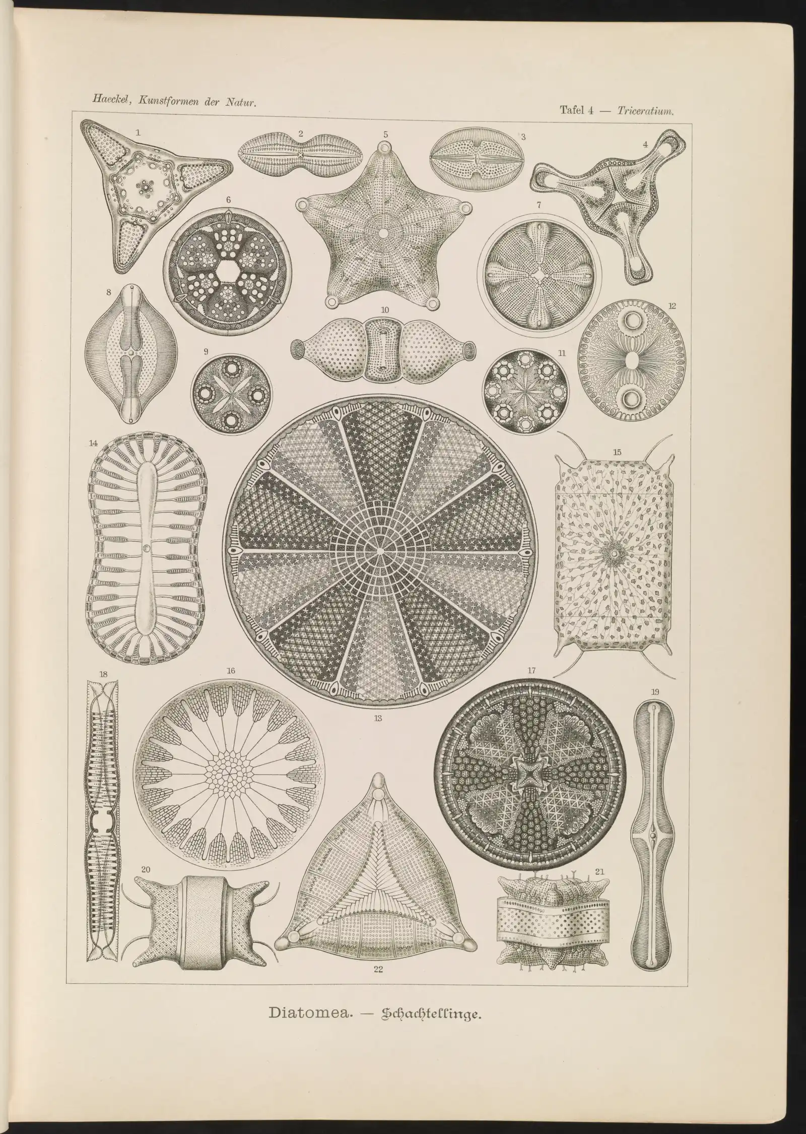
Image: “Diatomea. - Schachtellinge” by Ernst Haeckel (artist) and Adolf Giltsch (lithographer) (1904) · Public domain
"Diatomea. - Schachtellinge" by Ernst Haeckel (artist) and Adolf Giltsch (lithographer) (1904) — https://commons.wikimedia.org/wiki/File:Diatomea._-_Schachtellinge_LCCN2015647735.jpg — Public domain (artist Ernst Haeckel died 1919 and lithographer Adolf Giltsch died 1911, so the plate is out of copyright in the EU (life plus 70 years) and in the US (published 1904, before 1930); the scan is from the Library of Congress Prints and Photographs division (lccn.loc.gov/2015647735), which lists no rights restriction)

 Illustration
Volvox: Each Embryo Turns Inside Out Before It Can Swim
Image: Illustration: Ants, Cells & Atoms, all rights reserved
Illustration: Ants, Cells & Atoms. Generated schematic illustration — not a photograph, micrograph or specimen record. © Ants, Cells & Atoms, all rights reserved.