How Do Insects Breathe? Through Air Tubes, Not Lungs

Slide the ladder sideways to read its labels, or open it full size.
Tracheoles, the finest air tubes: about 0.45 µm to under 1 µm across [10−7 m] in a katydid's gut.
Log scale from 10 cm to 0.1 nm; dashed lines are the naked-eye and light-microscope limits explained on the Zoom ladder page. Data: how-insects-breathe-sizes.csv (sources, version and license, JSON).
"Zoom ladder: how insects breathe" by Ants, Cells & Atoms, https://antscellsandatoms.com/insects/how-insects-breathe/ — CC BY 4.0 (https://creativecommons.org/licenses/by/4.0/)
Data sources for this figure
Data: Li K et al. 2018, PLoS ONE 13(7):e0200405, CC BY 4.0, https://doi.org/10.1371/journal.pone.0200405 (tracheoles under 1 µm; cross-sections measured for this page against the Fig 7 scale bars), retrieved 2026-10-24; Dunn KN et al. 2022, PLoS ONE 17(6):e0268112, CC BY 4.0, https://doi.org/10.1371/journal.pone.0268112 (spiracle opening measured for this page against the Fig 4A scale bar), retrieved 2026-10-24; Socha JJ et al. 2007, BMC Biology 5:6, CC BY 2.0, https://doi.org/10.1186/1741-7007-5-6 (tracheae followed by x-ray video), retrieved 2026-10-24; Sauerwald J et al. 2019, eLife 8:e48857, CC BY 4.0, https://doi.org/10.7554/eLife.48857 (fruit-fly flight muscle width), retrieved 2026-10-24; Clapham ME, Karr JA 2012, PNAS 109(27):10927-10930, https://doi.org/10.1073/pnas.1204026109 (wingspans up to 70 cm; abstract), retrieved 2026-10-24
Data sources for this figure
Data: Li K et al. 2018, PLoS ONE 13(7):e0200405, CC BY 4.0, https://doi.org/10.1371/journal.pone.0200405, retrieved 2026-10-24; Dunn KN et al. 2022, PLoS ONE 17(6):e0268112, CC BY 4.0, https://doi.org/10.1371/journal.pone.0268112, retrieved 2026-10-24; Socha JJ et al. 2007, BMC Biology 5:6, CC BY 2.0, https://doi.org/10.1186/1741-7007-5-6, retrieved 2026-10-24; Sauerwald J et al. 2019, eLife 8:e48857, CC BY 4.0, https://doi.org/10.7554/eLife.48857, retrieved 2026-10-24; Clapham ME, Karr JA 2012, PNAS 109(27):10927-10930, https://doi.org/10.1073/pnas.1204026109 (abstract), retrieved 2026-10-24; Preuss A et al. 2025, Communications Biology 8:852, CC BY 4.0, https://doi.org/10.1038/s42003-025-08285-4 (plastron hair density; spacing computed 2026-10-24), retrieved 2026-10-24
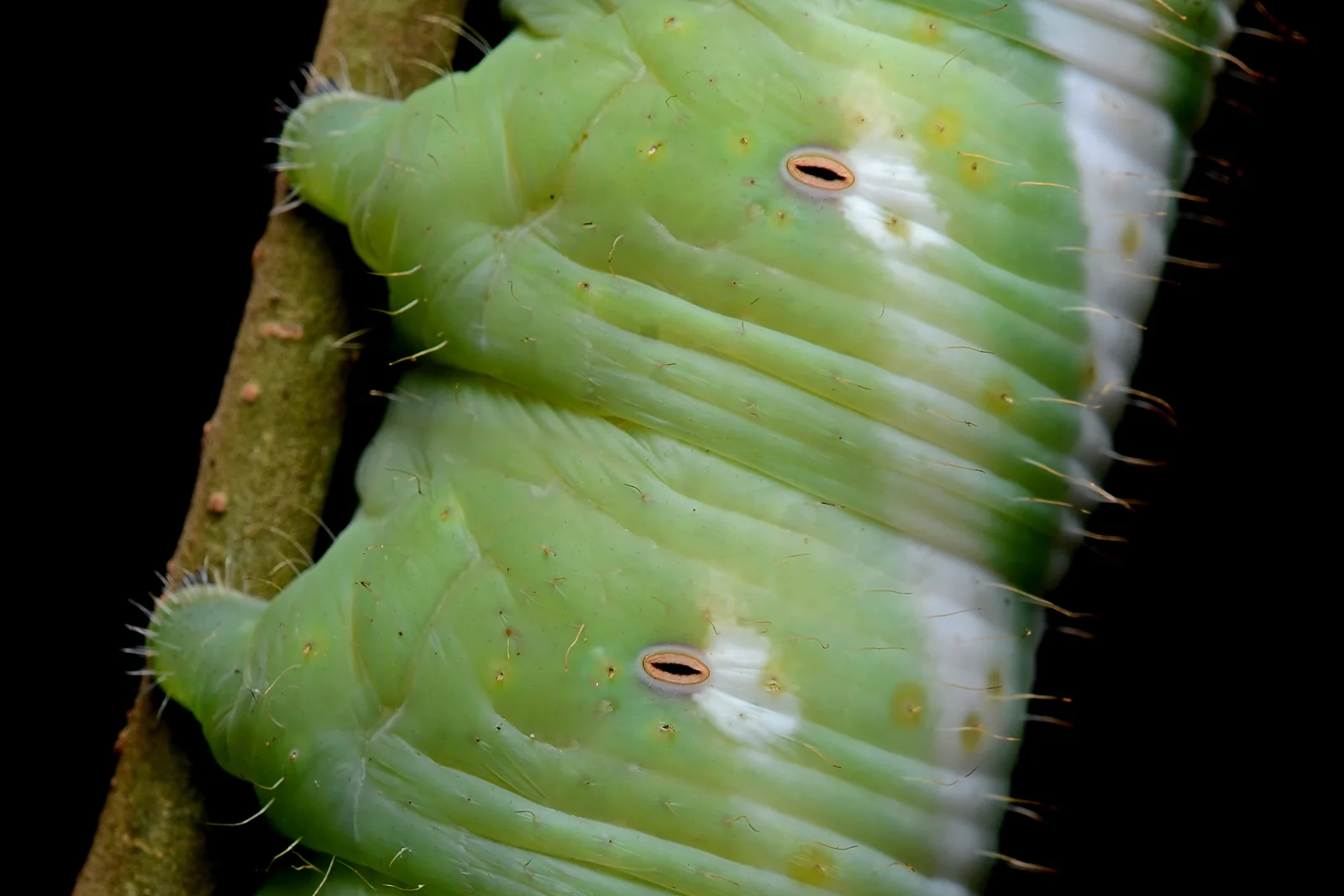
Spiracles on a hawk-moth caterpillar (family Sphingidae), each a doorway to air tubes whose finest branches measure under 1 µm across in a katydid [10−7 m].
Color: native
"Flickr - ggallice - Spiracles" by Geoff Gallice — https://commons.wikimedia.org/wiki/File:Flickr_-_ggallice_-_Spiracles.jpg — CC BY 2.0 (https://creativecommons.org/licenses/by/2.0/)

Where does the air go in?
Spiracles are valve-like openings on the sides of the thorax and abdomen. From them, a network of cuticle-lined tubes, the tracheae, branches through the body, as Lehnert and Myers describe it in a 2026 study of antlion larvae. A 2025 study of seal lice by Preuss and colleagues adds that spiracles can be opened and closed, and that spiral thickenings called taenidia keep the tubes from collapsing.
A spiracle is more than a hole. Dunn and colleagues imaged the abdominal spiracles of a last-instar larva of the firefly Photinus pyralis in 2022. Each has a two-chambered slit fringed with branched bars, a branched filter behind it, a bulb-shaped chamber and a closing muscle at its base. In their figure of one of them, the opening is about 48 µm long [10−5 m], measured for this page against the 50 µm scale bar. That is about half the roughly 0.1 mm the naked eye can make out.
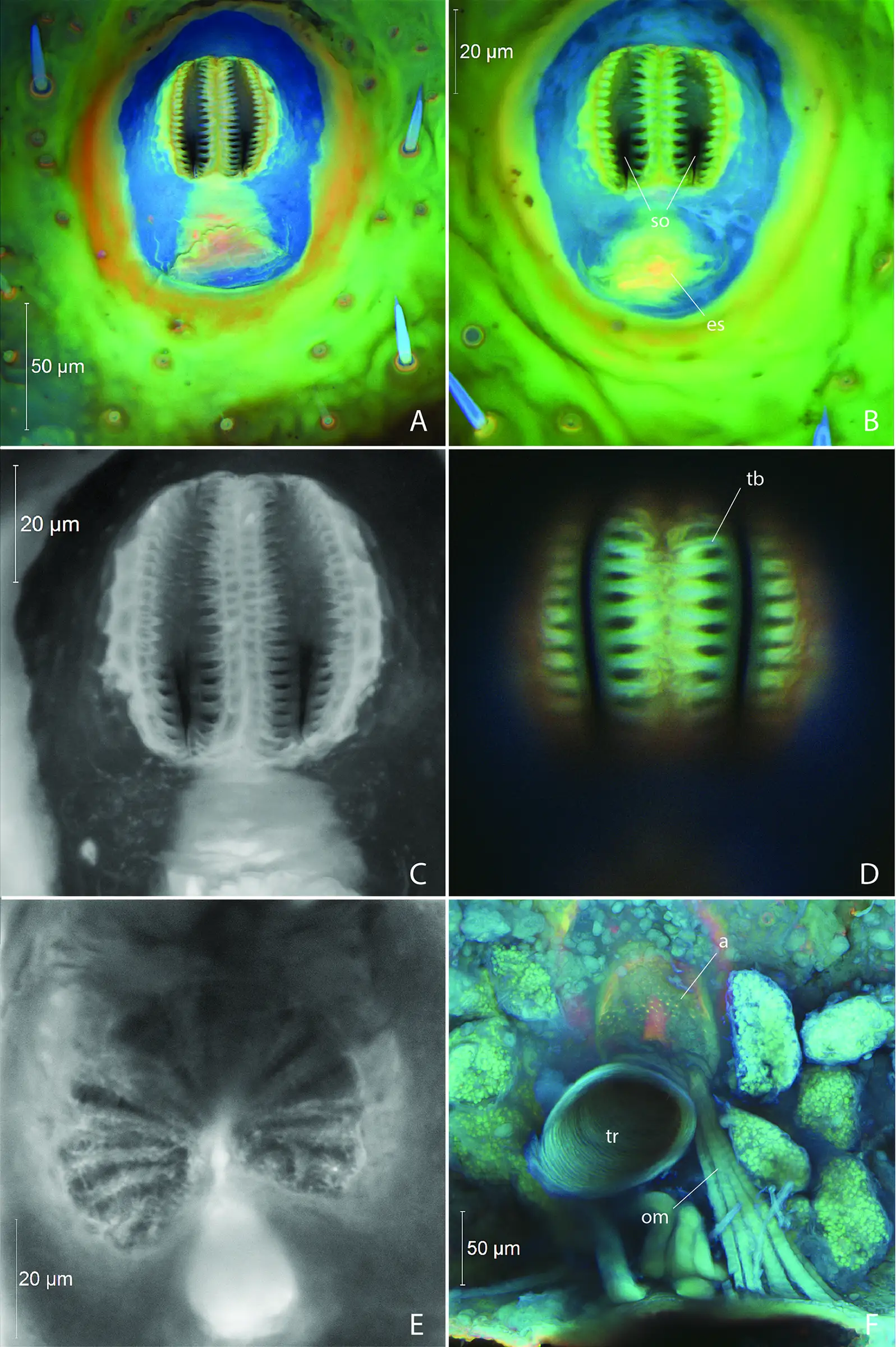
Abdominal spiracles of a last-instar firefly larva, Photinus pyralis: a two-chambered opening about 48 µm long [10−5 m] fringed with branched bars (A–D), the branched filter behind it (E) and, inside, the chamber, trachea and closing muscle (F). Size in this image: 50 µm and 20 µm scale bars.
Color: fluorescence (autofluorescence of the cuticle, recorded in blue, green and red; panels C and E in grey)
"Fig 4. Larval abdominal spiracles in Photinus pyralis" by Dunn KN, Davis SR, Herhold HW, Stanger-Hall KF, Bybee SM, Branham MA — https://doi.org/10.1371/journal.pone.0268112 — CC BY 4.0 (https://creativecommons.org/licenses/by/4.0/)
This is where insects part ways with lungs. In a lung, oxygen crosses from inhaled air into the blood of the capillaries, Maina writes in a 2012 comparison of lungs and tracheal systems. In insects the circulatory and tracheal systems are "totally disengaged", he writes, and oxygen diffuses from the air in the tubes to the tissues and cells.
How fine do the tubes get?
Behind the spiracle the tubes branch and narrow. In x-ray video of living insects they stand out as air-filled tubes. Socha and colleagues give a trachea 100 µm across as their example and describe earlier work that followed tubes down to 10 µm in diameter (2007). The rungs from 1 to 10 µm, between those tubes and the finest ones, are where the spider silk page places its threads.
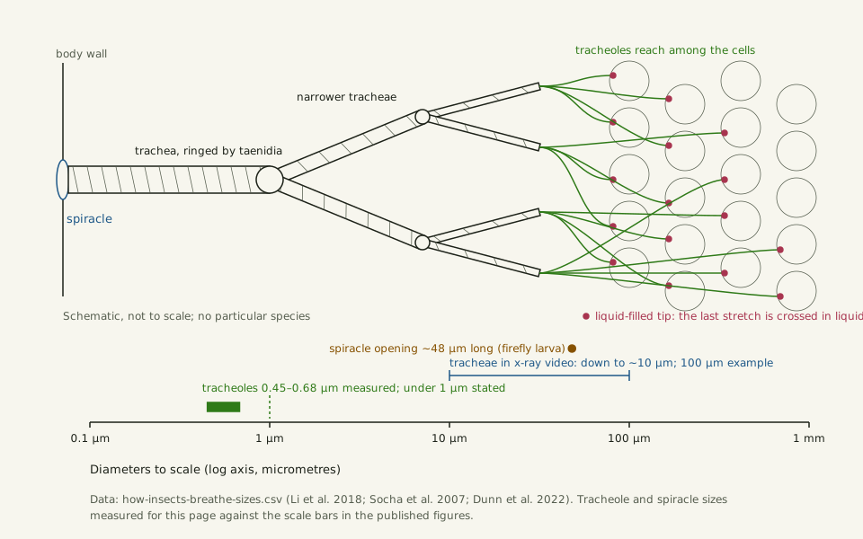
Slide the diagram sideways to read its labels, or open it full size.
From spiracle to tracheole: tubes narrow from about 100 µm to tracheoles 0.45–0.68 µm across [10−7 m] in a katydid's gut, whose liquid-filled tips reach among the cells.
Schematic above, not to scale; diameters below to scale on a log axis. Data: how-insects-breathe-sizes.csv (sources, version and license, JSON).
"The tracheal tree, from spiracle to tracheole" by Ants, Cells & Atoms, https://antscellsandatoms.com/insects/how-insects-breathe/ — CC BY 4.0 (https://creativecommons.org/licenses/by/4.0/)
Data sources for this figure
Data: Li K et al. 2018, PLoS ONE 13(7):e0200405, CC BY 4.0, https://doi.org/10.1371/journal.pone.0200405, retrieved 2026-10-24; Socha JJ et al. 2007, BMC Biology 5:6, CC BY 2.0, https://doi.org/10.1186/1741-7007-5-6, retrieved 2026-10-24; Dunn KN et al. 2022, PLoS ONE 17(6):e0268112, CC BY 4.0, https://doi.org/10.1371/journal.pone.0268112, retrieved 2026-10-24; Lehnert MS, Myers KO 2026, iScience 29(10):117638, CC BY 4.0, https://doi.org/10.1016/j.isci.2026.117638 (liquid-filled tracheole tips; principle only), retrieved 2026-10-24; Preuss A et al. 2025, Communications Biology 8:852, CC BY 4.0, https://doi.org/10.1038/s42003-025-08285-4 (spiral taenidia; principle only), retrieved 2026-10-24
Data sources for this figure
Data: Li K et al. 2018, PLoS ONE 13(7):e0200405, CC BY 4.0, https://doi.org/10.1371/journal.pone.0200405, retrieved 2026-10-24; Dunn KN et al. 2022, PLoS ONE 17(6):e0268112, CC BY 4.0, https://doi.org/10.1371/journal.pone.0268112, retrieved 2026-10-24; Socha JJ et al. 2007, BMC Biology 5:6, CC BY 2.0, https://doi.org/10.1186/1741-7007-5-6, retrieved 2026-10-24; Sauerwald J et al. 2019, eLife 8:e48857, CC BY 4.0, https://doi.org/10.7554/eLife.48857, retrieved 2026-10-24; Clapham ME, Karr JA 2012, PNAS 109(27):10927-10930, https://doi.org/10.1073/pnas.1204026109 (abstract), retrieved 2026-10-24; Preuss A et al. 2025, Communications Biology 8:852, CC BY 4.0, https://doi.org/10.1038/s42003-025-08285-4 (plastron hair density; spacing computed 2026-10-24), retrieved 2026-10-24
The tubes end in tracheoles. Their tips are filled with liquid, and they deliver oxygen directly to the tissues, Lehnert and Myers write. In the gut wall of the katydid Gampsocleis gratiosa, Li and colleagues found tracheoles less than 1 µm in diameter reaching into the gut cells themselves (2018).
In their electron micrograph, two labelled cross-sections measure about 0.45–0.5 µm on the short axis and 0.6–0.7 µm on the long one. Both were measured for this page against the 1 µm scale bars. That is about two to three and a half times the roughly 0.2 µm a light microscope can resolve, the second dashed line on the ladder.
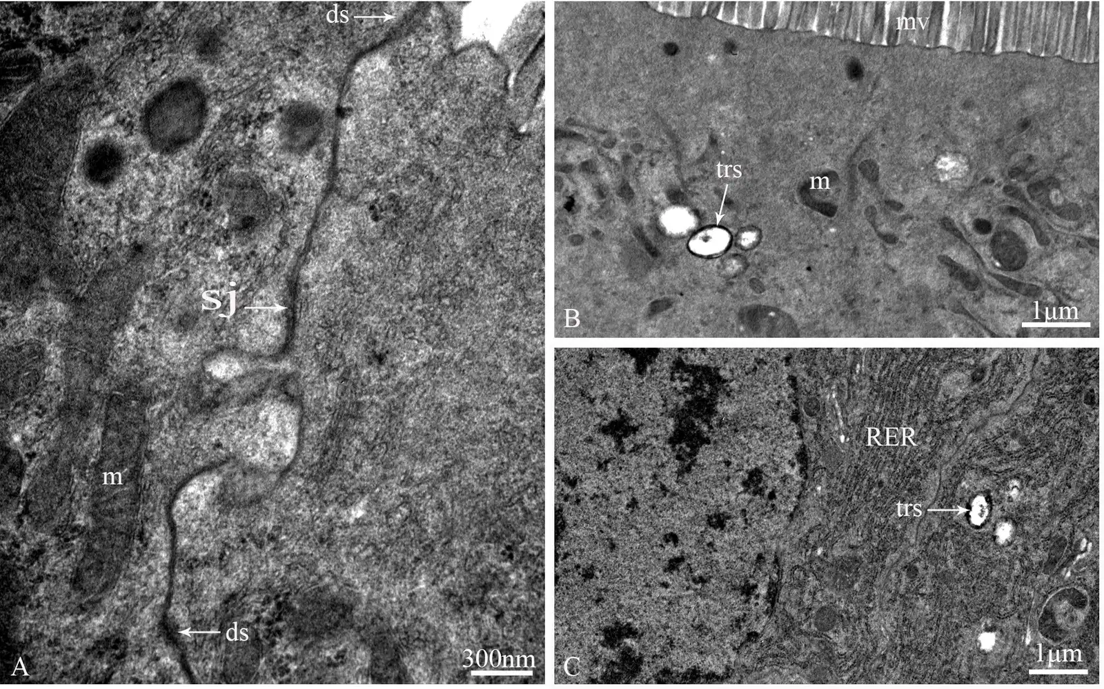
Tracheoles (trs) inside gut cells of the katydid Gampsocleis gratiosa, about 0.45–0.7 µm across [10−7 m] in panels B and C. Size in this image: 1 µm scale bars (B, C) and 300 nm (A).
Color: native (electron micrograph, no color)
"Fig 7. Cell junctions of principal cells and the distribution of tracheoles in the midgut of G. gratiosa, TEM" by Li K, Zhang JH, Yang YJ, Han W, Yin H — https://doi.org/10.1371/journal.pone.0200405 — CC BY 4.0 (https://creativecommons.org/licenses/by/4.0/)
Some tracheoles go inside cells. Each fruit-fly flight muscle is about 1 mm long and 100 µm wide, by the account of Sauerwald and colleagues (2019). They describe tracheal branches that wrap its surface and then grow into it, shortening the path oxygen must diffuse to the mitochondria. The indirect flight muscles of flying insects have "the highest known metabolic rates in the animal kingdom", they write, citing work from 1964; the bumblebee flight page looks at what such muscles drive.
In many tissues, Maina adds, the tracheoles may contact practically every cell, and the distance still shows. In the brains of fruit-fly larvae, Baccino-Calace and colleagues found that a cell's oxygen supply, read through a genetically built sensor, fell off exponentially with its distance from the nearest tracheole (2020). As of that study, they knew of no experiment that had measured oxygen diffusing from inside a tracheole into the cells around it.
How far can oxygen diffuse?
Diffusion is why the air goes so far in. Oxygen is nearly 10,000 times more diffusive in air than in water. Somerville and Proctor give its diffusion coefficient as 2.1 × 10⁻⁵ cm2 per second in water at 25 °C and about 0.2 cm2 per second in air, citing a standard handbook (2013). A 2019 lung model by Park and colleagues uses the same air value.
The time diffusion needs grows with the square of the distance, t ≈ x2/2D, which follows from how a diffusing cloud spreads, as set out by Vestergaard and colleagues in 2012. Computed for this page with those coefficients, oxygen crosses 1 cm of air in about 2.5 s but 1 cm of water in about 6.6 hours. Across 10 µm of water it needs about 24 milliseconds.
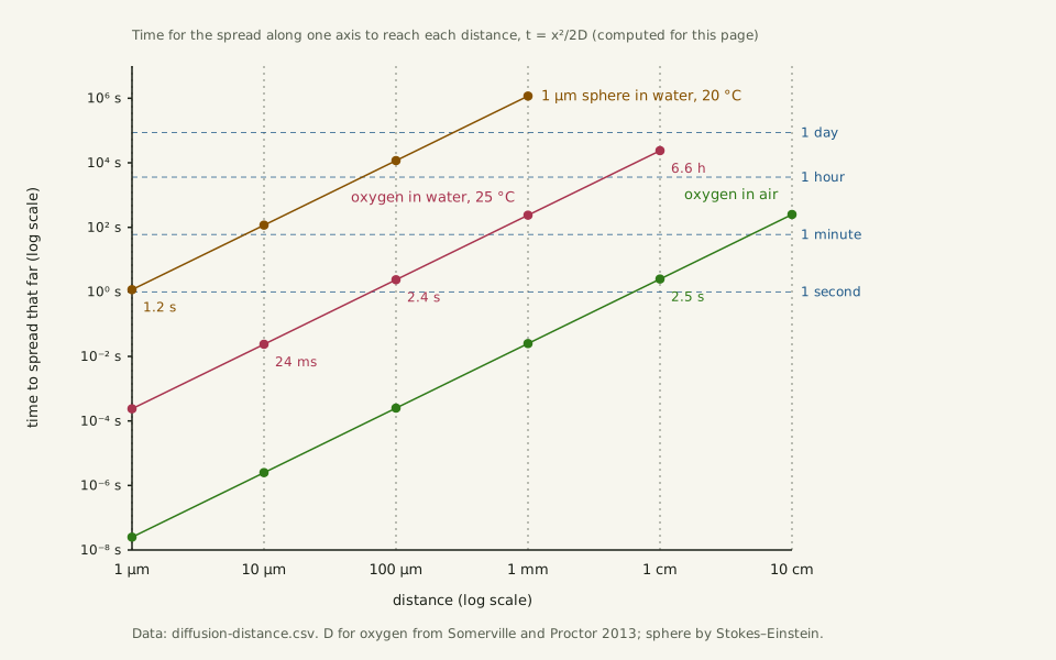
Slide the diagram sideways to read its labels, or open it full size.
Oxygen takes nearly 10,000 times longer to spread in water than in air; across 10 µm [10−5 m] of water it needs about 24 ms, across 1 cm about 6.6 h (computed).
Both axes are logarithmic: on a linear time axis running to the 13.5 days a 1 µm bead needs to spread 1 mm, every time under an hour would sit within about 0.3% of zero (computed). Data: diffusion-distance.csv (sources, version and license, JSON).
"Diffusion time against distance: oxygen in air and in water" by Ants, Cells & Atoms, https://antscellsandatoms.com/insects/how-insects-breathe/ — CC BY 4.0 (https://creativecommons.org/licenses/by/4.0/)
Data sources for this figure
Data: Somerville GA, Proctor RA 2013, BMC Microbiology 13:9, CC BY 2.0, https://doi.org/10.1186/1471-2180-13-9, retrieved 2026-10-24; Vestergaard CL, Flyvbjerg H, Møller IM 2012, Frontiers in Plant Science 3:295, CC BY 3.0, https://doi.org/10.3389/fpls.2012.00295, retrieved 2026-10-24; CODATA 2022 recommended values, NIST SRD 121, Boltzmann constant, retrieved 2026-10-24; IAPWS R12-08 viscosity of ordinary water substance, attribution to the International Association for the Properties of Water and Steam; value computed 2026-10-24
Data sources for this figure
Data: Somerville GA, Proctor RA 2013, BMC Microbiology 13:9, CC BY 2.0, https://doi.org/10.1186/1471-2180-13-9 (citing the CRC Handbook of Chemistry and Physics), retrieved 2026-10-24; Park K et al. 2019, PLoS ONE 14(1):e0204191, CC BY 4.0, https://doi.org/10.1371/journal.pone.0204191, retrieved 2026-10-24; Vestergaard CL, Flyvbjerg H, Møller IM 2012, Frontiers in Plant Science 3:295, CC BY 3.0, https://doi.org/10.3389/fpls.2012.00295, retrieved 2026-10-24; CODATA 2022 recommended values, NIST SRD 121, Boltzmann constant, retrieved 2026-10-24; IAPWS R12-08 viscosity of ordinary water substance, attribution to the International Association for the Properties of Water and Steam; value computed 2026-10-24 (shared file water-properties.csv)
So the long haul happens in air, and only the last stretch is liquid. As Wikipedia's article on the insect respiratory system puts it (read 2026-10-24), oxygen in the tube first dissolves in the liquid of the tracheole. Then it diffuses across the membrane of the neighbouring cell. The physics is the same for an insect and for a person; what changes is how far oxygen must travel through liquid.
Single cells face the same arithmetic. A cultured Escherichia coli cell, like those on the human body's bacteria page, is 1.0–1.5 µm wide, by Volkmer and Heinemann's measurements (2011). Oxygen crosses that width of water at 25 °C in about 0.2–0.5 ms (computed). The same square law runs through How Big Is a Cell. A bead 1 µm across in water at 20 °C, by contrast, wanders 1 µm in about 1.2 s (computed), the jiggle described in Brownian motion.
How do insects breathe underwater?
Water makes the problem harder. It holds less oxygen than air, and oxygen diffuses through it more slowly, as Verberk and Bilton note for insects with aquatic larvae (2011). By the figures Preuss and colleagues cite, only about 75,000 of 1 million named insect species can survive in water.
Those that do have several workarounds, as the same study summarizes. Some keep a closed tracheal system and take oxygen through the skin, as larvae of the midge Chironomus do, or through tracheal gills, as caddisfly larvae and damselfly nymphs do. Some fly larvae breathe through a siphon, a tube to the surface, and some shore flies and crane flies pierce plants to reach the air spaces inside them.
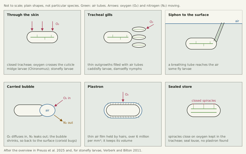
Slide the diagram sideways to read its labels, or open it full size.
Six ways to get oxygen under water, from skin breathing to a bubble that shrinks as it trades nitrogen for oxygen; plastron hairs, which can number over 6 million per mm2, stand under about 0.4 µm apart [10−7 m] (computed).
Plain shapes, not particular species, and not to scale.
"Six ways insects get oxygen under water" by Ants, Cells & Atoms, https://antscellsandatoms.com/insects/how-insects-breathe/ — CC BY 4.0 (https://creativecommons.org/licenses/by/4.0/)
Data: Preuss A et al. 2025, Communications Biology 8:852, CC BY 4.0, https://doi.org/10.1038/s42003-025-08285-4, retrieved 2026-10-24; Verberk WCEP, Bilton DT 2011, PLoS ONE 6(7):e22610, CC BY 4.0, https://doi.org/10.1371/journal.pone.0022610, retrieved 2026-10-24
Corixid bugs carry a bubble that works as a gill for a while. As the insect uses its oxygen, more diffuses in from the water and nitrogen diffuses out, so the bubble shrinks and the insect must surface to renew it.
A plastron is a thin air film held by water-repellent hairs that can number more than 6 million per square millimetre, under about 0.4 µm apart on a square grid (computed). The film keeps its volume, so it can draw oxygen from the water for long periods. The water strider page shows hair-held air at work on top of the surface.
The seal louse Echinophthirius horridus rides on seals through dives of 20–35 minutes to depths of up to 600 m. Preuss and colleagues found no plastron on it; instead, a closing apparatus at the spiracles keeps oxygen stored in its tracheal system (2025).
Do bigger insects need more tubes?
Diffusion alone has a reach, and many insects add pumping. Beetles, crickets and ants squeeze and re-expand tracheae in the head and thorax in rapid cycles, Westneat and colleagues reported in 2003 (abstract). They likened it to the inflation and deflation of vertebrate lungs. In x-ray video of a live ground beetle, Platynus decentis, Socha and colleagues timed one collapse and reinflation at 1.0 s (2007).
Size changes which mode matters. In the grasshopper Schistocerca americana, the youngest stages lack air sacs, rely more on diffusion and are the most sensitive to low oxygen. Larger individuals tend to move gas by bulk flow, Verberk and colleagues write in a 2021 review.
Bigger bodies also carry more tubing. Across four darkling beetle species spanning about three orders of magnitude in mass, Kaiser and colleagues found that tracheal volume scaled with mass to the power 1.29 (2007, abstract). Over a thousandfold range of mass, that makes the share of the body given to tubes about 7.4 times larger (computed for this page).
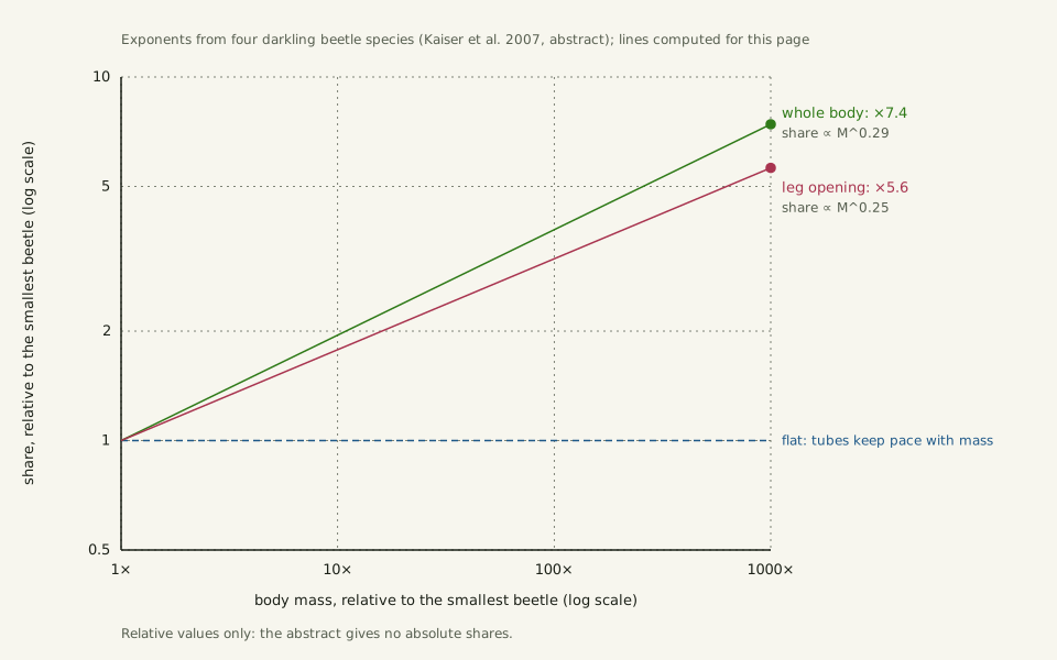
Slide the diagram sideways to read its labels, or open it full size.
Across beetles spanning a 1000-fold range of mass, the share of the body given to air tubes grows about 7.4-fold and their share of the leg opening about 5.6-fold (computed from the exponents in Kaiser and colleagues' abstract).
Relative values only; the abstract gives no absolute shares. Data: how-insects-breathe-tracheal-scaling.csv (sources, version and license, JSON).
"Bigger beetles give more of their body to air tubes" by Ants, Cells & Atoms, https://antscellsandatoms.com/insects/how-insects-breathe/ — CC BY 4.0 (https://creativecommons.org/licenses/by/4.0/)
Data: Kaiser A, Klok CJ, Socha JJ, Lee WK, Quinlan MC, Harrison JF 2007, PNAS 104(32):13198-13203, https://doi.org/10.1073/pnas.0611544104 (exponents from the abstract; multipliers computed 2026-10-24)
Data: Kaiser A, Klok CJ, Socha JJ, Lee WK, Quinlan MC, Harrison JF 2007, PNAS 104(32):13198-13203, https://doi.org/10.1073/pnas.0611544104 (abstract), retrieved 2026-10-24
In the legs, tracheal cross-section in the leg opening scaled with mass to the 1.02 power, the opening's own to the 0.77 power. Their share of the opening therefore grows about 5.6 times over the same range (computed), and the authors suggest that leg space may limit how big living beetles can get.
Larger species seem to pay in tubing rather than in oxygen shortage, Verberk and colleagues suggest. Across a range of insect species, the critical oxygen level for metabolism does not depend on adult size, though limits to tracheal expansion may still set an upper size. In a growing tobacco hornworm caterpillar, Manduca sexta, the tracheae can only be enlarged at a molt, so oxygen demand rises between molts while supply cannot.
How big did insects get?
The largest fossil insects lived around 300 million years ago. Wingspans reached as much as 70 cm in the Carboniferous and Permian, Clapham and Karr write in a study of more than 10,500 fossil wing lengths (2012, abstract). That is seven times the 10 cm hand at the top of the Zoom ladder.
The best known is Meganeura monyi. Wikipedia's article places it in the Late Carboniferous coal measures of Commentry, France (read 2026-10-24). The article gives a wing up to 32 cm long and a wingspan of about 65–75 cm, and describes griffenflies, the extinct order Meganisoptera, as resembling dragonflies. The GBIF Backbone lists the species under its original genus, as Dictyoneura monyi.
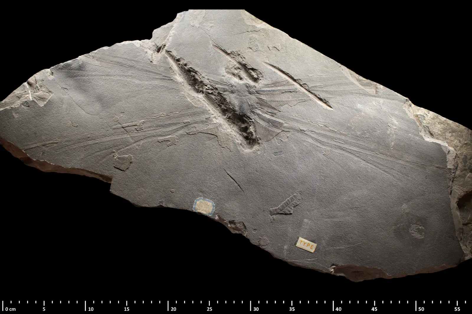
Lectotype of Meganeura monyi (MNHN R51142), a griffenfly about 65–75 cm across the wings [10−1 m]. Size in this image: centimetre ruler printed along the bottom edge.
Color: native
"Meganeura monyi MNHN R51142 2" by Gaëlle Doitteau & Sandra Daillie — https://commons.wikimedia.org/wiki/File:Meganeura_monyi_MNHN_R51142_2.jpg — CC BY 4.0 (https://creativecommons.org/licenses/by/4.0/)

Did extra oxygen make giant insects?
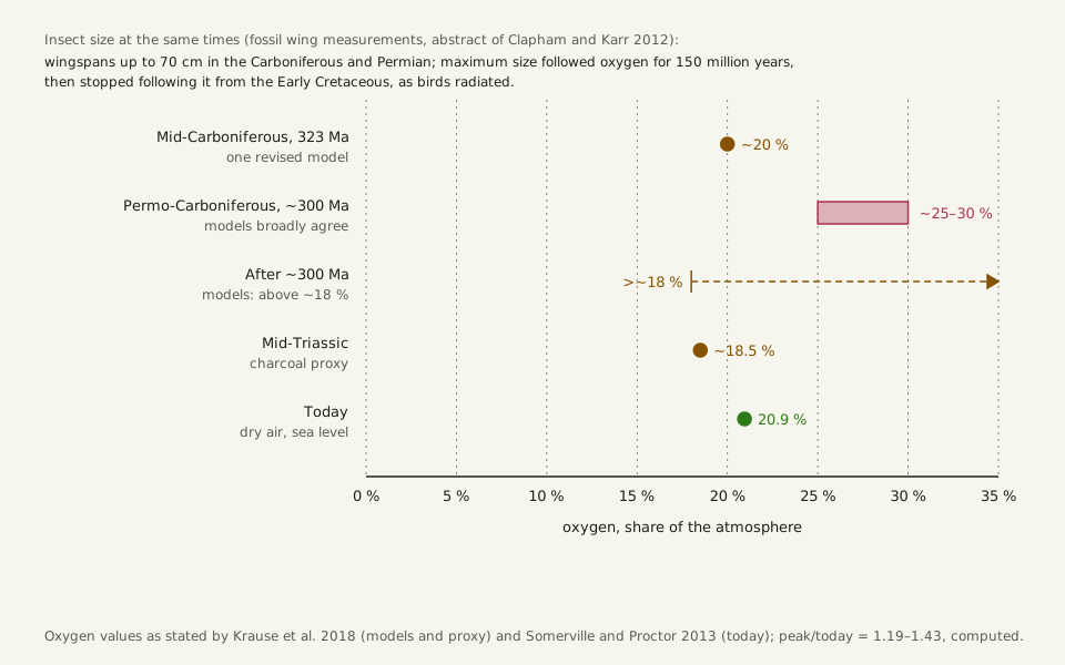
The giants lived when there was probably more oxygen in the air. Three models broadly agree that oxygen peaked at around 25–30% of the atmosphere about 300 million years ago and stayed above about 18% afterwards, Krause and colleagues write (2018). Dry air at sea level holds 20.946% today, by Somerville and Proctor's figure, so the peak was about 1.2–1.4 times today's level (computed).
Slide the diagram sideways to read its labels, or open it full size.
Oxygen then and now: three models put the peak near 300 million years ago at about 25–30%, when fossil wingspans reached 70 cm [10−1 m]; today's dry air holds 20.9%.
Values as stated in the cited studies; no model was run for this page. Data: how-insects-breathe-oxygen-and-size.csv (sources, version and license, JSON).
"Oxygen then and now" by Ants, Cells & Atoms, https://antscellsandatoms.com/insects/how-insects-breathe/ — CC BY 4.0 (https://creativecommons.org/licenses/by/4.0/)
Data: Krause AJ et al. 2018, Nature Communications 9:4081, CC BY 4.0, https://doi.org/10.1038/s41467-018-06383-y (model and proxy oxygen values as stated), retrieved 2026-10-24; Somerville GA, Proctor RA 2013, BMC Microbiology 13:9, CC BY 2.0, https://doi.org/10.1186/1471-2180-13-9 (oxygen in dry air today), retrieved 2026-10-24; Clapham ME, Karr JA 2012, PNAS 109(27):10927-10930, https://doi.org/10.1073/pnas.1204026109 (insect size; abstract), retrieved 2026-10-24
Data: Krause AJ et al. 2018, Nature Communications 9:4081, CC BY 4.0, https://doi.org/10.1038/s41467-018-06383-y, retrieved 2026-10-24; Somerville GA, Proctor RA 2013, BMC Microbiology 13:9, CC BY 2.0, https://doi.org/10.1186/1471-2180-13-9, retrieved 2026-10-24; Clapham ME, Karr JA 2012, PNAS 109(27):10927-10930, https://doi.org/10.1073/pnas.1204026109 (abstract), retrieved 2026-10-24
 Illustration
Large-winged insects flying over a fern swamp; not a reconstruction of any species or habitat.
Illustration — not to scale
Illustration: Ants, Cells & Atoms. Generated schematic illustration — not a photograph, micrograph or specimen record. © Ants, Cells & Atoms, all rights reserved.
Dudley argued in 1998 that the oxygen-rich Carboniferous air helped arthropods grow giant and that a late Permian fall in oxygen ended it (abstract). Harrison and colleagues' 2010 review adds support from experiments: most insects develop smaller in low oxygen, and some develop or evolve larger in high oxygen. Larger insects also invest more of the body in tracheae (abstract).
The record does not line up perfectly. Maximum insect size followed oxygen only for the first 150 million years of insect history, Clapham and Karr found. In the Early Cretaceous it fell even as oxygen rose, after early birds evolved and spread (abstract). Large griffenflies also lived on into the late Permian, when oxygen was putatively lower, Verberk and Bilton note (2011).
Verberk and Bilton look at the larvae instead. Many Paleozoic giants apparently had aquatic larvae, though larval habits are hard to read from fossils.
In their test, active larvae of the stonefly Dinocras cephalotes were warmed in water from 10 °C by 0.25 °C a minute until they stopped moving. They stopped 2.9 °C lower in oxygen-poor water (14 kPa) and 1.5 °C higher in oxygen-rich water (36 kPa), and recovered in cool water. In water, the authors suggest, oxygen supply set the tighter limit on larval size, and too much oxygen may even have favoured larger larvae, less at risk of oxygen poisoning.
Why is the question still open?
The size side of the debate rests on fossil wings. Charles Brongniart described the type specimen of Meganeura monyi in 1884, Wikipedia notes.
Slide the diagram sideways to read its labels, or open it full size.
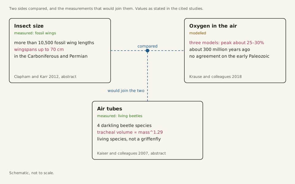
What the oxygen–size comparison rests on: measured fossil wingspans up to 70 cm [10−1 m], modelled oxygen, and air tubes measured in four living beetle species.
Schematic; the last paragraph of this section gives the same points in words. Data: how-insects-breathe-oxygen-and-size.csv (sources, version and license, JSON) and how-insects-breathe-tracheal-scaling.csv (sources, version and license, JSON).
"What the oxygen–size comparison rests on" by Ants, Cells & Atoms, https://antscellsandatoms.com/insects/how-insects-breathe/ — CC BY 4.0 (https://creativecommons.org/licenses/by/4.0/)
Data: Clapham ME, Karr JA 2012, PNAS 109(27):10927-10930, https://doi.org/10.1073/pnas.1204026109 (fossil wing lengths and wingspans; abstract), retrieved 2026-10-24; Krause AJ et al. 2018, Nature Communications 9:4081, CC BY 4.0, https://doi.org/10.1038/s41467-018-06383-y (model agreement on the oxygen peak), retrieved 2026-10-24; Kaiser A, Klok CJ, Socha JJ, Lee WK, Quinlan MC, Harrison JF 2007, PNAS 104(32):13198-13203, https://doi.org/10.1073/pnas.0611544104 (tracheal volume exponent; abstract), retrieved 2026-10-24
Data: Krause AJ et al. 2018, Nature Communications 9:4081, CC BY 4.0, https://doi.org/10.1038/s41467-018-06383-y, retrieved 2026-10-24; Somerville GA, Proctor RA 2013, BMC Microbiology 13:9, CC BY 2.0, https://doi.org/10.1186/1471-2180-13-9, retrieved 2026-10-24; Clapham ME, Karr JA 2012, PNAS 109(27):10927-10930, https://doi.org/10.1073/pnas.1204026109 (abstract), retrieved 2026-10-24
Data: Kaiser A, Klok CJ, Socha JJ, Lee WK, Quinlan MC, Harrison JF 2007, PNAS 104(32):13198-13203, https://doi.org/10.1073/pnas.0611544104 (abstract), retrieved 2026-10-24
The explanations in play, side by side:
| Idea | Who, year | Evidence | Bearing on the oxygen link |
|---|---|---|---|
| Oxygen-rich air seems to have enabled the giants, and a late Permian drop to have ended them | Dudley 1998 (abstract) | Geophysical models and physiology | For |
| Tracheal oxygen delivery may help set insect size | Harrison and colleagues 2010 (abstract) | Rearing experiments, tracheal measurements | For |
| Leg space for tracheae may limit beetle size | Kaiser and colleagues 2007 (abstract) | X-ray measurements of four living beetle species | For |
| Size followed oxygen at first; later, predation and competition, coinciding with the spread of early birds, may have become the tighter limit | Clapham and Karr 2012 (abstract) | More than 10,500 fossil wing lengths | For until the end of the Jurassic, then against |
| The aquatic larva may have been the limited stage | Verberk and Bilton 2011 | Stonefly larvae in low and high oxygen | Modifies it |
| Large griffenflies persisted when oxygen was putatively lower | Verberk and Bilton 2011, citing fossil work | Fossil occurrences | Against a simple ceiling |
The full table, with qualifiers and reading levels, is in how-insects-breathe-giant-insect-hypotheses.csv (sources, version and license, JSON) Data: Dudley R 1998, Journal of Experimental Biology 201(8):1043-1050, https://doi.org/10.1242/jeb.201.8.1043 (abstract), retrieved 2026-10-24; Harrison JF, Kaiser A, VandenBrooks JM 2010, Proceedings of the Royal Society B 277(1690):1937-1946, https://doi.org/10.1098/rspb.2010.0001 (abstract), retrieved 2026-10-24; Kaiser A et al. 2007, PNAS 104(32):13198-13203, https://doi.org/10.1073/pnas.0611544104 (abstract), retrieved 2026-10-24; Clapham ME, Karr JA 2012, PNAS 109(27):10927-10930, https://doi.org/10.1073/pnas.1204026109 (abstract), retrieved 2026-10-24; Verberk WCEP, Bilton DT 2011, PLoS ONE 6(7):e22610, CC BY 4.0, https://doi.org/10.1371/journal.pone.0022610, retrieved 2026-10-24; Verberk WCEP et al. 2021, Biological Reviews 96(1):247-268, CC BY 4.0, https://doi.org/10.1111/brv.12653, retrieved 2026-10-24.
With the fossil record as it is, Harrison and colleagues call it impossible to definitively accept or reject the oxygen-size link (abstract). One side of the comparison is measured: more than 10,500 fossil wings. The other comes from models that agree on the peak near 300 million years ago but not on the early Paleozoic, by Krause and colleagues' account. And the tube measurements that would join the two, like the 1.29 exponent, come from four living beetle species, not from a griffenfly.
Sources
- Lehnert MS, Myers KO. "Adaptive respiration patterns by antlion larvae support the chthonic hypothesis for discontinuous gas exchange cycles." iScience 29(10):117638 (2026). https://doi.org/10.1016/j.isci.2026.117638 Read: full text. Text license: CC BY 4.0 (https://creativecommons.org/licenses/by/4.0/).
- Preuss A, Schwaha T, Kovalev A, Ebmer D, Herzog I, Lehnert K, Grass C, Sandberg F, Hamann E, Zuber M, van de Kamp T, Gorb SN. "The ectoparasitic seal louse, Echinophthirius horridus, relies on a sealed tracheal system and spiracle closing apparatus for underwater respiration." Communications Biology 8:852 (2025). https://doi.org/10.1038/s42003-025-08285-4 Read: full text. Text license: CC BY 4.0 (https://creativecommons.org/licenses/by/4.0/).
- Dunn KN, Davis SR, Herhold HW, Stanger-Hall KF, Bybee SM, Branham MA. "Morphological changes in the tracheal system associated with light organs of the firefly Photinus pyralis (Coleoptera: Lampyridae) across life stages." PLoS ONE 17(6):e0268112 (2022). https://doi.org/10.1371/journal.pone.0268112 Read: full text. Text license: CC BY 4.0 (https://creativecommons.org/licenses/by/4.0/).
- Maina JN. "Comparative molecular developmental aspects of the mammalian- and the avian lungs, and the insectan tracheal system by branching morphogenesis: recent advances and future directions." Frontiers in Zoology 9:16 (2012). https://doi.org/10.1186/1742-9994-9-16 Read: full text. Text license: CC BY 2.0 (https://creativecommons.org/licenses/by/2.0/).
- Socha JJ, Westneat MW, Harrison JF, Waters JS, Lee WK. "Real-time phase-contrast x-ray imaging: a new technique for the study of animal form and function." BMC Biology 5:6 (2007). https://doi.org/10.1186/1741-7007-5-6 Read: full text. Text license: CC BY 2.0 (https://creativecommons.org/licenses/by/2.0/).
- Li K, Zhang JH, Yang YJ, Han W, Yin H. "Morphology and fine organization of the midgut of Gampsocleis gratiosa (Orthoptera: Tettigoniidae)." PLoS ONE 13(7):e0200405 (2018). https://doi.org/10.1371/journal.pone.0200405 Read: full text. Text license: CC BY 4.0 (https://creativecommons.org/licenses/by/4.0/).
- Sauerwald J, Backer W, Matzat T, Schnorrer F, Luschnig S. "Matrix metalloproteinase 1 modulates invasive behavior of tracheal branches during entry into Drosophila flight muscles." eLife 8:e48857 (2019). https://doi.org/10.7554/eLife.48857 Read: full text. Text license: CC BY 4.0 (https://creativecommons.org/licenses/by/4.0/).
- Baccino-Calace M, Prieto D, Cantera R, Egger B. "Compartment and cell-type specific hypoxia responses in the developing Drosophila brain." Biology Open 9(8):bio053629 (2020). https://doi.org/10.1242/bio.053629 Read: full text. Text license: CC BY 4.0 (https://creativecommons.org/licenses/by/4.0/).
- Somerville GA, Proctor RA. "Cultivation conditions and the diffusion of oxygen into culture media: the rationale for the flask-to-medium ratio in microbiology." BMC Microbiology 13:9 (2013). https://doi.org/10.1186/1471-2180-13-9 Read: full text. Text license: CC BY 2.0 (https://creativecommons.org/licenses/by/2.0/).
- Park K, Jung Y, Son T, Cho YJ, Jeon NL, Kim W, Kim HY. "Optimal diameter reduction ratio of acinar airways in human lungs." PLoS ONE 14(1):e0204191 (2019). https://doi.org/10.1371/journal.pone.0204191 Read: full text. Text license: CC BY 4.0 (https://creativecommons.org/licenses/by/4.0/).
- Vestergaard CL, Flyvbjerg H, Møller IM. "Intracellular signaling by diffusion: can waves of hydrogen peroxide transmit intracellular information in plant cells?" Frontiers in Plant Science 3:295 (2012). https://doi.org/10.3389/fpls.2012.00295 Read: full text. Text license: CC BY 3.0 (https://creativecommons.org/licenses/by/3.0/).
- Wikipedia contributors. "Respiratory system of insects." Wikipedia (read 2026-10-24). https://en.wikipedia.org/wiki/Respiratory_system_of_insects Read: full text. Text license: CC BY-SA 4.0 (https://creativecommons.org/licenses/by-sa/4.0/).
- Volkmer B, Heinemann M. "Condition-dependent cell volume and concentration of Escherichia coli to facilitate data conversion for systems biology modeling." PLoS ONE 6(7):e23126 (2011). https://doi.org/10.1371/journal.pone.0023126 Read: full text. Text license: CC BY 4.0 (https://creativecommons.org/licenses/by/4.0/).
- CODATA 2022 recommended values, NIST Standard Reference Database 121. "Boltzmann constant." https://physics.nist.gov/cgi-bin/cuu/Value?k Read: dataset record.
- International Association for the Properties of Water and Steam. IAPWS R12-08, Release on the IAPWS Formulation 2008 for the Viscosity of Ordinary Water Substance (2008). http://www.iapws.org/relguide/viscosity.html Read: values as computed from the release equations in this site's water-properties table.
- Verberk WCEP, Bilton DT. "Can oxygen set thermal limits in an insect and drive gigantism?" PLoS ONE 6(7):e22610 (2011). https://doi.org/10.1371/journal.pone.0022610 Read: full text. Text license: CC BY 4.0 (https://creativecommons.org/licenses/by/4.0/).
- Westneat MW, Betz O, Blob RW, Fezzaa K, Cooper WJ, Lee WK. "Tracheal respiration in insects visualized with synchrotron x-ray imaging." Science 299(5606):558–560 (2003). https://doi.org/10.1126/science.1078008 Read: abstract.
- Verberk WCEP, Atkinson D, Hoefnagel KN, Hirst AG, Horne CR, Siepel H. "Shrinking body sizes in response to warming: explanations for the temperature–size rule with special emphasis on the role of oxygen." Biological Reviews 96(1):247–268 (2021). https://doi.org/10.1111/brv.12653 Read: full text. Text license: CC BY 4.0 (https://creativecommons.org/licenses/by/4.0/).
- Kaiser A, Klok CJ, Socha JJ, Lee WK, Quinlan MC, Harrison JF. "Increase in tracheal investment with beetle size supports hypothesis of oxygen limitation on insect gigantism." Proceedings of the National Academy of Sciences of the USA 104(32):13198–13203 (2007). https://doi.org/10.1073/pnas.0611544104 Read: abstract.
- Clapham ME, Karr JA. "Environmental and biotic controls on the evolutionary history of insect body size." Proceedings of the National Academy of Sciences of the USA 109(27):10927–10930 (2012). https://doi.org/10.1073/pnas.1204026109 Read: abstract.
- Wikipedia contributors. "Meganeura." Wikipedia (read 2026-10-24). https://en.wikipedia.org/wiki/Meganeura Read: full text. Text license: CC BY-SA 4.0 (https://creativecommons.org/licenses/by-sa/4.0/).
- GBIF Backbone Taxonomy. Meganeura monyi (Brongniart, 1884), taxon key 8596724, synonym of Dictyoneura monyi (Brongniart, 1884), key 8725491. https://doi.org/10.15468/39omei https://www.gbif.org/species/8596724 Read: dataset record. Text license: CC BY 4.0 (https://creativecommons.org/licenses/by/4.0/).
- Krause AJ, Mills BJW, Zhang S, Planavsky NJ, Lenton TM, Poulton SW. "Stepwise oxygenation of the Paleozoic atmosphere." Nature Communications 9:4081 (2018). https://doi.org/10.1038/s41467-018-06383-y Read: full text. Text license: CC BY 4.0 (https://creativecommons.org/licenses/by/4.0/).
- Dudley R. "Atmospheric oxygen, giant Paleozoic insects and the evolution of aerial locomotor performance." Journal of Experimental Biology 201(8):1043–1050 (1998). https://doi.org/10.1242/jeb.201.8.1043 Read: abstract.
- Harrison JF, Kaiser A, VandenBrooks JM. "Atmospheric oxygen level and the evolution of insect body size." Proceedings of the Royal Society B 277(1690):1937–1946 (2010). https://doi.org/10.1098/rspb.2010.0001 Read: abstract.
Cite this page
Ants, Cells & Atoms. “How Do Insects Breathe? Through Air Tubes, Not Lungs.” Published 2026-10-25. https://antscellsandatoms.com/insects/how-insects-breathe/. Accessed (add the date you read it).
No DOI is issued for entries. Figures made here are CC BY 4.0; see each caption for the license of photographs.
Related entries
Water Strider: The Dimples Under Its Feet Hold Its Weight
Image: “Aquarius remigis 140810489” by Jason Swanson (2021) · CC0 1.0
"Aquarius remigis 140810489" by Jason Swanson — https://commons.wikimedia.org/wiki/File:Aquarius_remigis_140810489.jpg — CC0 1.0 (https://creativecommons.org/publicdomain/zero/1.0/)

Ants: Strong for Their Weight, Mostly Because They're Small
Image: “Rote Waldameisen mit Beute 01” by Stephan Sprinz (2026) · CC BY 4.0
"Rote Waldameisen mit Beute 01" by Stephan Sprinz — https://commons.wikimedia.org/wiki/File:Rote_Waldameisen_mit_Beute_01.jpg — CC BY 4.0 (https://creativecommons.org/licenses/by/4.0/)

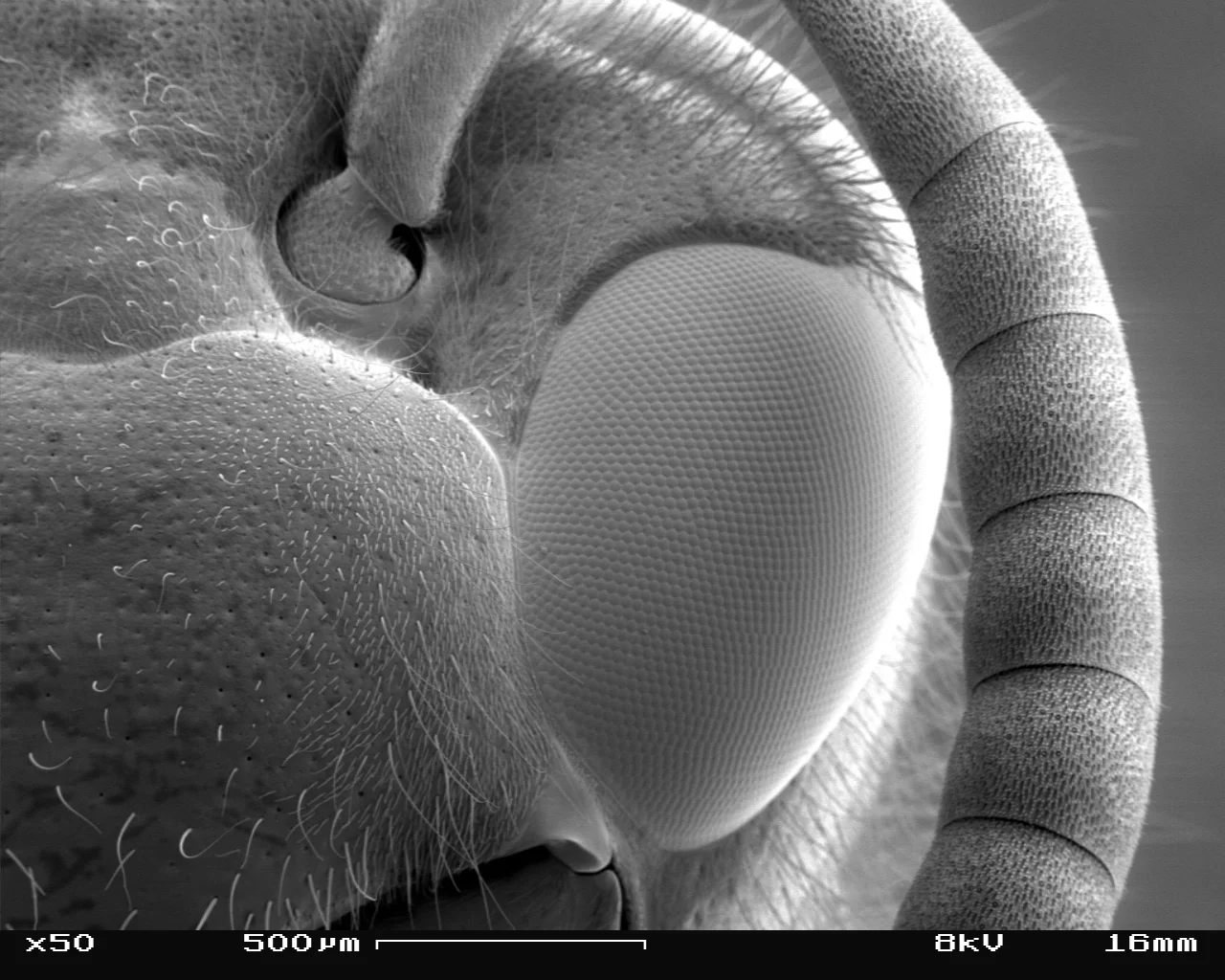
Compound Eye: One as Sharp as Yours Would Span About 7–12 m
Image: “Vespula germanica SEM Eye 01” by SecretDisc (2007) · CC BY 2.5
"Vespula germanica SEM Eye 01" by SecretDisc — https://commons.wikimedia.org/wiki/File:Vespula_germanica_SEM_Eye_01.jpg — CC BY 2.5 (https://creativecommons.org/licenses/by/2.5/)
