Compound Eye: One as Sharp as Yours Would Span About 7–12 m

Slide the ladder sideways to read its labels, or open it full size.
One facet of a compound eye, 15–29 µm wide in fruit flies, honeybees and the German wasp [10−5 m], with a lens of Megaphragma viggianii, about 7–8 µm, one rung smaller.
Log scale from 10 cm to 0.1 nm, each rung ten times smaller; dashed lines: naked eye about 0.1 mm, light microscope about 0.2 µm (How to read the Zoom ladder). The arrow points to the radius of an eye as sharp as a human eye, computed below. Data: compound-eye-ommatidia-counts.csv (sources, version and license, JSON).
"Zoom ladder: one facet of a compound eye" by Ants, Cells & Atoms, https://antscellsandatoms.com/insects/compound-eye/ — CC BY 4.0 (https://creativecommons.org/licenses/by/4.0/)
Data sources for this figure
Data: Fruit-fly facet widths: Tran H, Dostatni N, Ramaekers A 2025, Biology Open 14(6):bio061962, CC BY 4.0, https://doi.org/10.1242/bio.061962, retrieved 2026-10-25; German wasp facet widths: Gutiérrez D, Rigosi E, Nagloo N, O'Carroll D, Warrant EJ 2024, Journal of Experimental Biology 227(16):jeb246670, CC BY 4.0, https://doi.org/10.1242/jeb.246670, retrieved 2026-10-25; Megaphragma viggianii lens and eye: Makarova AA et al. 2025, eLife 14:RP103247, CC BY 4.0, https://doi.org/10.7554/eLife.103247, retrieved 2026-10-25; German wasp eye outline: measured against the printed scale bar of SecretDisc, Vespula germanica SEM Eye 01, CC BY 2.5, https://commons.wikimedia.org/wiki/File:Vespula_germanica_SEM_Eye_01.jpg; computed 2026-10-25; Eye radius for one minute of arc: computed 2026-10-25 from the sources listed in compound-eye-eye-size.csv

Data sources for this figure
Data: Makarova AA et al. 2025, eLife 14:RP103247, CC BY 4.0, https://doi.org/10.7554/eLife.103247, retrieved 2026-10-25; Buffry AD et al. 2024, BMC Biology 22:67, CC BY 4.0, https://doi.org/10.1186/s12915-024-01864-7, retrieved 2026-10-25; Tran H, Dostatni N, Ramaekers A 2025, Biology Open 14(6):bio061962, CC BY 4.0, https://doi.org/10.1242/bio.061962, retrieved 2026-10-25; Juusola M et al. 2017, eLife 6:e26117, CC BY 4.0, https://doi.org/10.7554/eLife.26117, retrieved 2026-10-25; Taylor GJ, Tichit P, Schmidt MD, Bodey AJ, Rau C, Baird E 2019, eLife 8:e40613, CC BY 4.0, https://doi.org/10.7554/eLife.40613, retrieved 2026-10-25; Gutiérrez D, Rigosi E, Nagloo N, O'Carroll D, Warrant EJ 2024, Journal of Experimental Biology 227(16):jeb246670, CC BY 4.0, https://doi.org/10.1242/jeb.246670, retrieved 2026-10-25; Scale-bar measurements: SecretDisc, Vespula germanica SEM Eye 01 and Eye 02, CC BY 2.5, https://commons.wikimedia.org/wiki/File:Vespula_germanica_SEM_Eye_01.jpg; computed 2026-10-25; GBIF Secretariat 2023, GBIF Backbone Taxonomy, checklist dataset https://doi.org/10.15468/39omei, CC BY 4.0, taxon keys in the gbif_key column, retrieved 2026-10-25
Bench card
- Name
- compound eye, an array of ommatidia, each with its own facet lens; examples on this page: fruit flies, honeybees, wasps and dragonfliesSource: Gutiérrez et al. 2024, https://doi.org/10.1242/jeb.246670
- Actual size
- one facet 15–29 µm wide in fruit flies, honeybees and the German wasp [10−5 m]; 6.9–8.0 µm on average in Megaphragma viggianii, one of the smallest insectsSource: Tran et al. 2025, https://doi.org/10.1242/bio.061962; Taylor et al. 2019; Gutiérrez et al. 2024; Makarova et al. 2025
- Size in this image
- facets about 23–25 µm apart in the upper middle of the eye, computed for this page from the printed 500 µm bar; narrower where the eye curves awaySource: computed for this page from the scale bar printed on the opening micrograph
- Lives in
- —
- Seen with
- naked eye for the whole eye, about 1 mm across in the opening micrograph; a light or electron microscope for single facets; all micrographs here are scanning electron imagesSource: figure captions on this page
- Color in images
- native: the four micrographs on this page are scanning electron images, without colorSource: figure captions on this page
compound-eye-bench.csv (CSV) · where each value comes from, and its license (JSON)
Data credits for this card
Data: GBIF Secretariat 2023, GBIF Backbone Taxonomy, Insecta (taxon key 216), checklist dataset https://doi.org/10.15468/39omei, CC BY 4.0, retrieved 2026-10-25; Gutiérrez D, Rigosi E, Nagloo N, O'Carroll D, Warrant EJ 2024, Journal of Experimental Biology 227(16):jeb246670, CC BY 4.0, https://doi.org/10.1242/jeb.246670, retrieved 2026-10-25; Tran H, Dostatni N, Ramaekers A 2025, Biology Open 14(6):bio061962, CC BY 4.0, https://doi.org/10.1242/bio.061962, retrieved 2026-10-25; Taylor GJ, Tichit P, Schmidt MD, Bodey AJ, Rau C, Baird E 2019, eLife 8:e40613, CC BY 4.0, https://doi.org/10.7554/eLife.40613, retrieved 2026-10-25; Makarova AA et al. 2025, eLife 14:RP103247, CC BY 4.0, https://doi.org/10.7554/eLife.103247, retrieved 2026-10-25; Heras FJH, Laughlin SB, eLife 13:RP96517 (2024; version of record 2026), CC BY 4.0, https://doi.org/10.7554/eLife.96517, retrieved 2026-10-25; Size in the opening micrograph: SecretDisc, Vespula germanica SEM Eye 01, CC BY 2.5, https://commons.wikimedia.org/wiki/File:Vespula_germanica_SEM_Eye_01.jpg; computed 2026-10-25
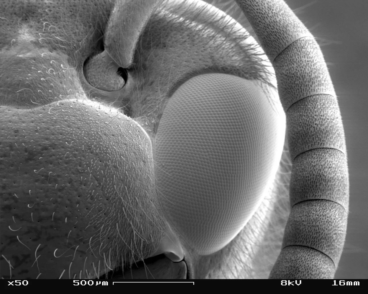
Head and compound eye of a German wasp, Vespula germanica, its facets about 23–25 µm apart against the printed 500 µm bar [10−5 m].
Color: native (electron micrograph, no color)
"Vespula germanica SEM Eye 01" by SecretDisc — https://commons.wikimedia.org/wiki/File:Vespula_germanica_SEM_Eye_01.jpg — CC BY 2.5 (https://creativecommons.org/licenses/by/2.5/)
What is a compound eye made of?
Each ommatidium is a narrow column. At the surface sits the corneal facet, a lens; beneath it, a crystalline cone; deeper still, the rhabdom, a light-catching rod built by photoreceptor cells, which reaches down to the basement membrane. Those are the labels Gutiérrez and colleagues gave a section through the eye of the German wasp, Vespula germanica, in 2024. In the wasp's sharpest region the rhabdom alone is 227 µm long, about eight times the width of the widest facets there, 29 µm.
The German wasp and the honeybee have apposition eyes of the fused-rhabdom type: the photoreceptors under one facet join into one rhabdom and together report a single pixel of the image, Heras and Laughlin explain. Flies wire theirs differently, but their photoreceptor spacing still matches the spacing of the ommatidia, Juusola and colleagues note. Either way, the angle between the viewing directions of neighbouring ommatidia, the interommatidial angle Δφ, sets how fine a grid the eye samples.
Geometry ties that angle to the eye's size. For a patch of eye with facets of width D, the radius of curvature is R = D/Δφ, in Heras and Laughlin's notation. In the German wasp's acute zone, its sharpest region, low on the front of the eye, facets 26–29 µm wide sit about 1.0° apart; higher up, the spacing widens to 2.0–3.0°. Applied to the acute zone, the formula gives a radius of 1.5–1.7 mm, computed for this page.
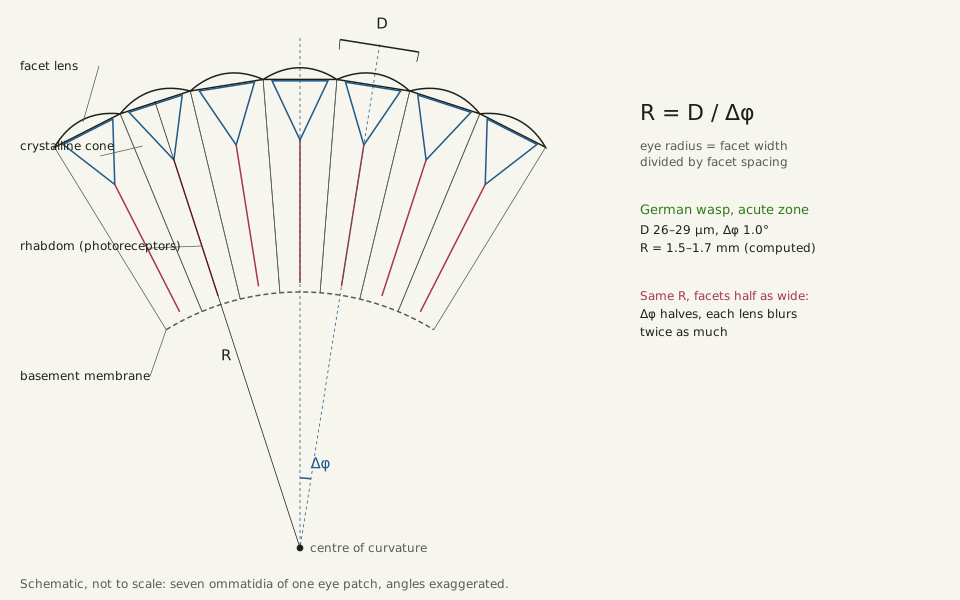
Slide the diagram sideways to read its labels, or open it full size.
Schematic of an apposition eye patch: each ommatidium has its own lens, 26–29 µm wide in the German wasp's acute zone [10−5 m], and the patch curves with radius R = D/Δφ.
Not to scale; part names as labelled by Gutiérrez and colleagues (2024), the relation as given by Heras and Laughlin.
"Compound eye: one ommatidium per facet, and the radius set by facet width and spacing" by Ants, Cells & Atoms, https://antscellsandatoms.com/insects/compound-eye/ — CC BY 4.0 (https://creativecommons.org/licenses/by/4.0/)
Data: Gutiérrez D, Rigosi E, Nagloo N, O'Carroll D, Warrant EJ 2024, Journal of Experimental Biology 227(16):jeb246670, CC BY 4.0, https://doi.org/10.1242/jeb.246670, retrieved 2026-10-25; Heras FJH, Laughlin SB, eLife 13:RP96517 (2024; version of record 2026), CC BY 4.0, https://doi.org/10.7554/eLife.96517, retrieved 2026-10-25; Local eye radius computed 2026-10-25
 Illustration
A fly on a sunny windowsill.
Illustration — not to scale
Illustration: Ants, Cells & Atoms. Generated schematic illustration — not a photograph, micrograph or specimen record. © Ants, Cells & Atoms, all rights reserved.
How many facets does an insect eye have?
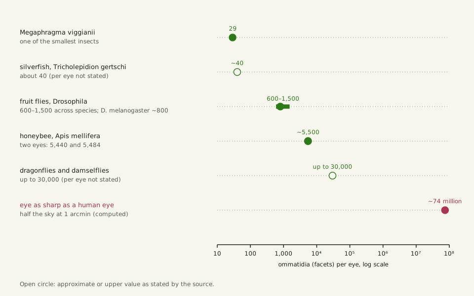
In the insects on this page, counts run from 29 to tens of thousands. Each eye of a female Megaphragma viggianii, a parasitic wasp about 290 µm long and one of the smallest insects, has 29 facets with lenses averaging 6.9–8.0 µm wide, Makarova and colleagues report (2025). The whole eye, about 51 µm tall, is built from 478 cells, they add. A silverfish, Tricholepidion gertschi, has about 40 ommatidia, while dragonflies and damselflies carry up to 30,000, by the account of Buffry and colleagues (2024).
Fruit flies have about 600 to 1,500 ommatidia per eye across Drosophila species, and about 800 in D. melanogaster, with facets about 15–22 µm wide, Tran and colleagues note (2025). Honeybees have more. In 2019 Taylor and colleagues counted 5,440 and 5,484 facets on two honeybee eyes, close to an earlier study's average of 5,375, with the widest facets 25.4–25.6 µm.
Within one species, eye size matters too. Larger workers of the bumblebee Bombus terrestris have bigger eyes with more facets, and wider ones, Taylor and colleagues found. In the close-up below, the German wasp's facets fit together as hexagons, the grid that counting software such as Tran and colleagues' EyeHex follows as it maps ommatidia one by one. Another six-sided pattern on this site is the snowflake.
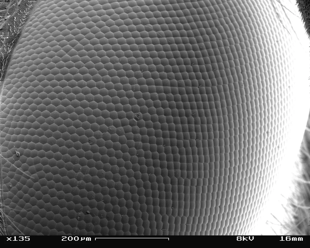
Facets of a German wasp's eye, each about 25 µm wide against the printed 200 µm bar, computed for this page [10−5 m].
Color: native (electron micrograph, no color)
"Vespula germanica SEM Eye 02" by SecretDisc — https://commons.wikimedia.org/wiki/File:Vespula_germanica_SEM_Eye_02.jpg — CC BY 2.5 (https://creativecommons.org/licenses/by/2.5/)

Like every micrograph on this page, it is a gray scanning electron image with no borrowed color added.
Slide the diagram sideways to read its labels, or open it full size.
Ommatidia counts on a log scale, per eye where the source says so: 29 in Megaphragma viggianii, 600–1,500 in fruit flies, about 5,500 in honeybees, up to 30,000 in dragonflies, and about 74 million to cover half the sky at one-minute spacing, computed for this page.
Open circles: approximate or upper values as stated. Data: compound-eye-ommatidia-counts.csv (sources, version and license, JSON); the last value from compound-eye-eye-size.csv (sources, version and license, JSON).
"Facet counts in the insects on this page: 29 to tens of thousands, per eye where stated" by Ants, Cells & Atoms, https://antscellsandatoms.com/insects/compound-eye/ — CC BY 4.0 (https://creativecommons.org/licenses/by/4.0/)
Data sources for this figure
Data: Makarova AA et al. 2025, eLife 14:RP103247, CC BY 4.0, https://doi.org/10.7554/eLife.103247, retrieved 2026-10-25; Buffry AD et al. 2024, BMC Biology 22:67, CC BY 4.0, https://doi.org/10.1186/s12915-024-01864-7, retrieved 2026-10-25; Tran H, Dostatni N, Ramaekers A 2025, Biology Open 14(6):bio061962, CC BY 4.0, https://doi.org/10.1242/bio.061962, retrieved 2026-10-25; Taylor GJ, Tichit P, Schmidt MD, Bodey AJ, Rau C, Baird E 2019, eLife 8:e40613, CC BY 4.0, https://doi.org/10.7554/eLife.40613, retrieved 2026-10-25; Half-sky facet count computed 2026-10-25
Data sources for this figure
Data: Makarova AA et al. 2025, eLife 14:RP103247, CC BY 4.0, https://doi.org/10.7554/eLife.103247, retrieved 2026-10-25; Buffry AD et al. 2024, BMC Biology 22:67, CC BY 4.0, https://doi.org/10.1186/s12915-024-01864-7, retrieved 2026-10-25; Tran H, Dostatni N, Ramaekers A 2025, Biology Open 14(6):bio061962, CC BY 4.0, https://doi.org/10.1242/bio.061962, retrieved 2026-10-25; Juusola M et al. 2017, eLife 6:e26117, CC BY 4.0, https://doi.org/10.7554/eLife.26117, retrieved 2026-10-25; Taylor GJ, Tichit P, Schmidt MD, Bodey AJ, Rau C, Baird E 2019, eLife 8:e40613, CC BY 4.0, https://doi.org/10.7554/eLife.40613, retrieved 2026-10-25; Gutiérrez D, Rigosi E, Nagloo N, O'Carroll D, Warrant EJ 2024, Journal of Experimental Biology 227(16):jeb246670, CC BY 4.0, https://doi.org/10.1242/jeb.246670, retrieved 2026-10-25; Scale-bar measurements: SecretDisc, Vespula germanica SEM Eye 01 and Eye 02, CC BY 2.5, https://commons.wikimedia.org/wiki/File:Vespula_germanica_SEM_Eye_01.jpg; computed 2026-10-25; GBIF Secretariat 2023, GBIF Backbone Taxonomy, checklist dataset https://doi.org/10.15468/39omei, CC BY 4.0, taxon keys in the gbif_key column, retrieved 2026-10-25
Data sources for this figure
Data: Heras FJH, Laughlin SB, eLife 13:RP96517 (2024; version of record 2026), CC BY 4.0, https://doi.org/10.7554/eLife.96517, retrieved 2026-10-25; Taylor GJ, Tichit P, Schmidt MD, Bodey AJ, Rau C, Baird E 2019, eLife 8:e40613, CC BY 4.0, https://doi.org/10.7554/eLife.40613, retrieved 2026-10-25; Gutiérrez D, Rigosi E, Nagloo N, O'Carroll D, Warrant EJ 2024, Journal of Experimental Biology 227(16):jeb246670, CC BY 4.0, https://doi.org/10.1242/jeb.246670, retrieved 2026-10-25; Tran H, Dostatni N, Ramaekers A 2025, Biology Open 14(6):bio061962, CC BY 4.0, https://doi.org/10.1242/bio.061962, retrieved 2026-10-25; Juusola M et al. 2017, eLife 6:e26117, CC BY 4.0, https://doi.org/10.7554/eLife.26117, retrieved 2026-10-25; One-minute target: de Jong PTVM 2024, Eye 38(1):13-24, CC BY 4.0, https://doi.org/10.1038/s41433-022-02180-6, as listed in this site's reference-objects.csv, retrieved 2026-10-25; Facet widths, radii and the half-sky facet count computed 2026-10-25
Why do narrow lenses blur?
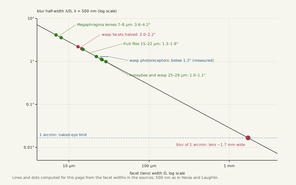
Light passing through a lens spreads out by diffraction, and the narrower the lens, the wider the spread. For a facet of width D, the blur spot has a half-width of about λ/D radians, where λ is the wavelength of the light; Heras and Laughlin take λ as 500 nm. By that formula a 25 µm facet blurs a point of light over about 1.1°, and an 8 µm Megaphragma lens over about 3.6°, computed for this page.
The German wasp shows the limit at work. For its acute zone, Gutiérrez and colleagues took D ≈ 26 µm and green light of 530 nm, which gives a blur of 1.17°. The narrowest acceptance angles they recorded there, the width of sky each photoreceptor takes in, were below 1.3°. With facets about 1.0° apart, the spacing and the blur of each lens are about the same size.
That is why, past a point, packing more facets into the same eye does not help. Keep the acute zone's curvature and halve the facet width: Δφ halves to about 0.5°, but each lens now blurs over about 2.0–2.2°, roughly four times the new spacing (computed for this page). By that arithmetic the eye samples a finer grid but sees no finer. Taylor and colleagues, citing Snyder (1979), put the threshold at a facet width times spacing, D × Δφ, of about 0.29 µm: there, resolution is limited by the diffraction of green light.
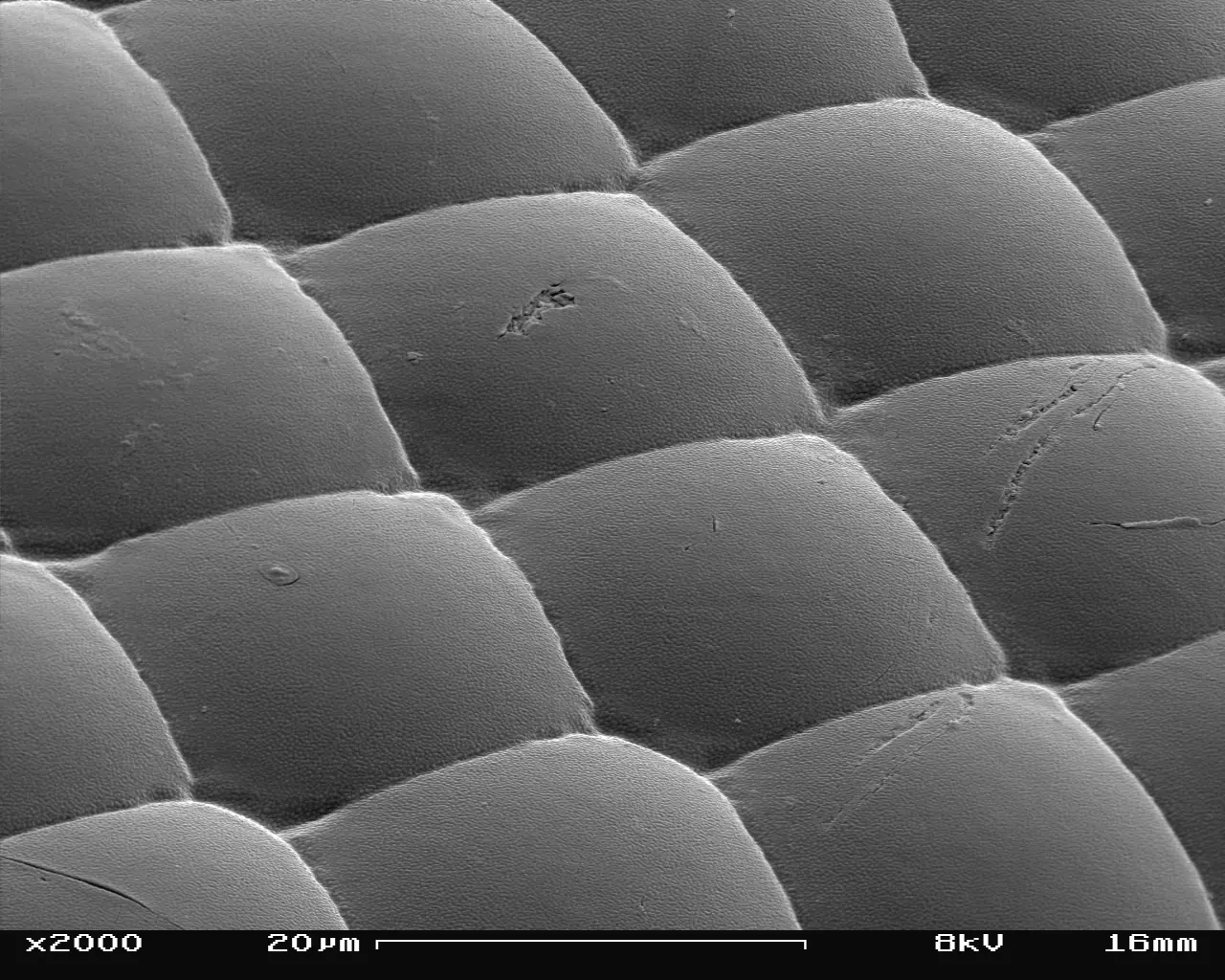
Facet lenses of a German wasp, each about as wide as the printed 20 µm bar, seen at a slant [10−5 m].
Color: native (electron micrograph, no color)
"Vespula germanica SEM Eye 04" by SecretDisc — https://commons.wikimedia.org/wiki/File:Vespula_germanica_SEM_Eye_04.jpg — CC BY 2.5 (https://creativecommons.org/licenses/by/2.5/)

Slide the diagram sideways to read its labels, or open it full size.
Diffraction blur of facet lenses 7–29 µm wide [10−6–10−5 m]: 3.6–4.2° in Megaphragma, about 1.0–1.1° in honeybees and wasps; a blur of 1 arcmin needs a lens about 1.7 mm wide.
Half-width λ/D with λ = 500 nm, computed for this page. Data: compound-eye-blur.csv (sources, version and license, JSON).
"Diffraction blur of a facet lens against its width" by Ants, Cells & Atoms, https://antscellsandatoms.com/insects/compound-eye/ — CC BY 4.0 (https://creativecommons.org/licenses/by/4.0/)
Data sources for this figure
Data: Blur half-width lambda/D and lambda = 500 nm: Heras FJH, Laughlin SB, eLife 13:RP96517 (2024; version of record 2026), CC BY 4.0, https://doi.org/10.7554/eLife.96517, retrieved 2026-10-25; German wasp facets, stated blur and acceptance angles: Gutiérrez D, Rigosi E, Nagloo N, O'Carroll D, Warrant EJ 2024, Journal of Experimental Biology 227(16):jeb246670, CC BY 4.0, https://doi.org/10.1242/jeb.246670, retrieved 2026-10-25; Facet widths: Makarova AA et al. 2025, eLife 14:RP103247, CC BY 4.0, https://doi.org/10.7554/eLife.103247; Tran H, Dostatni N, Ramaekers A 2025, Biology Open 14(6):bio061962, CC BY 4.0, https://doi.org/10.1242/bio.061962; Taylor GJ et al. 2019, eLife 8:e40613, CC BY 4.0, https://doi.org/10.7554/eLife.40613; retrieved 2026-10-25; Blur values computed 2026-10-25
Data sources for this figure
Data: Heras FJH, Laughlin SB, eLife 13:RP96517 (2024; version of record 2026), CC BY 4.0, https://doi.org/10.7554/eLife.96517, retrieved 2026-10-25; Gutiérrez D, Rigosi E, Nagloo N, O'Carroll D, Warrant EJ 2024, Journal of Experimental Biology 227(16):jeb246670, CC BY 4.0, https://doi.org/10.1242/jeb.246670, retrieved 2026-10-25; Makarova AA et al. 2025, eLife 14:RP103247, CC BY 4.0, https://doi.org/10.7554/eLife.103247, retrieved 2026-10-25; Tran H, Dostatni N, Ramaekers A 2025, Biology Open 14(6):bio061962, CC BY 4.0, https://doi.org/10.1242/bio.061962, retrieved 2026-10-25; Taylor GJ, Tichit P, Schmidt MD, Bodey AJ, Rau C, Baird E 2019, eLife 8:e40613, CC BY 4.0, https://doi.org/10.7554/eLife.40613, retrieved 2026-10-25; Blur values computed 2026-10-25
How big would an eye as sharp as yours be?
A human eye makes a fair target. Robert Hooke put the naked eye's limit at about one minute of arc, 1/60 of a degree, as a 2024 history of acuity testing recounts, and later work confirmed his figure. Two routes lead from there to the size of a compound eye that sharp, both computed for this page.
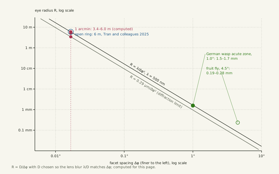
Route one starts from the optics. A lens that blurs no more than one minute at 500 nm must be about 1.7 mm wide, and facets that wide, one minute apart, form an eye with a radius of about 5.9 m. Using the 0.29 µm diffraction limit for D × Δφ from Taylor and colleagues instead gives facets 1.0 mm wide and a radius of about 3.4 m.
Route two scales up a real eye. Resolution in an apposition eye improves only with the square root of its radius, Heras and Laughlin note, citing Kirschfeld (1976). To make the wasp's 1.0° spacing 60 times finer, its 1.5–1.7 mm radius must grow 3,600 times, to 5.4–6.0 m.
| Route | Inputs | Facet width | Eye radius |
|---|---|---|---|
| Optics, blur of 1 arcmin | λ = 500 nm; R = D/Δφ | 1.7 mm | 5.9 m |
| Optics, diffraction limit | D × Δφ = 0.29 µm; R = D/Δφ | 1.0 mm | 3.4 m |
| German wasp, scaled up | D 26–29 µm at Δφ 1.0°; R grows as 1/Δφ2 | 1.6–1.7 mm | 5.4–6.0 m |
The routes share two inputs, the one-minute target and R = D/Δφ. They agree within a factor of two: a radius of 3.4–6.0 m, an eye roughly 7–12 m across. Tran and colleagues give the same order, writing that human-level resolution would take millions of ommatidia and a radius of 6 m, in a passage that cites Kirschfeld, Land and Mallock on the lower acuity of compound eyes.
Covering half the sky at one-minute spacing would take about 74 million facets, computed for this page. That eye would be sharp in every direction, while human vision is sharp only in a small central region, the fovea, Taylor and colleagues point out.
Slide the diagram sideways to read its labels, or open it full size.
Eye radius against facet spacing, from a fruit-fly eye region of 0.19–0.28 mm to 3.4–6.0 m for one minute of arc [10−4–100 m], computed for this page.
The wasp's acute zone sits on the line R = λ/Δφ2; the open ring marks the 6 m in Tran and colleagues (2025). Data: compound-eye-eye-size.csv (sources, version and license, JSON).
"Eye radius needed for a given facet spacing: the square law" by Ants, Cells & Atoms, https://antscellsandatoms.com/insects/compound-eye/ — CC BY 4.0 (https://creativecommons.org/licenses/by/4.0/)
Data sources for this figure
Data: Heras FJH, Laughlin SB, eLife 13:RP96517 (2024; version of record 2026), CC BY 4.0, https://doi.org/10.7554/eLife.96517, retrieved 2026-10-25; Taylor GJ, Tichit P, Schmidt MD, Bodey AJ, Rau C, Baird E 2019, eLife 8:e40613, CC BY 4.0, https://doi.org/10.7554/eLife.40613, retrieved 2026-10-25; Gutiérrez D, Rigosi E, Nagloo N, O'Carroll D, Warrant EJ 2024, Journal of Experimental Biology 227(16):jeb246670, CC BY 4.0, https://doi.org/10.1242/jeb.246670, retrieved 2026-10-25; Tran H, Dostatni N, Ramaekers A 2025, Biology Open 14(6):bio061962, CC BY 4.0, https://doi.org/10.1242/bio.061962, retrieved 2026-10-25; Juusola M et al. 2017, eLife 6:e26117, CC BY 4.0, https://doi.org/10.7554/eLife.26117, retrieved 2026-10-25; One-minute target: de Jong PTVM 2024, Eye 38(1):13-24, CC BY 4.0, https://doi.org/10.1038/s41433-022-02180-6, as listed in this site's reference-objects.csv; radii computed 2026-10-25
Data sources for this figure
Data: Heras FJH, Laughlin SB, eLife 13:RP96517 (2024; version of record 2026), CC BY 4.0, https://doi.org/10.7554/eLife.96517, retrieved 2026-10-25; Taylor GJ, Tichit P, Schmidt MD, Bodey AJ, Rau C, Baird E 2019, eLife 8:e40613, CC BY 4.0, https://doi.org/10.7554/eLife.40613, retrieved 2026-10-25; Gutiérrez D, Rigosi E, Nagloo N, O'Carroll D, Warrant EJ 2024, Journal of Experimental Biology 227(16):jeb246670, CC BY 4.0, https://doi.org/10.1242/jeb.246670, retrieved 2026-10-25; Tran H, Dostatni N, Ramaekers A 2025, Biology Open 14(6):bio061962, CC BY 4.0, https://doi.org/10.1242/bio.061962, retrieved 2026-10-25; Juusola M et al. 2017, eLife 6:e26117, CC BY 4.0, https://doi.org/10.7554/eLife.26117, retrieved 2026-10-25; One-minute target: de Jong PTVM 2024, Eye 38(1):13-24, CC BY 4.0, https://doi.org/10.1038/s41433-022-02180-6, as listed in this site's reference-objects.csv, retrieved 2026-10-25; Facet widths, radii and the half-sky facet count computed 2026-10-25
Why are dragonfly eyes so big?
Insects whose photoreceptors take in patches narrower than 1° are often predators, dragonflies among them, Gutiérrez and colleagues note, and such hunters typically have a forward-pointing acute zone for tracking prey. In the sharp zones of houseflies, praying mantises, dragonflies and robber flies, the facet lenses are enlarged to reduce the diffraction limit and the ommatidia are packed more densely, Heras and Laughlin write. By the square law above, both moves take eye area.
The bill comes in other forms too. In Heras and Laughlin's models, an apposition eye the size of a dragonfly's is 100 times less efficient than a single-lens, camera-type eye of the same cost, and one the size of a fruit fly's 10 times. Eyes also use oxygen. An isolated blowfly eye, Calliphora vicina, consumed 6.48 mm3 of oxygen an hour in bright light and a third of that in darkness, figures Heras and Laughlin take from Pangršič and colleagues (2005).
Some fast-flying insects with large facets lighten the load by wrapping each ommatidium in air-filled tracheae, the insect's air tubes. In a dragonfly's upper eye, air fills at least half the cross-section of the photoreceptor layer, by Heras and Laughlin's reading of published micrographs.
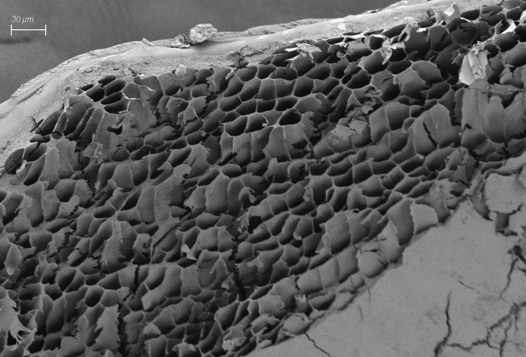
Inside a dragonfly's compound eye: walled channels, one per ommatidium in the photographers' description, beside a printed 30 µm bar [10−5 m].
Color: native (electron micrograph, no color)
"Ojo compuesto visto por dentro, el detalle de los canales de los oamtidios de la libélula SEM" by Brandon Antonio Segura Torres & Priscilla Vieto Bonilla — https://commons.wikimedia.org/wiki/File:Ojo_compuesto_visto_por_dentro,_el_detalle_de_los_canales_de_los_oamtidios_de_la_lib%C3%A9lula_SEM.jpg — CC BY-SA 4.0 (https://creativecommons.org/licenses/by-sa/4.0/)

Do insects see a mosaic?
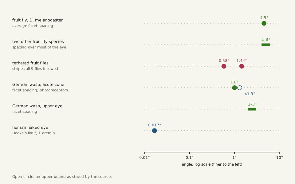
The classic picture of a fly's view is a coarse grid. In Drosophila melanogaster, about 750 ommatidia per eye serve as its sampling points, an average 4.5° apart, Juusola and colleagues wrote in 2017; that spacing was long held to set the limit of what the fly can see.
Their experiments complicated that picture. The fly's photoreceptors contract in response to light, moving their receptive fields, and all nine tethered flies tested steered to follow rotating stripes with gaps of 1.44° and 0.58° between bars, finer than the facet spacing. By the authors' estimate, the flies saw detail at least four times finer than their eyes' sampling limit. As of that study, whether photoreceptors in other animals, humans included, behave the same way was still to be determined.
Slide the diagram sideways to read its labels, or open it full size.
Angles on a log scale: fruit-fly facets 4.5° apart, stripes the flies followed 0.58–1.44°, German wasp facets 1.0° apart, and the naked-eye limit, 0.017°.
Data: compound-eye-angles.csv (sources, version and license, JSON).
"Angles: facet spacing, stripes fruit flies followed, and the naked-eye limit" by Ants, Cells & Atoms, https://antscellsandatoms.com/insects/compound-eye/ — CC BY 4.0 (https://creativecommons.org/licenses/by/4.0/)
Data sources for this figure
Data: Juusola M et al. 2017, eLife 6:e26117, CC BY 4.0, https://doi.org/10.7554/eLife.26117, retrieved 2026-10-25; Buffry AD et al. 2024, BMC Biology 22:67, CC BY 4.0, https://doi.org/10.1186/s12915-024-01864-7, retrieved 2026-10-25; Gutiérrez D, Rigosi E, Nagloo N, O'Carroll D, Warrant EJ 2024, Journal of Experimental Biology 227(16):jeb246670, CC BY 4.0, https://doi.org/10.1242/jeb.246670, retrieved 2026-10-25; One-minute naked-eye limit: de Jong PTVM 2024, Eye 38(1):13-24, CC BY 4.0, https://doi.org/10.1038/s41433-022-02180-6, as listed in this site's reference-objects.csv, retrieved 2026-10-25; converted to degrees 2026-10-25
Data sources for this figure
Data: Juusola M et al. 2017, eLife 6:e26117, CC BY 4.0, https://doi.org/10.7554/eLife.26117, retrieved 2026-10-25; Buffry AD et al. 2024, BMC Biology 22:67, CC BY 4.0, https://doi.org/10.1186/s12915-024-01864-7, retrieved 2026-10-25; Gutiérrez D, Rigosi E, Nagloo N, O'Carroll D, Warrant EJ 2024, Journal of Experimental Biology 227(16):jeb246670, CC BY 4.0, https://doi.org/10.1242/jeb.246670, retrieved 2026-10-25; de Jong PTVM 2024, Eye 38(1):13-24, CC BY 4.0, https://doi.org/10.1038/s41433-022-02180-6, as listed in this site's reference-objects.csv, retrieved 2026-10-25
Sources
- Gutiérrez D, Rigosi E, Nagloo N, O'Carroll D, Warrant EJ. "Spatial resolution and optical sensitivity in the compound eyes of two common European wasps, Vespula germanica and Vespula vulgaris." Journal of Experimental Biology 227(16):jeb246670 (2024). https://doi.org/10.1242/jeb.246670 Read: full text. Text license: CC BY 4.0 (https://creativecommons.org/licenses/by/4.0/).
- Heras FJH, Laughlin SB. "Investments in photoreceptors compete with investments in optics to determine eye design." eLife 13:RP96517 (2024; version of record 2026). https://doi.org/10.7554/eLife.96517 Read: full text. Text license: CC BY 4.0 (https://creativecommons.org/licenses/by/4.0/).
- Juusola M, Dau A, Song Z, Solanki N, Rien D, Jaciuch D, Dongre SA, Blanchard F, de Polavieja GG, Hardie RC, Takalo J. "Microsaccadic sampling of moving image information provides Drosophila hyperacute vision." eLife 6:e26117 (2017). https://doi.org/10.7554/eLife.26117 Read: full text. Text license: CC BY 4.0 (https://creativecommons.org/licenses/by/4.0/).
- Makarova AA, Chua NJ, Diakova AV, Desyatirkina IA, Gunn P, Pang S, Xu CS, Hess HF, Chklovskii DB, Polilov AA. "The first complete 3D reconstruction and morphofunctional mapping of an insect eye." eLife 14:RP103247 (2025). https://doi.org/10.7554/eLife.103247 Read: full text. Text license: CC BY 4.0 (https://creativecommons.org/licenses/by/4.0/).
- Buffry AD, Currea JP, Franke-Gerth FA, Palavalli-Nettimi R, Bodey AJ, Rau C, Samadi N, Gstöhl SJ, Schlepütz CM, McGregor AP, Sumner-Rooney L, Theobald J, Kittelmann M. "Evolution of compound eye morphology underlies differences in vision between closely related Drosophila species." BMC Biology 22:67 (2024). https://doi.org/10.1186/s12915-024-01864-7 Read: full text. Text license: CC BY 4.0 (https://creativecommons.org/licenses/by/4.0/).
- Tran H, Dostatni N, Ramaekers A. "EyeHex toolbox for complete segmentation of ommatidia in fruit fly eyes." Biology Open 14(6):bio061962 (2025). https://doi.org/10.1242/bio.061962 Read: full text. Text license: CC BY 4.0 (https://creativecommons.org/licenses/by/4.0/).
- Taylor GJ, Tichit P, Schmidt MD, Bodey AJ, Rau C, Baird E. "Bumblebee visual allometry results in locally improved resolution and globally improved sensitivity." eLife 8:e40613 (2019). https://doi.org/10.7554/eLife.40613 Read: full text. Text license: CC BY 4.0 (https://creativecommons.org/licenses/by/4.0/).
- de Jong PTVM. "A history of visual acuity testing and optotypes." Eye 38(1):13–24 (2024). https://doi.org/10.1038/s41433-022-02180-6 Read: full text. Text license: CC BY 4.0 (https://creativecommons.org/licenses/by/4.0/).
- Piegari G, De Biase D, d'Aquino I, Prisco F, Fico R, Ilsami R, Pozzato N, Genovese A, Paciello O. "Diagnosis of Drowning and the Value of the Diatom Test in Veterinary Forensic Pathology." Frontiers in Veterinary Science 6:404 (2019). https://doi.org/10.3389/fvets.2019.00404 Read: full text. Text license: CC BY 4.0 (https://creativecommons.org/licenses/by/4.0/).
Cite this page
Ants, Cells & Atoms. “Compound Eye: One as Sharp as Yours Would Span About 7–12 m.” Published 2026-10-25. https://antscellsandatoms.com/insects/compound-eye/. Accessed (add the date you read it).
No DOI is issued for entries. Figures made here are CC BY 4.0; see each caption for the license of photographs.
Related entries
Water Strider: The Dimples Under Its Feet Hold Its Weight
Image: “Aquarius remigis 140810489” by Jason Swanson (2021) · CC0 1.0
"Aquarius remigis 140810489" by Jason Swanson — https://commons.wikimedia.org/wiki/File:Aquarius_remigis_140810489.jpg — CC0 1.0 (https://creativecommons.org/publicdomain/zero/1.0/)

How Do Insects Breathe? Through Air Tubes, Not Lungs
Image: “Flickr - ggallice - Spiracles” by Geoff Gallice (2011) · CC BY 2.0
"Flickr - ggallice - Spiracles" by Geoff Gallice — https://commons.wikimedia.org/wiki/File:Flickr_-_ggallice_-_Spiracles.jpg — CC BY 2.0 (https://creativecommons.org/licenses/by/2.0/)

Ants: Strong for Their Weight, Mostly Because They're Small
Image: “Rote Waldameisen mit Beute 01” by Stephan Sprinz (2026) · CC BY 4.0
"Rote Waldameisen mit Beute 01" by Stephan Sprinz — https://commons.wikimedia.org/wiki/File:Rote_Waldameisen_mit_Beute_01.jpg — CC BY 4.0 (https://creativecommons.org/licenses/by/4.0/)
