Diatoms: Why Most Glass-Walled Algae Shrink as They Multiply

Slide the ladder sideways to read its labels, or open it full size.
Diatoms: 2–2,000 µm across species, most 20–200 µm [10−5–10−4 m].
Dashed lines: naked eye about 0.1 mm and light microscope about 0.2 µm, explained on the Zoom ladder page. Data: diatoms-size-records.csv (sources, version and license, JSON).
"Zoom ladder: diatoms" by Ants, Cells & Atoms, https://antscellsandatoms.com/microscopic-life/diatoms/ — CC BY 4.0 (https://creativecommons.org/licenses/by/4.0/)
Data sources for this figure
Data: Terracciano M, De Stefano L, Rea I 2018, Pharmaceutics 10(4):242, CC BY 4.0, https://doi.org/10.3390/pharmaceutics10040242, retrieved 2026-10-24; Piegari G et al. 2019, Frontiers in Veterinary Science 6:404, CC BY 4.0, https://doi.org/10.3389/fvets.2019.00404, retrieved 2026-10-24; Gastineau R et al. 2021, Biology 10(4):328, CC BY 4.0, https://doi.org/10.3390/biology10040328, retrieved 2026-10-24; Hesgrove C, Boothby TC 2020, Cell Communication and Signaling 18:178, CC BY 4.0, https://doi.org/10.1186/s12964-020-00670-2, retrieved 2026-10-24; Li C et al. 2023, Journal of Biomedical Optics 28(11):116501, CC BY 4.0, https://doi.org/10.1117/1.JBO.28.11.116501, retrieved 2026-10-24; Valli J et al. 2021, Journal of Biological Chemistry 297(1):100791, CC BY 4.0, https://doi.org/10.1016/j.jbc.2021.100791, retrieved 2026-10-24
Data sources for this figure
Data: Terracciano M, De Stefano L, Rea I 2018, Pharmaceutics 10(4):242, CC BY 4.0, https://doi.org/10.3390/pharmaceutics10040242, retrieved 2026-10-24; Piegari G et al. 2019, Frontiers in Veterinary Science 6:404, CC BY 4.0, https://doi.org/10.3389/fvets.2019.00404, retrieved 2026-10-24; Gastineau R et al. 2021, Biology 10(4):328, CC BY 4.0, https://doi.org/10.3390/biology10040328, retrieved 2026-10-24; Moore ER et al. 2017, PLoS ONE 12(7):e0181098, CC BY 4.0, https://doi.org/10.1371/journal.pone.0181098, retrieved 2026-10-24; Hesgrove C, Boothby TC 2020, Cell Communication and Signaling 18:178, CC BY 4.0, https://doi.org/10.1186/s12964-020-00670-2, retrieved 2026-10-24; Li C et al. 2023, Journal of Biomedical Optics 28(11):116501, CC BY 4.0, https://doi.org/10.1117/1.JBO.28.11.116501, retrieved 2026-10-24; Valli J et al. 2021, Journal of Biological Chemistry 297(1):100791, CC BY 4.0, https://doi.org/10.1016/j.jbc.2021.100791, retrieved 2026-10-24; Velho Rodrigues MF, Lisicki M, Lauga E 2021, PLOS ONE 16(6):e0252291, Table 9, CC BY 4.0, https://doi.org/10.1371/journal.pone.0252291, retrieved 2026-10-24; Yang E et al. 2016, Nature Communications 7:13698, CC BY 4.0, https://doi.org/10.1038/ncomms13698, retrieved 2026-10-24
Bench card
- Actual size
- 2–2,000 µm across species; most 20–200 µm [10−5–10−4 m]Source: Terracciano et al. 2018 (2 µm to 2 mm across species); Piegari et al. 2019 (usually 20–200 µm)
- Size in this image
- not measurable from this image
- Lives in
- salt water and fresh water
- Color in images
- native (hero: living cells, unstained)

diatoms-bench.csv (CSV) · where each value comes from, and its license (JSON)
Data credits for this card
Data: GBIF Backbone Taxonomy, Bacillariophyceae (taxon key 7947184), checklist dataset https://doi.org/10.15468/39omei, CC BY 4.0, retrieved 2026-10-24; Terracciano M, De Stefano L, Rea I 2018, Pharmaceutics 10(4):242, CC BY 4.0, https://doi.org/10.3390/pharmaceutics10040242, retrieved 2026-10-24; Piegari G et al. 2019, Frontiers in Veterinary Science 6:404, CC BY 4.0, https://doi.org/10.3389/fvets.2019.00404, retrieved 2026-10-24; Gastineau R et al. 2021, Biology 10(4):328, CC BY 4.0, https://doi.org/10.3390/biology10040328, retrieved 2026-10-24; Fuhrmann-Lieker T et al. 2021, Journal of the Royal Society Interface 18(179):20210146, CC BY 4.0, https://doi.org/10.1098/rsif.2021.0146, retrieved 2026-10-24
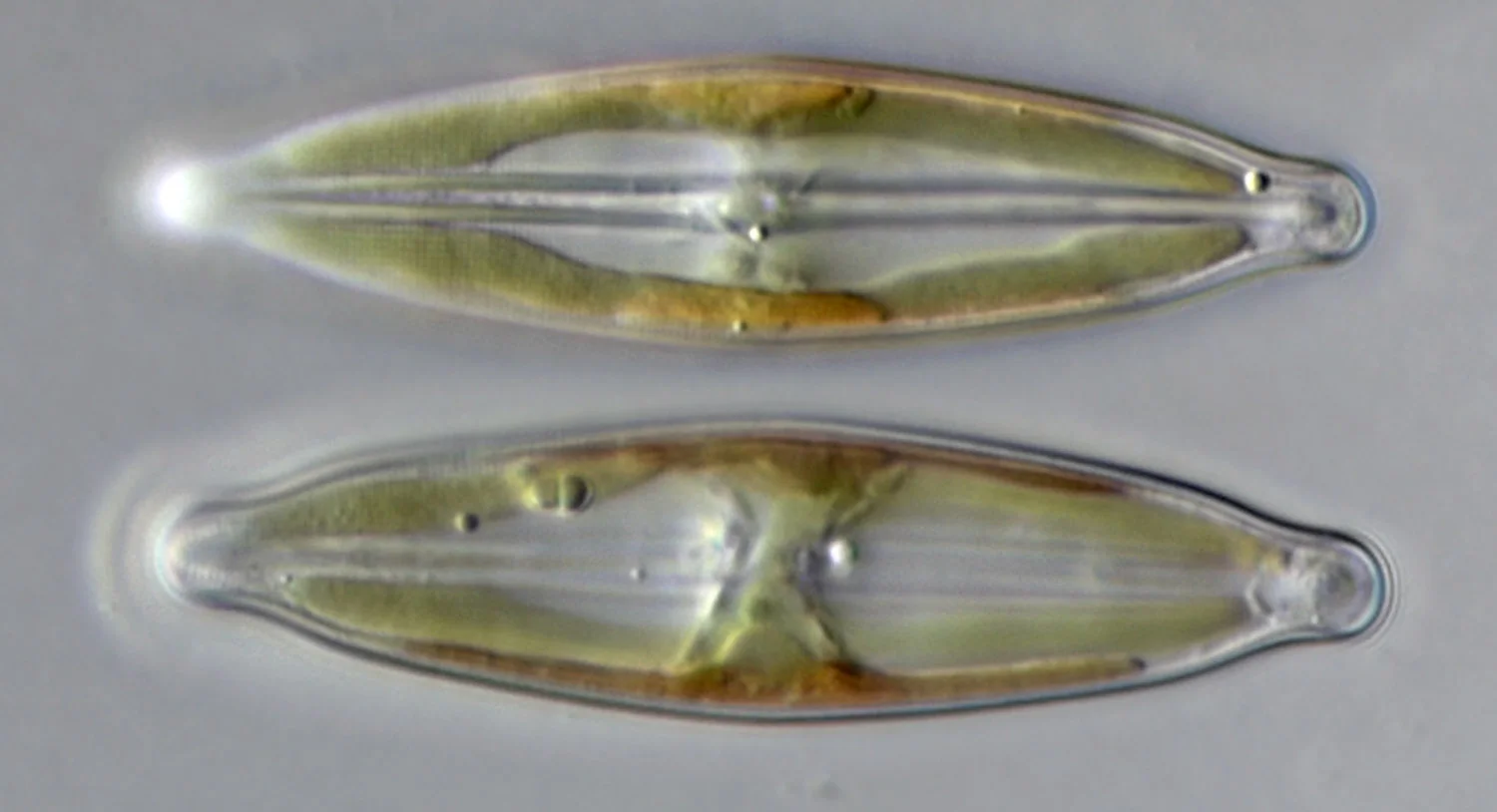
Two living naviculoid (boat-shaped) diatoms side by side; most diatoms are 20–200 µm across [10−5–10−4 m], and this photo has no scale bar.
Color: native (living cells, unstained; Nomarski optics)
"Naviculoid diatom" by Djpmapfer — https://commons.wikimedia.org/wiki/File:Naviculoid_diatom.jpg — CC BY-SA 4.0 (https://creativecommons.org/licenses/by-sa/4.0/)
Diatoms live in oceans, fresh waters and soils. Their walls are made of opal, a hydrated form of silica, and because the finished wall is rigid, the new halves produced during cell division have to fit inside the old ones.
 Illustration
Spindle-shaped diatom cells drifting at different depths of focus, two of them sharp; no particular species is shown.
Illustration: Ants, Cells & Atoms. Generated schematic illustration — not a photograph, micrograph or specimen record. © Ants, Cells & Atoms, all rights reserved.
How big are diatoms?
Single diatom cells range from 2 µm to 2,000 µm (about 0.00008–0.08 in) across species, a thousandfold spread. Most fall between 20 and 200 µm. The largest species pass the roughly 0.1 mm naked-eye line on the ladder, about the width of a water strider's leg, while the common 20–200 µm range straddles it.
Small species stay small even at their peak. In a 2017 experiment, newly formed initial cells of Thalassiosira pseudonana measured 7–12 µm, the largest size reported for that species. Tardigrades, at 50–1,000 µm, occupy these same rungs, and so does a typical human hair, 50–100 µm wide. A single cell of Paramecium lands there too: in a 2021 database of swimming microbes, Paramecium caudatum cells measure 140–311 µm long.
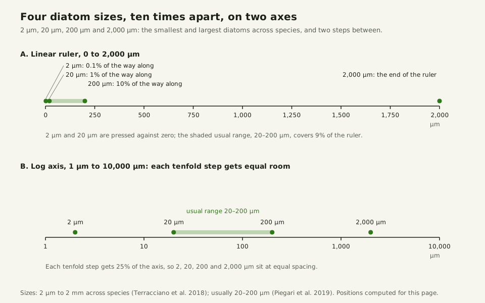
Look at the axis, too. On an ordinary linear ruler running to 2,000 µm, a 2 µm diatom would sit 0.1% of the way along and a 20 µm one at 1%, both pressed against zero. The ladder is logarithmic, so every tenfold step gets equal room, and for sizes that span this many powers of ten, a logarithmic axis is the only honest choice.
Slide the diagram sideways to read its labels, or open it full size.
Diatom sizes from 2 µm to 2,000 µm [10−6–10−3 m] on a linear ruler and on a log axis; only the log axis keeps them apart.
Data: diatoms-size-records.csv (sources, version and license, JSON); positions computed for this page.
"Diatom sizes on a linear ruler and on a log axis" by Ants, Cells & Atoms, https://antscellsandatoms.com/microscopic-life/diatoms/ — CC BY 4.0 (https://creativecommons.org/licenses/by/4.0/)
Data: Terracciano M, De Stefano L, Rea I 2018, Pharmaceutics 10(4):242, CC BY 4.0, https://doi.org/10.3390/pharmaceutics10040242, retrieved 2026-10-24; Piegari G et al. 2019, Frontiers in Veterinary Science 6:404, CC BY 4.0, https://doi.org/10.3389/fvets.2019.00404, retrieved 2026-10-24; Positions on both axes computed 2026-10-24
Data sources for this figure
Data: Terracciano M, De Stefano L, Rea I 2018, Pharmaceutics 10(4):242, CC BY 4.0, https://doi.org/10.3390/pharmaceutics10040242, retrieved 2026-10-24; Piegari G et al. 2019, Frontiers in Veterinary Science 6:404, CC BY 4.0, https://doi.org/10.3389/fvets.2019.00404, retrieved 2026-10-24; Gastineau R et al. 2021, Biology 10(4):328, CC BY 4.0, https://doi.org/10.3390/biology10040328, retrieved 2026-10-24; Moore ER et al. 2017, PLoS ONE 12(7):e0181098, CC BY 4.0, https://doi.org/10.1371/journal.pone.0181098, retrieved 2026-10-24; Hesgrove C, Boothby TC 2020, Cell Communication and Signaling 18:178, CC BY 4.0, https://doi.org/10.1186/s12964-020-00670-2, retrieved 2026-10-24; Li C et al. 2023, Journal of Biomedical Optics 28(11):116501, CC BY 4.0, https://doi.org/10.1117/1.JBO.28.11.116501, retrieved 2026-10-24; Valli J et al. 2021, Journal of Biological Chemistry 297(1):100791, CC BY 4.0, https://doi.org/10.1016/j.jbc.2021.100791, retrieved 2026-10-24; Velho Rodrigues MF, Lisicki M, Lauga E 2021, PLOS ONE 16(6):e0252291, Table 9, CC BY 4.0, https://doi.org/10.1371/journal.pone.0252291, retrieved 2026-10-24; Yang E et al. 2016, Nature Communications 7:13698, CC BY 4.0, https://doi.org/10.1038/ncomms13698, retrieved 2026-10-24
Why does a diatom get smaller when it divides?
When a diatom divides, each daughter inherits one half of the parent's frustule and uses it as its own epitheca. Then it deposits a fresh hypotheca inside that half. The daughter that inherited the parent's epitheca therefore comes out the same size as its parent.
The other daughter inherited the parent's hypotheca, which now serves as its epitheca, and its new hypotheca must fit inside that smaller piece, so the whole cell is one size step smaller. Diatom biologists call this predictable pattern the MacDonald–Pfitzer rule. Over many divisions, the average size of the population falls while the spread of sizes widens.
After 10 divisions, how many cells are still full size?
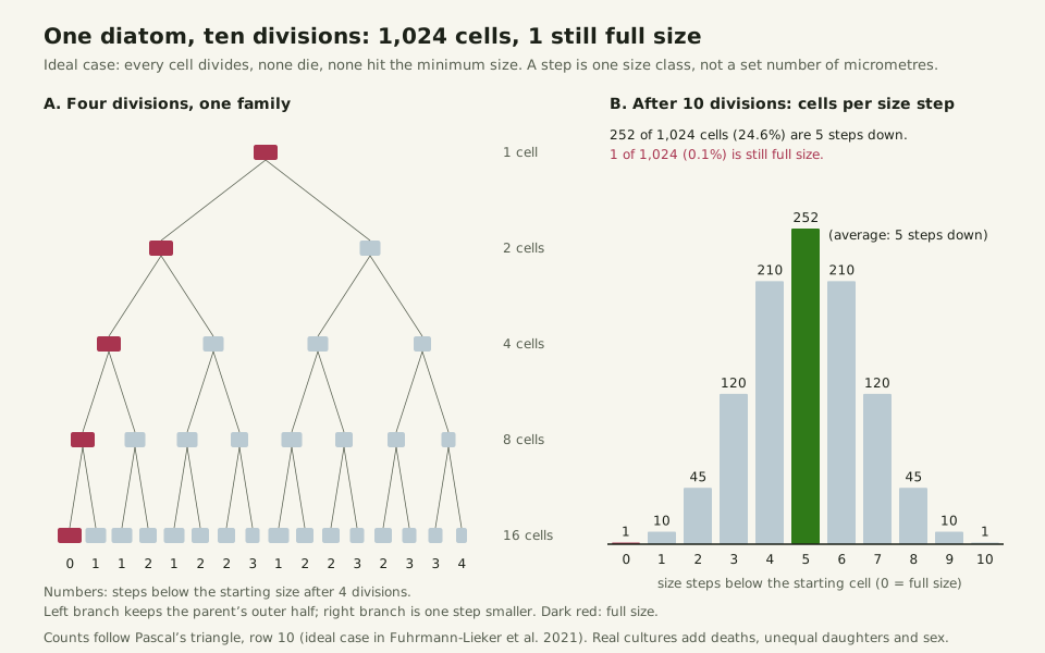
Take one full-size cell, let every cell divide, and let none die. That is the ideal case in a 2021 mathematical model by Thomas Fuhrmann-Lieker and colleagues, in which the number of cells in each size class follows Pascal's triangle until the smallest class is reached. After t divisions there are 2t cells, and exactly one of them is still full size.
After 10 divisions, that is 1 cell of 1,024, or about 0.1%. The most common size is five steps down, held by 252 cells (24.6%), and 65.6% of all cells sit within one step of it. A species with room for at least 20 steps would have just 1 full-size cell in 1,048,576 after 20 divisions. Until the smallest class is reached, the average falls by half a step per division, and the full-size line never disappears; it thins to one cell in a million.
Slide the diagram sideways to read its labels, or open it full size.
Ideal size-step model: 1 diatom, 10 divisions, 1,024 cells, 1 still full size.
Steps are size classes, not micrometres. Data: diatoms-size-steps.csv (sources, version and license, JSON).
"One diatom, ten divisions" by Ants, Cells & Atoms, https://antscellsandatoms.com/microscopic-life/diatoms/ — CC BY 4.0 (https://creativecommons.org/licenses/by/4.0/)
Data: Model: Fuhrmann-Lieker T et al. 2021, Journal of the Royal Society Interface 18(179):20210146, CC BY 4.0, https://doi.org/10.1098/rsif.2021.0146; cell counts per size step are the binomial coefficients C(t, k), computed 2026-10-24 for t = 0–12
Does the shrinking show up in real cultures?
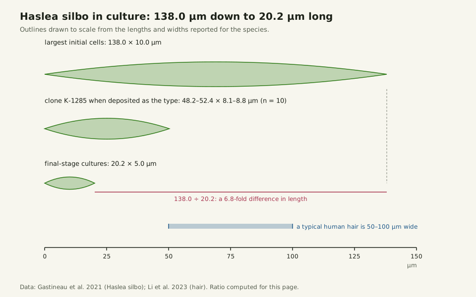
Laboratory cultures show the same pattern. In cultures of the blue diatom Haslea silbo, cell length ranged from 138.0 µm in the largest initial cells, longer than a typical human hair is wide (50–100 µm), down to 20.2 µm in final-stage cultures, a 6.8-fold difference. Photographs of one clone, K-1285, document it getting shorter between July 2009 and January 2012, and as the cells shrank, their glass walls frequently became deformed.
Slide the diagram sideways to read its labels, or open it full size.
Haslea silbo cells drawn to scale, from 138.0 µm initial cells to 20.2 µm in final-stage cultures [10−5–10−4 m].
Data: diatoms-size-records.csv (sources, version and license, JSON); the 6.8-fold ratio is computed for this page.
"Haslea silbo in culture: 138.0 to 20.2 micrometres long" by Ants, Cells & Atoms, https://antscellsandatoms.com/microscopic-life/diatoms/ — CC BY 4.0 (https://creativecommons.org/licenses/by/4.0/)
Data: Gastineau R et al. 2021, Biology 10(4):328, CC BY 4.0, https://doi.org/10.3390/biology10040328, retrieved 2026-10-24; Li C et al. 2023, Journal of Biomedical Optics 28(11):116501, CC BY 4.0, https://doi.org/10.1117/1.JBO.28.11.116501, retrieved 2026-10-24; Length ratio 138.0 / 20.2 computed 2026-10-24
Data sources for this figure
Data: Terracciano M, De Stefano L, Rea I 2018, Pharmaceutics 10(4):242, CC BY 4.0, https://doi.org/10.3390/pharmaceutics10040242, retrieved 2026-10-24; Piegari G et al. 2019, Frontiers in Veterinary Science 6:404, CC BY 4.0, https://doi.org/10.3389/fvets.2019.00404, retrieved 2026-10-24; Gastineau R et al. 2021, Biology 10(4):328, CC BY 4.0, https://doi.org/10.3390/biology10040328, retrieved 2026-10-24; Moore ER et al. 2017, PLoS ONE 12(7):e0181098, CC BY 4.0, https://doi.org/10.1371/journal.pone.0181098, retrieved 2026-10-24; Hesgrove C, Boothby TC 2020, Cell Communication and Signaling 18:178, CC BY 4.0, https://doi.org/10.1186/s12964-020-00670-2, retrieved 2026-10-24; Li C et al. 2023, Journal of Biomedical Optics 28(11):116501, CC BY 4.0, https://doi.org/10.1117/1.JBO.28.11.116501, retrieved 2026-10-24; Valli J et al. 2021, Journal of Biological Chemistry 297(1):100791, CC BY 4.0, https://doi.org/10.1016/j.jbc.2021.100791, retrieved 2026-10-24; Velho Rodrigues MF, Lisicki M, Lauga E 2021, PLOS ONE 16(6):e0252291, Table 9, CC BY 4.0, https://doi.org/10.1371/journal.pone.0252291, retrieved 2026-10-24; Yang E et al. 2016, Nature Communications 7:13698, CC BY 4.0, https://doi.org/10.1038/ncomms13698, retrieved 2026-10-24
Haslea silbo cells have blue tips, and that blue is native color: a marennine-like pigment the cells make themselves and hold in vacuoles at their ends. Blue from a laboratory stain would be borrowed color, and every micrograph caption on this site states which kind it shows.
Other laboratories report the same drift. A 2015 study of Pseudo-nitzschia notes that clonal strains become shorter over time, so its cultured cells were small compared with the same species observed in the field. According to a 2022 study, the shrinking phase can last months or years, depending on growth conditions.
How do diatoms get big again?
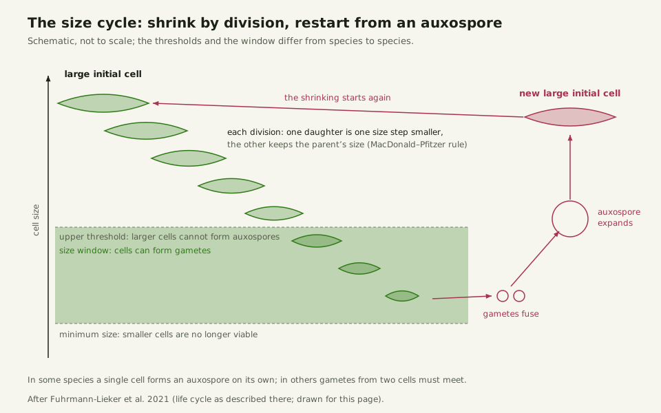
Usually through sexual reproduction. Once cells shrink into a size window specific to their species, they can produce gametes, the sex cells, which fuse into a zygote. That zygote, called an auxospore, expands and builds a large new "initial cell," which then starts shrinking all over again. In some species, a single cell can form an auxospore on its own.
Slide the diagram sideways to read its labels, or open it full size.
The diatom size cycle for cells most often 20–200 µm [10−5–10−4 m]: shrinking divisions, a size window for sex, then a large initial cell from the auxospore.
Schematic, not to scale; thresholds differ by species.
"The diatom size cycle" by Ants, Cells & Atoms, https://antscellsandatoms.com/microscopic-life/diatoms/ — CC BY 4.0 (https://creativecommons.org/licenses/by/4.0/)
Data: Fuhrmann-Lieker T et al. 2021, Journal of the Royal Society Interface 18(179):20210146, CC BY 4.0, https://doi.org/10.1098/rsif.2021.0146, retrieved 2026-10-24
Size, not age, decides whether a cell can go sexual; von Stosch showed this in experiments that changed cell size artificially, according to the 2021 paper's summary. Cells above an upper size threshold cannot form auxospores at all.
Some species also have a lower threshold. There is always a minimum size below which cells are no longer viable. In many species, a lineage that never reproduces sexually keeps shrinking until it is too small to survive, the 2022 study notes. According to the 2021 paper, William M. Lewis of the University of Colorado called this whole cycle a "diatom sex clock" in 1984.
Does every diatom shrink?
Not every species follows the rule. The 2021 paper mentions species whose cells do not shrink during ordinary (vegetative) division, and some that even enlarge without sexual reproduction. Phaeodactylum tricornutum is one documented case: no size reduction has been observed in it so far. A 2018 study describes it as an atypical diatom that can take three forms, fusiform (spindle-shaped), triradiate (three-armed) and oval, with a poorly silicified wall, and suggests the missing size reduction may be connected to that weak silicification.
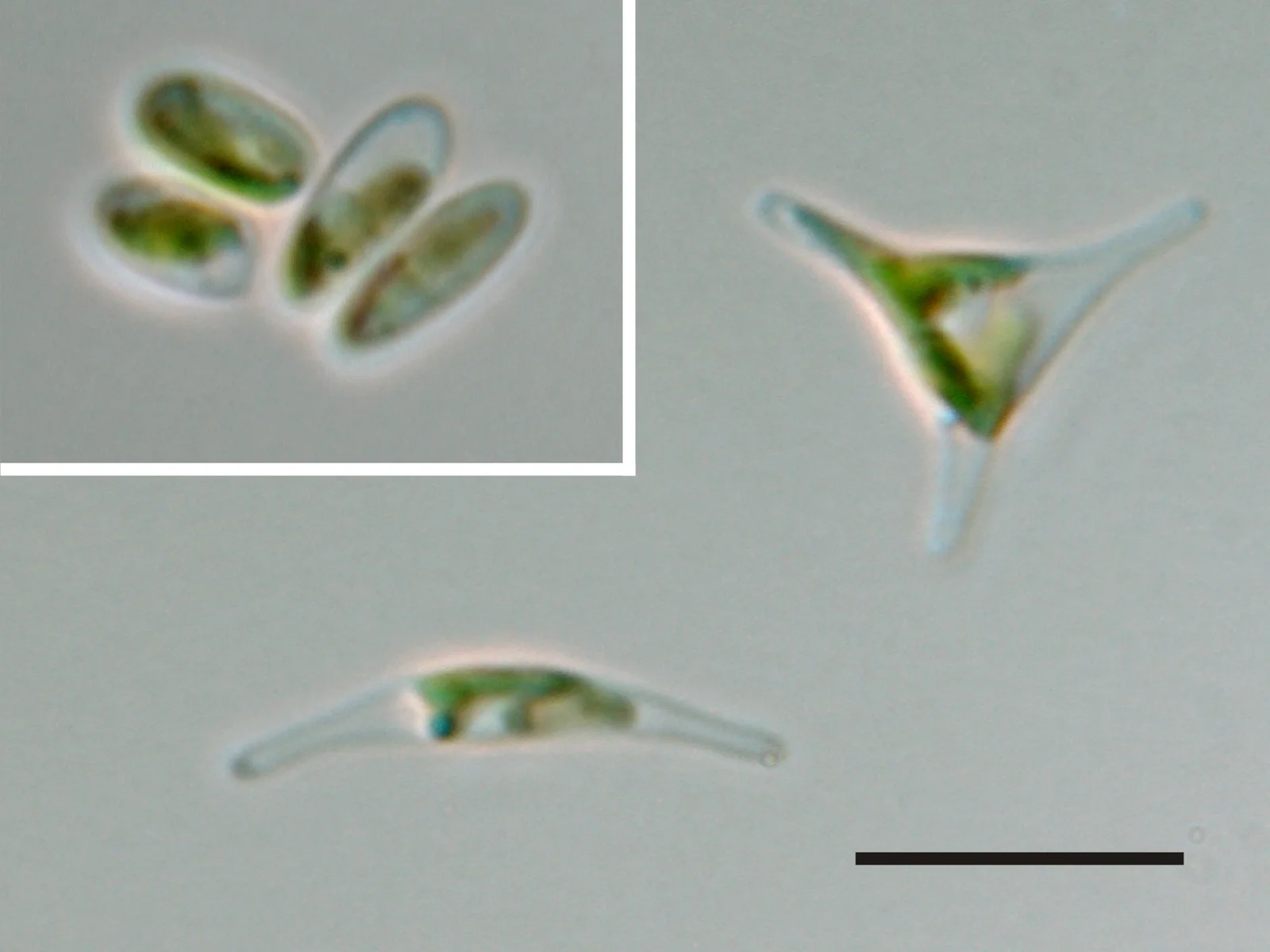
Phaeodactylum tricornutum in its three forms, triradiate (top right), fusiform (bottom) and oval (top left), with a 10 µm scale bar [10−5 m].
Color: native (no stain stated by the source)
"Phaeodactylum tricornutum" by Dr. Ansgar Gruber, Universität Konstanz — https://commons.wikimedia.org/wiki/File:Phaeodactylum_tricornutum.jpg — CC BY 4.0 (https://creativecommons.org/licenses/by/4.0/)

How much of Earth's oxygen do diatoms make?
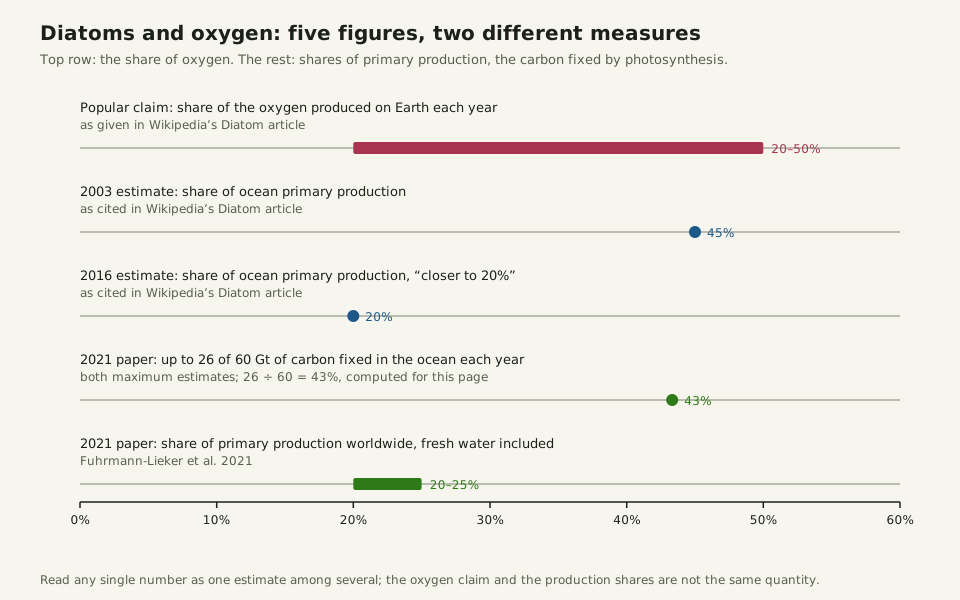
Popular claim: Diatoms make about 20–50% of the oxygen produced on Earth each year. That range appears in Wikipedia's article on diatoms.
What the cited studies say: The published figures are shares of primary production, the carbon fixed by photosynthesis, and they vary. A 2021 paper cites up to 26 of an estimated 60 gigatonnes of carbon fixed in the ocean each year for diatoms, both maximum estimates, and 20–25% of primary production worldwide once fresh water is added. For the ocean alone, Wikipedia cites a 2003 estimate of 45% and a 2016 estimate closer to 20%. Read any single number as one estimate among several.
Slide the diagram sideways to read its labels, or open it full size.
Five figures for diatoms' share: 20–50% of oxygen in the popular claim, 20–45% of primary production in the studies cited here.
The 2003 and 2016 figures are as cited by Wikipedia; 26 of 60 gigatonnes is 43%, computed for this page.
"Diatoms and oxygen: five figures on one axis" by Ants, Cells & Atoms, https://antscellsandatoms.com/microscopic-life/diatoms/ — CC BY 4.0 (https://creativecommons.org/licenses/by/4.0/)
Data: Fuhrmann-Lieker T et al. 2021, Journal of the Royal Society Interface 18(179):20210146, CC BY 4.0, https://doi.org/10.1098/rsif.2021.0146, retrieved 2026-10-24; Wikipedia contributors, "Diatom", Wikipedia, CC BY-SA 4.0, oldid 1381485354 (the oxygen range and the 2003 and 2016 estimates as cited there; text paraphrased, no data file), read 2026-10-24; Share 26 / 60 computed 2026-10-24
Is the diatom sex clock a real clock?
Slide the diagram sideways to read its labels, or open it full size.
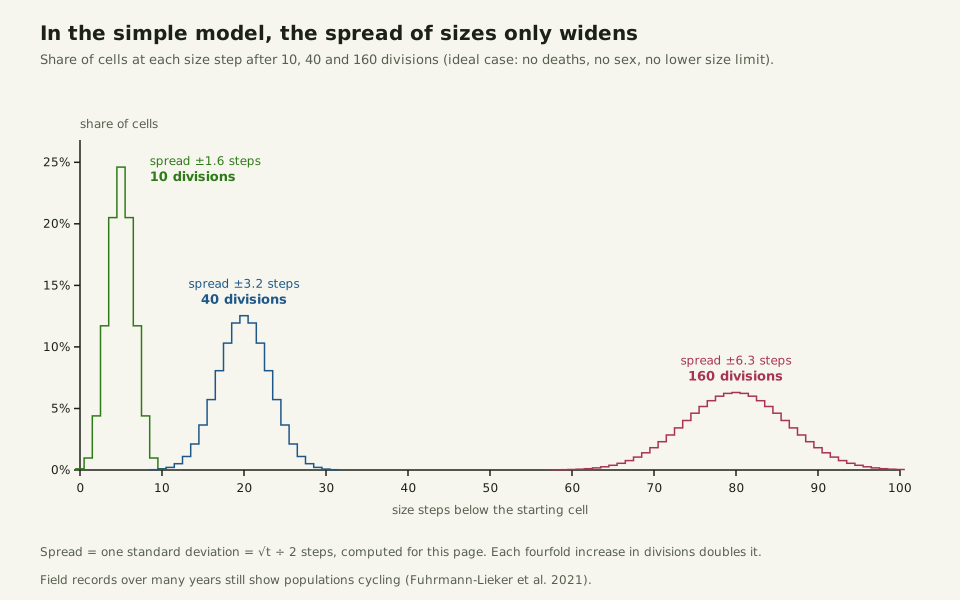
In the ideal model the spread of sizes keeps widening: about 1.6 size steps after 10 divisions, 3.2 after 40 and 6.3 after 160.
Spread is one standard deviation, √t ÷ 2 steps, computed for this page.
"The ideal model only widens" by Ants, Cells & Atoms, https://antscellsandatoms.com/microscopic-life/diatoms/ — CC BY 4.0 (https://creativecommons.org/licenses/by/4.0/)
Data: Model: Fuhrmann-Lieker T et al. 2021, Journal of the Royal Society Interface 18(179):20210146, CC BY 4.0, https://doi.org/10.1098/rsif.2021.0146; shares C(t, k) / 2^t and spreads computed 2026-10-24 for t = 10, 40 and 160
Field records spanning many years show diatom populations cycling roughly on the expected schedule, the 2021 paper notes, yet in the simple model the size distribution only keeps widening. What trims that spread back in real water, and how quickly? Answering it would require a cell-by-cell size census of one wild population across a complete cycle, and a single cycle can last many years.
Sources
- Fuhrmann-Lieker T, Kubetschek N, Ziebarth J, Klassen R, Seiler W. "Is the diatom sex clock a clock?" Journal of the Royal Society Interface 18(179):20210146 (2021). https://doi.org/10.1098/rsif.2021.0146 Read: full text. Text license: CC BY 4.0 (https://creativecommons.org/licenses/by/4.0/).
- Gastineau R, et al. "Haslea silbo, A Novel Cosmopolitan Species of Blue Diatoms." Biology 10(4):328 (2021). https://doi.org/10.3390/biology10040328 Read: full text. Text license: CC BY 4.0 (https://creativecommons.org/licenses/by/4.0/).
- Annunziata R, et al. "Trade-off between sex and growth in diatoms: Molecular mechanisms and demographic implications." Science Advances 8(3):eabj9466 (2022). https://doi.org/10.1126/sciadv.abj9466 Read: full text. Text license: CC BY 4.0 (https://creativecommons.org/licenses/by/4.0/).
- Ovide C, et al. "Comparative in depth RNA sequencing of P. tricornutum's morphotypes reveals specific features of the oval morphotype." Scientific Reports 8:14340 (2018). https://doi.org/10.1038/s41598-018-32519-7 Read: full text. Text license: CC BY 4.0 (https://creativecommons.org/licenses/by/4.0/).
- Harðardóttir S, et al. "Dangerous Relations in the Arctic Marine Food Web: Interactions between Toxin Producing Pseudo-nitzschia Diatoms and Calanus Copepodites." Marine Drugs 13(6):3809 (2015). https://doi.org/10.3390/md13063809 Read: full text. Text license: CC BY 4.0 (https://creativecommons.org/licenses/by/4.0/).
- Moore ER, et al. "Morphological and transcriptomic evidence for ammonium induction of sexual reproduction in Thalassiosira pseudonana and other centric diatoms." PLoS ONE 12(7):e0181098 (2017). https://doi.org/10.1371/journal.pone.0181098 Read: full text. Text license: CC BY 4.0 (https://creativecommons.org/licenses/by/4.0/).
- Wikipedia contributors. "Diatom." Wikipedia (read 2026-10-24). https://en.wikipedia.org/wiki/Diatom Read: full text; paraphrased only. Text license: CC BY-SA 4.0 (https://creativecommons.org/licenses/by-sa/4.0/).
- Hesgrove C, Boothby TC. "The biology of tardigrade disordered proteins in extreme stress tolerance." Cell Communication and Signaling 18:178 (2020). https://doi.org/10.1186/s12964-020-00670-2 Read: full text. Text license: CC BY 4.0 (https://creativecommons.org/licenses/by/4.0/).
- Li C, Du Y, Chen H, Han X, Wu W, Kong X, Zhang C, Yuan X. "Transmission infrared micro-spectroscopic study of individual human hair." Journal of Biomedical Optics 28(11):116501 (2023). https://doi.org/10.1117/1.JBO.28.11.116501 Read: full text. Text license: CC BY 4.0 (https://creativecommons.org/licenses/by/4.0/).
- Wikipedia contributors. "Naked eye." Wikipedia (read 2026-10-24). https://en.wikipedia.org/wiki/Naked_eye Read: full text; paraphrased only. Text license: CC BY-SA 4.0 (https://creativecommons.org/licenses/by-sa/4.0/).
- Valli J, Garcia-Burgos A, Rooney LM, Vale de Melo e Oliveira B, Duncan RR, Rickman C. "Seeing beyond the limit: A guide to choosing the right super-resolution microscopy technique." Journal of Biological Chemistry 297(1):100791 (2021). https://doi.org/10.1016/j.jbc.2021.100791 Read: full text. Text license: CC BY 4.0 (https://creativecommons.org/licenses/by/4.0/).
- GBIF Secretariat. "GBIF Backbone Taxonomy." Checklist dataset https://doi.org/10.15468/39omei (read 2026-10-24). Bacillariophyceae, taxon key 7947184 (Bacillariophyta listed as a synonym). https://www.gbif.org/species/7947184 Read: dataset record. Text license: CC BY 4.0 (https://creativecommons.org/licenses/by/4.0/).
- Piegari G, De Biase D, d'Aquino I, Prisco F, Fico R, Ilsami R, Pozzato N, Genovese A, Paciello O. "Diagnosis of Drowning and the Value of the Diatom Test in Veterinary Forensic Pathology." Frontiers in Veterinary Science 6:404 (2019). https://doi.org/10.3389/fvets.2019.00404 Read: full text. Text license: CC BY 4.0 (https://creativecommons.org/licenses/by/4.0/).
- Terracciano M, De Stefano L, Rea I. "Diatoms Green Nanotechnology for Biosilica-Based Drug Delivery Systems." Pharmaceutics 10(4):242 (2018). https://doi.org/10.3390/pharmaceutics10040242 Read: full text. Text license: CC BY 4.0 (https://creativecommons.org/licenses/by/4.0/).
- Velho Rodrigues MF, Lisicki M, Lauga E. "The bank of swimming organisms at the micron scale (BOSO-Micro)." PLOS ONE 16(6):e0252291 (2021). https://doi.org/10.1371/journal.pone.0252291 Read: full text. Text license: CC BY 4.0 (https://creativecommons.org/licenses/by/4.0/).
- Yang E, Son JH, Lee S, Jablonski PG, Kim H. "Water striders adjust leg movement speed to optimize takeoff velocity for their morphology." Nature Communications 7:13698 (2016). https://doi.org/10.1038/ncomms13698 Read: full text. Text license: CC BY 4.0 (https://creativecommons.org/licenses/by/4.0/).
Cite this page
Ants, Cells & Atoms. “Diatoms: Why Most Glass-Walled Algae Shrink as They Multiply.” Published 2026-10-25. https://antscellsandatoms.com/microscopic-life/diatoms/. Accessed (add the date you read it).
No DOI is issued for entries. Figures made here are CC BY 4.0; see each caption for the license of photographs.
Related entries
Plankton: From Bacteria to Jellyfish, Sorted in Size Steps
Image: “Diatomea. - Schachtellinge” by Ernst Haeckel (artist) and Adolf Giltsch (lithographer) (1904) · Public domain
"Diatomea. - Schachtellinge" by Ernst Haeckel (artist) and Adolf Giltsch (lithographer) (1904) — https://commons.wikimedia.org/wiki/File:Diatomea._-_Schachtellinge_LCCN2015647735.jpg — Public domain (artist Ernst Haeckel died 1919 and lithographer Adolf Giltsch died 1911, so the plate is out of copyright in the EU (life plus 70 years) and in the US (published 1904, before 1930); the scan is from the Library of Congress Prints and Photographs division (lccn.loc.gov/2015647735), which lists no rights restriction)

Paramecium: How a Nerve-Like Signal Sends One Cell Backward
Image: “Paramecium 1” by Nbnidhi (2017) · CC BY 4.0
"Paramecium 1" by Nbnidhi — https://commons.wikimedia.org/wiki/File:Paramecium_1.jpg — CC BY 4.0 (https://creativecommons.org/licenses/by/4.0/)

 Illustration
Volvox: Each Embryo Turns Inside Out Before It Can Swim
Image: Illustration: Ants, Cells & Atoms, all rights reserved
Illustration: Ants, Cells & Atoms. Generated schematic illustration — not a photograph, micrograph or specimen record. © Ants, Cells & Atoms, all rights reserved.