Baker's Yeast: Each Bud Leaves a Scar on the Mother Cell

Slide the ladder sideways to read its labels, or open it full size.
Saccharomyces cerevisiae cells of one lab strain: 3.6–6.0 µm before the first bud, up to 11.0 µm before the last [10−6–10−5 m], with a bud scar of about 1 µm for scale.
Log scale from 10 cm to 0.1 nm, each rung ten times smaller; dashed lines: naked eye about 0.1 mm, light microscope about 0.2 µm (How to read the Zoom ladder). Data: bakers-yeast-measurements.csv (sources, version and license, JSON).
"Zoom ladder: baker's yeast cell" by Ants, Cells & Atoms, https://antscellsandatoms.com/microscopic-life/bakers-yeast/ — CC BY 4.0 (https://creativecommons.org/licenses/by/4.0/)
Data: Cell cross-sections: Janssens GE, Veenhoff LM 2016, PLOS ONE 11(12):e0167394 and its S1 File, CC BY 4.0, https://doi.org/10.1371/journal.pone.0167394, retrieved 2026-10-25; equivalent diameters computed 2026-10-25; Bud scar size: Pillet F et al. 2014, BMC Biology 12:6, CC BY 2.0, https://doi.org/10.1186/1741-7007-12-6, retrieved 2026-10-25
Data sources for this figure
Data: Janssens GE, Veenhoff LM 2016, PLOS ONE 11(12):e0167394 and its S1 File, CC BY 4.0, https://doi.org/10.1371/journal.pone.0167394, retrieved 2026-10-25; Eigenfeld M, Kerpes R, Becker T 2021, Frontiers in Fungal Biology 2:665490, CC BY 4.0, https://doi.org/10.3389/ffunb.2021.665490, retrieved 2026-10-25; Pillet F et al. 2014, BMC Biology 12:6, CC BY 2.0, https://doi.org/10.1186/1741-7007-12-6, retrieved 2026-10-25; Zhang Y et al. 2012, PLOS ONE 7(11):e48275, CC BY 4.0, https://doi.org/10.1371/journal.pone.0048275, retrieved 2026-10-25; Steffen KK, Kennedy BK, Kaeberlein M 2009, Journal of Visualized Experiments 28:1209, CC BY 2.0, https://doi.org/10.3791/1209, retrieved 2026-10-25; Mortimer RK, Johnston JR 1959, Nature 183:1751-1752, https://doi.org/10.1038/1831751a0, numbers as reported in the Eigenfeld review above, retrieved 2026-10-25; Parapouli M, Vasileiadis A, Afendra AS, Hatziloukas E 2020, AIMS Microbiology 6(1):1-31, CC BY 4.0, https://doi.org/10.3934/microbiol.2020001, retrieved 2026-10-25; Engel SR et al. 2014, G3: Genes, Genomes, Genetics 4(3):389-398, CC BY 3.0, https://doi.org/10.1534/g3.113.008995, retrieved 2026-10-25; total computed 2026-10-25; Human hair 50-100 µm: Li C et al. 2023, Journal of Biomedical Optics 28(11):116501, CC BY 4.0, https://doi.org/10.1117/1.JBO.28.11.116501, as listed in this site's reference-objects.csv; cells per hair width computed 2026-10-25
Bench card
- Name
- Saccharomyces cerevisiae (Desm.) Meyen, baker's yeast; a single-celled fungus that multiplies by buddingSource: https://www.gbif.org/species/7440012; Zhang et al. 2012, https://doi.org/10.1371/journal.pone.0048275
- Actual size
- young cells 3.6–6.0 µm across (median 4.74 µm) and old mothers up to 11.0 µm in one lab strain [10−6–10−5 m]; bud scars about 1 µmSource: Janssens and Veenhoff 2016, https://doi.org/10.1371/journal.pone.0167394 (range computed for this page); Pillet et al. 2014, https://doi.org/10.1186/1741-7007-12-6
- Size in this image
- not measurable from this image
- Lives in
- oak bark and fruit in the wild; bakeries and breweriesSource: Liti 2015, https://doi.org/10.7554/eLife.05835
- Seen with
- light microscope; bud scars here with calcofluor white fluorescenceSource: Pillet et al. 2014, https://doi.org/10.1186/1741-7007-12-6
- Color in images
- native in the two light micrographs (no stain stated); fluorescence (calcofluor white) in the bud-scar micrograph; drawn colours in the diagramsSource: photo captions on this page
bakers-yeast-bench.csv (CSV) · where each value comes from, and its license (JSON)
Data credits for this card
Data: GBIF Secretariat, GBIF Backbone Taxonomy, checklist dataset https://doi.org/10.15468/39omei, CC BY 4.0, Saccharomyces cerevisiae (7440012), retrieved 2026-10-25; Janssens GE, Veenhoff LM 2016, PLOS ONE 11(12):e0167394, CC BY 4.0, https://doi.org/10.1371/journal.pone.0167394, retrieved 2026-10-25; Zhang Y et al. 2012, PLOS ONE 7(11):e48275, CC BY 4.0, https://doi.org/10.1371/journal.pone.0048275, retrieved 2026-10-25; Pillet F et al. 2014, BMC Biology 12:6, CC BY 2.0, https://doi.org/10.1186/1741-7007-12-6, retrieved 2026-10-25; Liti G 2015, eLife 4:e05835, CC BY 4.0, https://doi.org/10.7554/eLife.05835, retrieved 2026-10-25
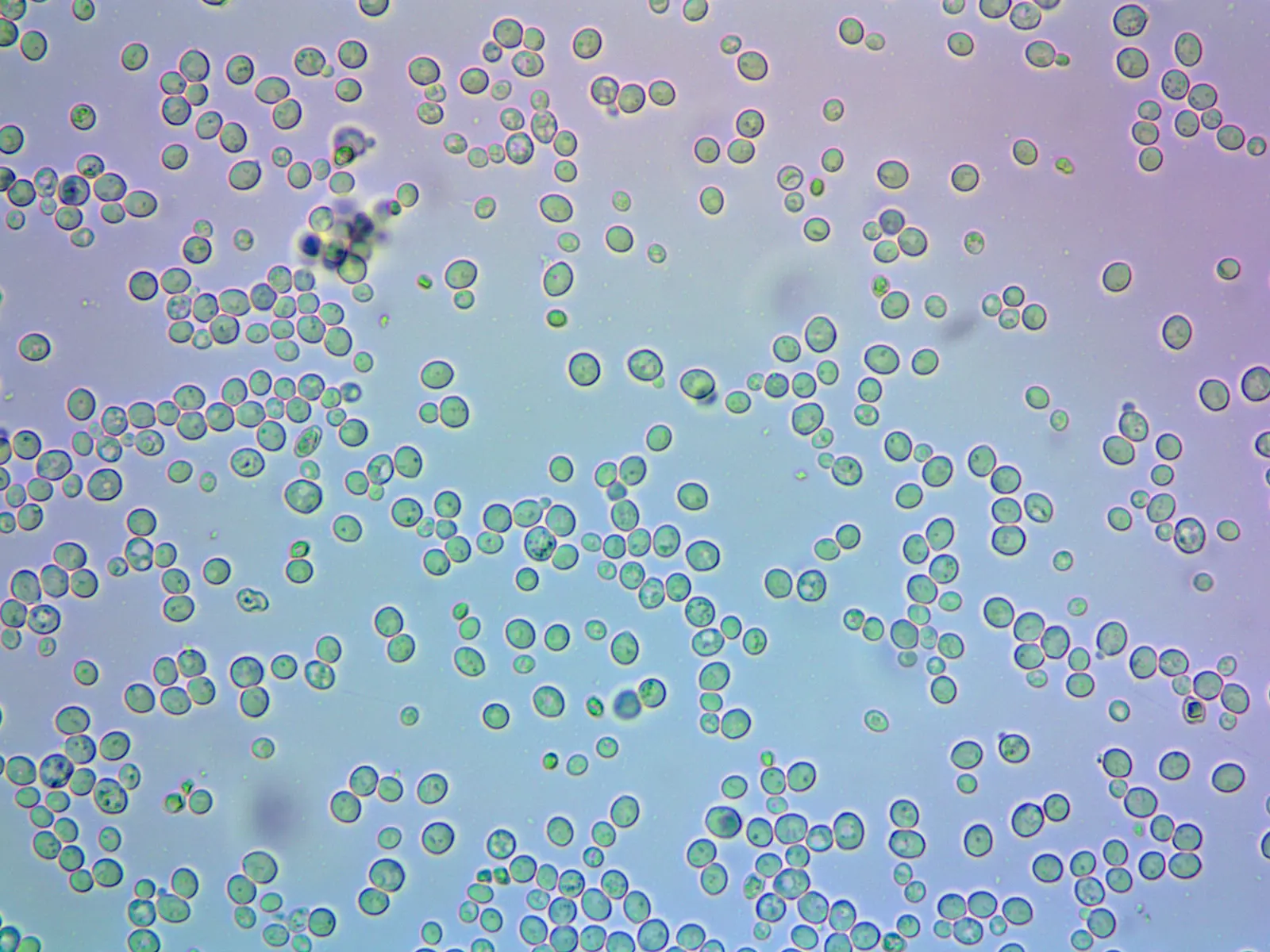
Cells of compressed baker's yeast, round to oval; in one lab strain, yeast cells run 3.6–11.0 µm across [10−6–10−5 m], and a few here carry small buds.
Color: native (no stain stated by the photographer)
"Дрожжевые клетки без воздействия различных веществ (Yeast cells without exposure to various substances)" by Marina taikevich — https://commons.wikimedia.org/wiki/File:Дрожжевые_клетки_без_воздействия_различных_веществ.jpg — CC BY 4.0 (https://creativecommons.org/licenses/by/4.0/)

How big is a baker's yeast cell?
The GBIF Backbone Taxonomy files baker's yeast under kingdom Fungi, phylum Ascomycota (as of October 2026), and each cell lives on its own. In a 2016 study, Janssens and Veenhoff followed 119 mother cells from six versions of one laboratory strain, held under soft pillars in a microfluidic chip at 30 °C, where a flow of fresh medium washed each daughter away. Just before its first bud there, the median mother measured 17.65 µm2 in outline, which the authors convert to a diameter of 4.74 µm. Across all 119, those first outlines match circles 3.6–6.0 µm wide, computed for this page from the paper's data file.
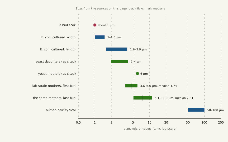
Mothers grow as they age. By the fifth bud the median outline had grown 1.2-fold, Janssens and Veenhoff report. Before its last bud the median mother matched a circle 7.3 µm wide, the largest 11.0 µm, computed for this page. Daughters start smaller: a 2021 review cites 2–4 µm for daughters and 6 µm for mothers (Eigenfeld and colleagues).
That makes a young yeast cell a few times wider than one well-studied bacterium. Cultured Escherichia coli cells averaged 1.0–1.5 µm wide and 1.6–3.9 µm long across 22 growth conditions (Volkmer and Heinemann, 2011), the kind of cell counted on the page about cells in the human body. A human hair, typically 50–100 µm across (Li and colleagues, 2023) and the yardstick of How Big Is a Cell?, would span about 10 to 21 young yeast cells laid side by side, computed for this page. Even the smallest of the 119 is more than three times the width of the 1 µm bead modelled on the page about Brownian motion.
Slide the diagram sideways to read its labels, or open it full size.
Baker's yeast on a log scale: lab-strain mothers 3.6–6.0 µm before the first bud and 5.1–11.0 µm before the last [10−6–10−5 m], beside cultured E. coli and a hair.
Bars are ranges, black ticks medians; the mother and daughter sizes from the 2021 review are as cited there. Data: bakers-yeast-measurements.csv (sources, version and license, JSON).
"Baker's yeast beside a bacterium and a hair, on a log scale" by Ants, Cells & Atoms, https://antscellsandatoms.com/microscopic-life/bakers-yeast/ — CC BY 4.0 (https://creativecommons.org/licenses/by/4.0/)
Data sources for this figure
Data: Yeast mothers: Janssens GE, Veenhoff LM 2016, PLOS ONE 11(12):e0167394 and its S1 File, CC BY 4.0, https://doi.org/10.1371/journal.pone.0167394, retrieved 2026-10-25; equivalent diameters computed 2026-10-25; Daughter and mother sizes as cited in Eigenfeld M, Kerpes R, Becker T 2021, Frontiers in Fungal Biology 2:665490, CC BY 4.0, https://doi.org/10.3389/ffunb.2021.665490, retrieved 2026-10-25; Bud scar: Pillet F et al. 2014, BMC Biology 12:6, CC BY 2.0, https://doi.org/10.1186/1741-7007-12-6, retrieved 2026-10-25; Escherichia coli: Volkmer B, Heinemann M 2011, PLoS ONE 6(7):e23126, Table 1, CC BY 4.0, https://doi.org/10.1371/journal.pone.0023126, as listed in this site's cell-sizes.csv, retrieved 2026-10-25; Human hair 50-100 µm: Li C et al. 2023, Journal of Biomedical Optics 28(11):116501, CC BY 4.0, https://doi.org/10.1117/1.JBO.28.11.116501, as listed in this site's reference-objects.csv, retrieved 2026-10-25
Data sources for this figure
Data: Janssens GE, Veenhoff LM 2016, PLOS ONE 11(12):e0167394 and its S1 File, CC BY 4.0, https://doi.org/10.1371/journal.pone.0167394, retrieved 2026-10-25; Eigenfeld M, Kerpes R, Becker T 2021, Frontiers in Fungal Biology 2:665490, CC BY 4.0, https://doi.org/10.3389/ffunb.2021.665490, retrieved 2026-10-25; Pillet F et al. 2014, BMC Biology 12:6, CC BY 2.0, https://doi.org/10.1186/1741-7007-12-6, retrieved 2026-10-25; Zhang Y et al. 2012, PLOS ONE 7(11):e48275, CC BY 4.0, https://doi.org/10.1371/journal.pone.0048275, retrieved 2026-10-25; Steffen KK, Kennedy BK, Kaeberlein M 2009, Journal of Visualized Experiments 28:1209, CC BY 2.0, https://doi.org/10.3791/1209, retrieved 2026-10-25; Mortimer RK, Johnston JR 1959, Nature 183:1751-1752, https://doi.org/10.1038/1831751a0, numbers as reported in the Eigenfeld review above, retrieved 2026-10-25; Parapouli M, Vasileiadis A, Afendra AS, Hatziloukas E 2020, AIMS Microbiology 6(1):1-31, CC BY 4.0, https://doi.org/10.3934/microbiol.2020001, retrieved 2026-10-25; Engel SR et al. 2014, G3: Genes, Genomes, Genetics 4(3):389-398, CC BY 3.0, https://doi.org/10.1534/g3.113.008995, retrieved 2026-10-25; total computed 2026-10-25; Human hair 50-100 µm: Li C et al. 2023, Journal of Biomedical Optics 28(11):116501, CC BY 4.0, https://doi.org/10.1117/1.JBO.28.11.116501, as listed in this site's reference-objects.csv; cells per hair width computed 2026-10-25
How does a yeast cell bud?
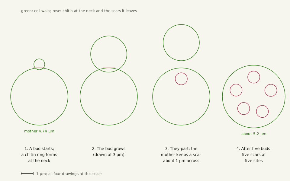
A yeast cell does not split down the middle. A bud swells out of one spot on the mother, and the daughter's wall is built new at that site, so the daughter does not inherit the mother's surface (Zhang and colleagues, 2012). The mother is usually the larger of the two, and Janssens and Veenhoff describe the daughter as somewhat smaller and reset in age; it grows before it buds in turn.
Chitin, a polysaccharide that makes up 1–2% of the yeast cell wall (Pillet and colleagues, 2014), marks the spot. By the account of a 2021 review (Eigenfeld and colleagues), a ring of it forms at the neck between mother and bud as the bud emerges, and once the two part, the patch where the bud grew stays covered by a chitin ring. That ring is the bud scar.
Slide the diagram sideways to read its labels, or open it full size.
Budding drawn to one scale: a 4.74 µm mother [10−6 m], a bud drawn at 3 µm, and ring scars about 1 µm across, one per daughter.
Schematic: scar positions are placed for clarity, not traced from an image; sizes from the sources in bakers-yeast-measurements.csv (sources, version and license, JSON).
"How a yeast bud leaves a scar on its mother" by Ants, Cells & Atoms, https://antscellsandatoms.com/microscopic-life/bakers-yeast/ — CC BY 4.0 (https://creativecommons.org/licenses/by/4.0/)
Data sources for this figure
Data: Mother size and growth: Janssens GE, Veenhoff LM 2016, PLOS ONE 11(12):e0167394, CC BY 4.0, https://doi.org/10.1371/journal.pone.0167394, retrieved 2026-10-25; Chitin ring at the neck and the scar it leaves: Eigenfeld M, Kerpes R, Becker T 2021, Frontiers in Fungal Biology 2:665490, CC BY 4.0, https://doi.org/10.3389/ffunb.2021.665490, retrieved 2026-10-25; Bud scar size: Pillet F et al. 2014, BMC Biology 12:6, CC BY 2.0, https://doi.org/10.1186/1741-7007-12-6, retrieved 2026-10-25; Daughter wall made new at the bud site: Zhang Y et al. 2012, PLOS ONE 7(11):e48275, CC BY 4.0, https://doi.org/10.1371/journal.pone.0048275, retrieved 2026-10-25
Data sources for this figure
Data: Janssens GE, Veenhoff LM 2016, PLOS ONE 11(12):e0167394 and its S1 File, CC BY 4.0, https://doi.org/10.1371/journal.pone.0167394, retrieved 2026-10-25; Eigenfeld M, Kerpes R, Becker T 2021, Frontiers in Fungal Biology 2:665490, CC BY 4.0, https://doi.org/10.3389/ffunb.2021.665490, retrieved 2026-10-25; Pillet F et al. 2014, BMC Biology 12:6, CC BY 2.0, https://doi.org/10.1186/1741-7007-12-6, retrieved 2026-10-25; Zhang Y et al. 2012, PLOS ONE 7(11):e48275, CC BY 4.0, https://doi.org/10.1371/journal.pone.0048275, retrieved 2026-10-25; Steffen KK, Kennedy BK, Kaeberlein M 2009, Journal of Visualized Experiments 28:1209, CC BY 2.0, https://doi.org/10.3791/1209, retrieved 2026-10-25; Mortimer RK, Johnston JR 1959, Nature 183:1751-1752, https://doi.org/10.1038/1831751a0, numbers as reported in the Eigenfeld review above, retrieved 2026-10-25; Parapouli M, Vasileiadis A, Afendra AS, Hatziloukas E 2020, AIMS Microbiology 6(1):1-31, CC BY 4.0, https://doi.org/10.3934/microbiol.2020001, retrieved 2026-10-25; Engel SR et al. 2014, G3: Genes, Genomes, Genetics 4(3):389-398, CC BY 3.0, https://doi.org/10.1534/g3.113.008995, retrieved 2026-10-25; total computed 2026-10-25; Human hair 50-100 µm: Li C et al. 2023, Journal of Biomedical Optics 28(11):116501, CC BY 4.0, https://doi.org/10.1117/1.JBO.28.11.116501, as listed in this site's reference-objects.csv; cells per hair width computed 2026-10-25
In height maps of the cell surface made with an atomic force microscope, a scar on cells grown at 30 °C was about 1 µm across at most: a raised centre, then a dip, then a rigid rim where chitin gathers (Pillet and colleagues, 2014). The same 2021 review repeats an old observation that no new bud grows where a scar sits, so a mother's unscarred surface was taken as one limit on how many buds it can make.
Diatoms also produce daughters of unequal size, for a different reason: a rigid glass wall. Each half of the old wall becomes the outer half of a daughter, which builds a new inner half inside it, so the mean size of a diatom population falls from one generation to the next (Fuhrmann-Lieker and colleagues, 2021).
How do you count a yeast cell's scars?
Count the rings and you have counted the daughters: the number of bud scars on a cell gives the number of daughters it has already made, Zhang and colleagues write (2012). The scars stand out once something binds their chitin and glows. Pillet's team stained chitin with calcofluor white, a fluorescent dye, and saw bud scars about 1 µm across. Zhang's team used wheat germ agglutinin, a chitin-binding protein, tagged with fluorescein.
That glow is borrowed color: the cyan in the photograph below comes from the dye, not from the yeast. In the pair at the centre, one cell shows two rings and the other at least three.
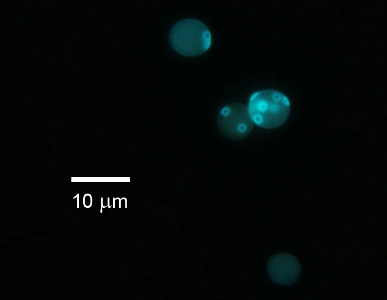
Baker's yeast cells stained with calcofluor white, about 6–7 µm across by the image's 10 µm scale bar [10−6 m]; the small bright rings are bud scars.
Color: fluorescence (calcofluor white)
"Yeast CalcofluorWhite1" by Kunetskiy — https://commons.wikimedia.org/wiki/File:Yeast_CalcofluorWhite1.jpg — CC BY-SA 3.0 (https://creativecommons.org/licenses/by-sa/3.0/)

Most cells in a growing culture should be young: more than half virgin daughters or first-time mothers, Zhang and colleagues note, and among 99 cells from such a culture, stained that way, they found a mean of 0.9 scars per cell. Lifespan experiments start instead from virgin daughters, cells that have never budded, and count each bud as it appears (Steffen and colleagues, 2009).
How many buds can one mother make?
By Zhang and colleagues' account, the first such count came from Mortimer and Johnston, who grew mother cells on agar and lifted each daughter away with a micromanipulator, a microscope fitted with a fine needle (1959, bibliographic record). A 2021 review gives their result: 36 mothers averaged 23.9 daughters, with a standard deviation of 8.3, the first sign that a yeast cell's divisions run out (Eigenfeld and colleagues).
Later counts of common lab strains land nearby. A 2009 methods paper puts typical laboratory strains at 20–30 daughters per mother on agar at 30 °C (Steffen and colleagues). In a 2012 microfluidic device, mothers of two lab strains averaged 23.6 and 26.0 daughters, about two generations short of the agar method by the authors' calibration, because the cells had already budded before loading (Zhang and colleagues).
| Count | Daughters per mother | Cells and method | Source |
|---|---|---|---|
| First count (1959) | mean 23.9, standard deviation 8.3 | 36 mothers, microdissection | Mortimer and Johnston, as reported in a 2021 review |
| Typical laboratory strains | 20–30 | microdissection on agar, 30 °C | Steffen and colleagues, 2009 |
| Lab strain BY4741 | mean 23.6 | 40 mothers, microfluidic device | Zhang and colleagues, 2012 |
| Lab strain BY4742 | mean 26.0 | 61 mothers, microfluidic device | Zhang and colleagues, 2012 |
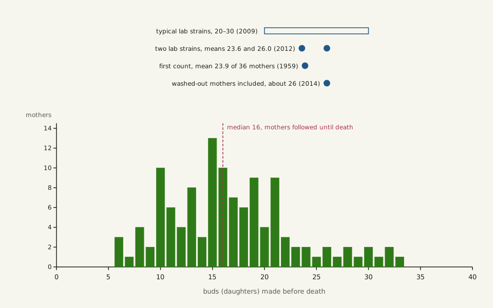
| BY4742-background strains | median 16 (range 6–33) | 119 mothers followed until death, chip, 30 °C | Janssens and Veenhoff, 2016 |
| BY4741, same chip, earlier data | median about 26 | 900 mothers, washed-out ones included | as reported by Janssens and Veenhoff, 2016 |
The chart below counts every bud of the 119 mothers in Janssens and Veenhoff's data file. The mothers made 6 to 33 buds in the chip before dying, median 16. That median runs short, the authors note, because only mothers that stayed in view until death are included; an earlier data set from the same device gave about 26 once washed-out mothers were counted too.
Slide the diagram sideways to read its labels, or open it full size.
Buds made in the chip before death by 119 mothers of one lab strain, 3.6–6.0 µm across at the start [10−6 m]: 6 to 33 buds, median 16.
Only mothers that stayed in view until death are counted, so the median runs short; reported means and ranges are marked above. Data: bakers-yeast-lifespans.csv (sources, version and license, JSON).
"Buds made before death by 119 yeast mothers of one lab strain" by Ants, Cells & Atoms, https://antscellsandatoms.com/microscopic-life/bakers-yeast/ — CC BY 4.0 (https://creativecommons.org/licenses/by/4.0/)
Data sources for this figure
Data: Buds per mother: Janssens GE, Veenhoff LM 2016, PLOS ONE 11(12):e0167394, S1 File, CC BY 4.0, https://doi.org/10.1371/journal.pone.0167394, retrieved 2026-10-25; Means of two lab strains: Zhang Y et al. 2012, PLOS ONE 7(11):e48275, CC BY 4.0, https://doi.org/10.1371/journal.pone.0048275, retrieved 2026-10-25; Typical laboratory strains: Steffen KK, Kennedy BK, Kaeberlein M 2009, Journal of Visualized Experiments 28:1209, CC BY 2.0, https://doi.org/10.3791/1209, retrieved 2026-10-25; First count: Mortimer RK, Johnston JR 1959, Nature 183:1751-1752, https://doi.org/10.1038/1831751a0, numbers as reported in Eigenfeld M, Kerpes R, Becker T 2021, Frontiers in Fungal Biology 2:665490, CC BY 4.0, https://doi.org/10.3389/ffunb.2021.665490, retrieved 2026-10-25
Data: Janssens GE, Veenhoff LM 2016, PLOS ONE 11(12):e0167394, S1 File, CC BY 4.0, https://doi.org/10.1371/journal.pone.0167394, retrieved 2026-10-25; equivalent diameters computed 2026-10-25
A yeast cell is alive in the plainest sense: it grows, buds and dies. In that chip, the authors counted a cell as dead when it suddenly shrank and stopped budding (Janssens and Veenhoff). Divisions slowed near the end, and the largest outline before a last bud in the data file matches a circle 11.0 µm wide, computed for this page.
How does yeast make bread rise?
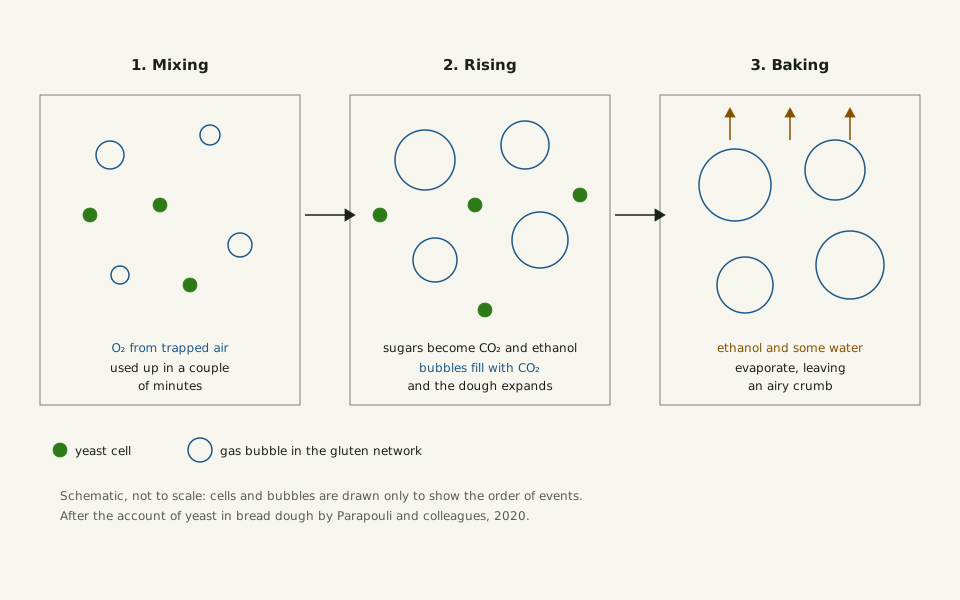
In dough the work is chemical. Air trapped during mixing gives the yeast some oxygen, which its respiration uses up within a couple of minutes, Parapouli and colleagues write (2020); after that, with no oxygen left, the cells ferment. They turn glucose and fructose, freed from the flour's starch and sugars, into carbon dioxide and ethanol.
The gas does the lifting. Bubbles held in the dough's gluten network grow as they fill with carbon dioxide, the dough expands, and in the oven the ethanol evaporates with some water, leaving the airy crumb (Parapouli and colleagues). The same review notes that S. cerevisiae turns sugar into ethanol even when oxygen is present, and that ethanol is toxic or growth-stopping for most microbes that compete with it.
Slide the diagram sideways to read its labels, or open it full size.
Bread dough in three stages, not to scale: yeast cells, 3.6–6.0 µm across when young in one lab strain [10−6 m], use up the trapped oxygen, then ferment sugars into carbon dioxide and ethanol.
Schematic of the order of events after Parapouli and colleagues (2020): bubbles grow as they fill with carbon dioxide, and in the oven the ethanol and some water leave.
"What yeast does in bread dough, in three stages" by Ants, Cells & Atoms, https://antscellsandatoms.com/microscopic-life/bakers-yeast/ — CC BY 4.0 (https://creativecommons.org/licenses/by/4.0/)
Data: Parapouli M, Vasileiadis A, Afendra AS, Hatziloukas E 2020, AIMS Microbiology 6(1):1-31, CC BY 4.0, https://doi.org/10.3934/microbiol.2020001, retrieved 2026-10-25
 Illustration
A slice of bread, its crumb full of holes, beside baker's yeast.
Illustration — not to scale
Illustration: Ants, Cells & Atoms. Generated schematic illustration — not a photograph, micrograph or specimen record. © Ants, Cells & Atoms, all rights reserved.
A yeast cell in dough carries on without oxygen. The page on how insects breathe follows the opposite problem, getting air almost to the cells of an animal. People have used S. cerevisiae for brewing and baking for over ten thousand years, and Louis Pasteur discovered its essential role in alcoholic fermentation in 1857, Liti writes (2015). In the wild it lives on oak bark and on fruit, where fruit flies eat it and carry it about.
Do bakery cells match the lab numbers?
The cell sizes and bud counts above come from laboratory strains. The reference strain S288C carries one set of 16 chromosomes, 12,071,326 base pairs in all, computed for this page from Engel and colleagues' 2014 table; how long that much DNA would stretch is worked out on its own page. Bakery strains are mostly polyploid, carrying extra chromosome sets: in one worldwide analysis of 330 bakery strains, 75% of the commercial ones were tetraploid, Parapouli and colleagues report.
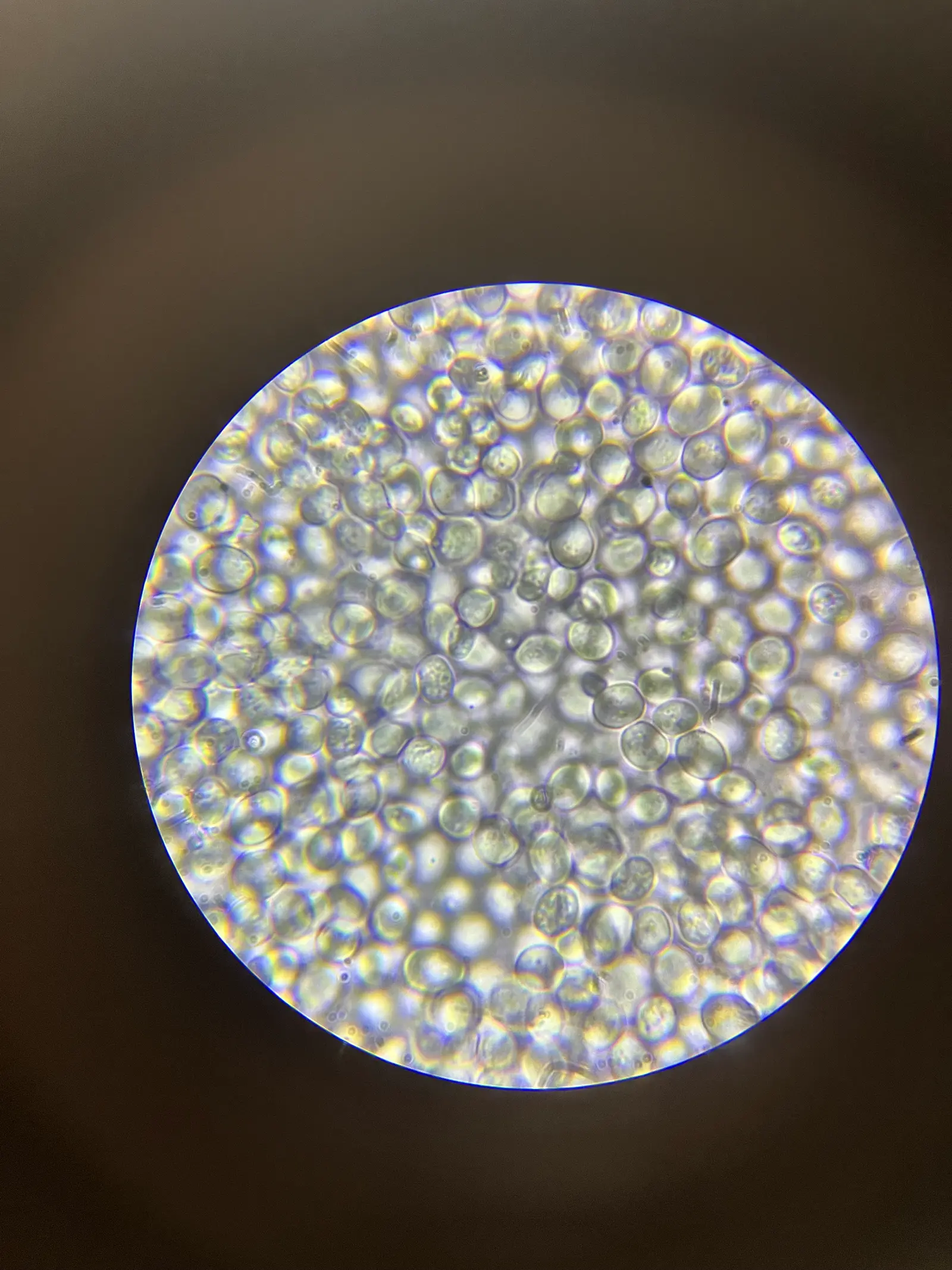
Baking yeast cells through a 40× objective, packed edge to edge; young lab-strain cells run 3.6–6.0 µm across [10−6 m], and this photo has no scale bar.
Color: native (no stain stated by the photographer)
"Baking yeast cells under 40x microscope objective" by ~delta — https://commons.wikimedia.org/wiki/File:Baking_yeast_cells_under_40x_microscope_objective.jpg — CC BY-SA 4.0 (https://creativecommons.org/licenses/by-sa/4.0/)

In a 2018 comparison of strains with one to four genome sets, mean cell size rose with the number of sets (Zadrag-Tecza and colleagues). None of the sources cited here measured a bakery strain's cells, counted the buds of a bakery mother, or counted the scars on the cells in a packet of dried yeast.
Sources
- GBIF Secretariat. GBIF Backbone Taxonomy. Checklist dataset https://doi.org/10.15468/39omei, accessed 2026-10-25; Saccharomyces cerevisiae (Desm.) Meyen, usage key 7440012. Read: dataset record (species match and species record). Text license: CC BY 4.0 (https://creativecommons.org/licenses/by/4.0/).
- Janssens GE, Veenhoff LM. "The Natural Variation in Lifespans of Single Yeast Cells Is Related to Variation in Cell Size, Ribosomal Protein, and Division Time." PLOS ONE 11(12):e0167394 (2016). https://doi.org/10.1371/journal.pone.0167394 Read: full text and its S1 data file. Text license: CC BY 4.0 (https://creativecommons.org/licenses/by/4.0/).
- Eigenfeld M, Kerpes R, Becker T. "Understanding the Impact of Industrial Stress Conditions on Replicative Aging in Saccharomyces cerevisiae." Frontiers in Fungal Biology 2:665490 (2021). https://doi.org/10.3389/ffunb.2021.665490 Read: full text. Text license: CC BY 4.0 (https://creativecommons.org/licenses/by/4.0/).
- Volkmer B, Heinemann M. "Condition-Dependent Cell Volume and Concentration of Escherichia coli to Facilitate Data Conversion for Systems Biology Modeling." PLoS ONE 6(7):e23126 (2011). https://doi.org/10.1371/journal.pone.0023126 Read: full text. Text license: CC BY 4.0 (https://creativecommons.org/licenses/by/4.0/).
- Li C, Du Y, Chen H, Han X, Wu W, Kong X, Zhang C, Yuan X. "Transmission infrared micro-spectroscopic study of individual human hair." Journal of Biomedical Optics 28(11):116501 (2023). https://doi.org/10.1117/1.JBO.28.11.116501 Read: full text. Text license: CC BY 4.0 (https://creativecommons.org/licenses/by/4.0/).
- Zhang Y, Luo C, Zou K, Xie Z, Brandman O, Ouyang Q, Li H. "Single Cell Analysis of Yeast Replicative Aging Using a New Generation of Microfluidic Device." PLoS ONE 7(11):e48275 (2012). https://doi.org/10.1371/journal.pone.0048275 Read: full text. Text license: CC BY 4.0 (https://creativecommons.org/licenses/by/4.0/).
- Pillet F, Lemonier S, Schiavone M, Formosa C, Martin-Yken H, Francois JM, Dague E. "Uncovering by Atomic Force Microscopy of an original circular structure at the yeast cell surface in response to heat shock." BMC Biology 12:6 (2014). https://doi.org/10.1186/1741-7007-12-6 Read: full text. Text license: CC BY 2.0 (https://creativecommons.org/licenses/by/2.0/).
- Fuhrmann-Lieker T, Kubetschek N, Ziebarth J, Klassen R, Seiler W. "Is the diatom sex clock a clock?" Journal of the Royal Society Interface 18(179):20210146 (2021). https://doi.org/10.1098/rsif.2021.0146 Read: full text. Text license: CC BY 4.0 (https://creativecommons.org/licenses/by/4.0/).
- Steffen KK, Kennedy BK, Kaeberlein M. "Measuring Replicative Life Span in the Budding Yeast." Journal of Visualized Experiments (28):1209 (2009). https://doi.org/10.3791/1209 Read: full text. Text license: CC BY 2.0 (https://creativecommons.org/licenses/by/2.0/).
- Mortimer RK, Johnston JR. "Life Span of Individual Yeast Cells." Nature 183(4677):1751–1752 (1959). https://doi.org/10.1038/1831751a0 Read: bibliographic record only.
- Parapouli M, Vasileiadis A, Afendra AS, Hatziloukas E. "Saccharomyces cerevisiae and its industrial applications." AIMS Microbiology 6(1):1–31 (2020). https://doi.org/10.3934/microbiol.2020001 Read: full text. Text license: CC BY 4.0 (https://creativecommons.org/licenses/by/4.0/).
- Liti G. "The fascinating and secret wild life of the budding yeast S. cerevisiae." eLife 4:e05835 (2015). https://doi.org/10.7554/eLife.05835 Read: full text. Text license: CC BY 4.0 (https://creativecommons.org/licenses/by/4.0/).
- Engel SR, Dietrich FS, Fisk DG, Binkley G, Balakrishnan R, Costanzo MC, Dwight SS, Hitz BC, Karra K, Nash RS, Weng S, Wong ED, Lloyd P, Skrzypek MS, Miyasato SR, Simison M, Cherry JM. "The Reference Genome Sequence of Saccharomyces cerevisiae: Then and Now." G3: Genes, Genomes, Genetics 4(3):389–398 (2014). https://doi.org/10.1534/g3.113.008995 Read: full text. Text license: CC BY 3.0 (https://creativecommons.org/licenses/by/3.0/).
- Zadrag-Tecza R, Kwolek-Mirek M, Alabrudzińska M, Skoneczna A. "Cell Size Influences the Reproductive Potential and Total Lifespan of the Saccharomyces cerevisiae Yeast as Revealed by the Analysis of Polyploid Strains." Oxidative Medicine and Cellular Longevity 2018:1898421 (2018). https://doi.org/10.1155/2018/1898421 Read: full text. Text license: CC BY 4.0 (https://creativecommons.org/licenses/by/4.0/).
Cite this page
Ants, Cells & Atoms. “Baker's Yeast: Each Bud Leaves a Scar on the Mother Cell.” Published 2026-10-25. https://antscellsandatoms.com/microscopic-life/bakers-yeast/. Accessed (add the date you read it).
No DOI is issued for entries. Figures made here are CC BY 4.0; see each caption for the license of photographs.
Related entries
 Illustration
Diatoms: Why Most Glass-Walled Algae Shrink as They Multiply
Image: Illustration: Ants, Cells & Atoms, all rights reserved
Illustration: Ants, Cells & Atoms. Generated schematic illustration — not a photograph, micrograph or specimen record. © Ants, Cells & Atoms, all rights reserved.
Plankton: From Bacteria to Jellyfish, Sorted in Size Steps
Image: “Diatomea. - Schachtellinge” by Ernst Haeckel (artist) and Adolf Giltsch (lithographer) (1904) · Public domain
"Diatomea. - Schachtellinge" by Ernst Haeckel (artist) and Adolf Giltsch (lithographer) (1904) — https://commons.wikimedia.org/wiki/File:Diatomea._-_Schachtellinge_LCCN2015647735.jpg — Public domain (artist Ernst Haeckel died 1919 and lithographer Adolf Giltsch died 1911, so the plate is out of copyright in the EU (life plus 70 years) and in the US (published 1904, before 1930); the scan is from the Library of Congress Prints and Photographs division (lccn.loc.gov/2015647735), which lists no rights restriction)

Paramecium: How a Nerve-Like Signal Sends One Cell Backward
Image: “Paramecium 1” by Nbnidhi (2017) · CC BY 4.0
"Paramecium 1" by Nbnidhi — https://commons.wikimedia.org/wiki/File:Paramecium_1.jpg — CC BY 4.0 (https://creativecommons.org/licenses/by/4.0/)
