Slime Mold: Not a Mold, and Its Body Can Be a Single Cell

Slide the ladder sideways to read its labels, or open it full size.
Physarum polycephalum plasmodium, one cell: 0.5 cm in small lab cultures to tens of centimetres [10−3–10−1 m], with its veins (10–140 µm wide in one study) and nuclei (about 5.65 µm) for scale.
Log scale from 10 cm to 0.1 nm, each rung ten times smaller; dashed lines: naked eye about 0.1 mm, light microscope about 0.2 µm (How to read the Zoom ladder); the arrow marks sizes past 10 cm. Data: slime-mold-measurements.csv (sources, version and license, JSON).
"Zoom ladder: Physarum polycephalum plasmodium" by Ants, Cells & Atoms, https://antscellsandatoms.com/microscopic-life/slime-mold/ — CC BY 4.0 (https://creativecommons.org/licenses/by/4.0/)
Data sources for this figure
Data: Plasmodium size and nucleus size: Gerber T et al. 2022, eLife 11:e69745, CC BY 4.0, https://doi.org/10.7554/eLife.69745, retrieved 2026-10-25; Plasmodium area under ideal conditions: Reid CR 2023, Animal Cognition 26(6):1783-1797, CC BY 4.0, https://doi.org/10.1007/s10071-023-01782-1, retrieved 2026-10-25; Vein radii: Marbach S, Ziethen N, Bastin L, Bäuerle FK, Alim K 2023, eLife 12:e78100, CC BY 4.0, https://doi.org/10.7554/eLife.78100, retrieved 2026-10-25; diameters and the 30 cm square side computed 2026-10-25
Data sources for this figure
Data: Gerber T et al. 2022, eLife 11:e69745, CC BY 4.0, https://doi.org/10.7554/eLife.69745, retrieved 2026-10-25; Schaap P et al. 2016, Genome Biology and Evolution 8(1):109-125, CC BY 4.0, https://doi.org/10.1093/gbe/evv237, retrieved 2026-10-25; Reid CR 2023, Animal Cognition 26(6):1783-1797, CC BY 4.0, https://doi.org/10.1007/s10071-023-01782-1, retrieved 2026-10-25; Marbach S, Ziethen N, Bastin L, Bäuerle FK, Alim K 2023, eLife 12:e78100, CC BY 4.0, https://doi.org/10.7554/eLife.78100, retrieved 2026-10-25; Oettmeier C, Döbereiner HG 2019, PLOS ONE 14(4):e0215622, CC BY 4.0, https://doi.org/10.1371/journal.pone.0215622, retrieved 2026-10-25; Computed rows computed 2026-10-25
Bench card
- Name
- Physarum polycephalum Schwein., 1822, a plasmodial slime mold; the feeding stage, the plasmodium, is one cell with many nucleiSource: https://www.gbif.org/species/3214606; Gerber et al. 2022, https://doi.org/10.7554/eLife.69745
- Group
- Amoebozoa (Schaap et al.); in GBIF: kingdom Protozoa, phylum Mycetozoa, class Myxomycetes, order Physarales; not a fungusSource: https://www.gbif.org/species/3214606; Schaap et al. 2016, https://doi.org/10.1093/gbe/evv237
- Actual size
- plasmodium 0.5–1.0 cm in small lab networks to tens of centimetres or more [10−3–10−1 m]; as large as 900 cm2 in area under ideal conditions in one review, square metres in another account; nuclei 5.65 µm on average; veins 10–140 µm wide in one imaging studySource: Gerber et al. 2022, https://doi.org/10.7554/eLife.69745; Reid 2023, https://doi.org/10.1007/s10071-023-01782-1; Marbach et al. 2023, https://doi.org/10.7554/eLife.78100
- Size in this image
- not measurable from this image
- Lives in
- decaying plant material; its prey includes bacteria and yeastsSource: Schaap et al. 2016, https://doi.org/10.1093/gbe/evv237; Reid 2023, https://doi.org/10.1007/s10071-023-01782-1
- Seen with
- naked eye for the plasmodium and its wider veins; light microscope for the nuclei and the flowSource: Schaap et al. 2016 (macroscopic plasmodial cells), https://doi.org/10.1093/gbe/evv237; Gerber et al. 2022 (nuclei imaged by microscope), https://doi.org/10.7554/eLife.69745
- Color in images
- native in the three photographs (no stain stated); drawn colours in the diagramsSource: photo captions on this page
slime-mold-bench.csv (CSV) · where each value comes from, and its license (JSON)
Data credits for this card
Data: GBIF Secretariat, GBIF Backbone Taxonomy, checklist dataset https://doi.org/10.15468/39omei, CC BY 4.0, Physarum polycephalum (3214606), retrieved 2026-10-25; Gerber T et al. 2022, eLife 11:e69745, CC BY 4.0, https://doi.org/10.7554/eLife.69745, retrieved 2026-10-25; Schaap P et al. 2016, Genome Biology and Evolution 8(1):109-125, CC BY 4.0, https://doi.org/10.1093/gbe/evv237, retrieved 2026-10-25; Reid CR 2023, Animal Cognition 26(6):1783-1797, CC BY 4.0, https://doi.org/10.1007/s10071-023-01782-1, retrieved 2026-10-25; Marbach S, Ziethen N, Bastin L, Bäuerle FK, Alim K 2023, eLife 12:e78100, CC BY 4.0, https://doi.org/10.7554/eLife.78100, retrieved 2026-10-25
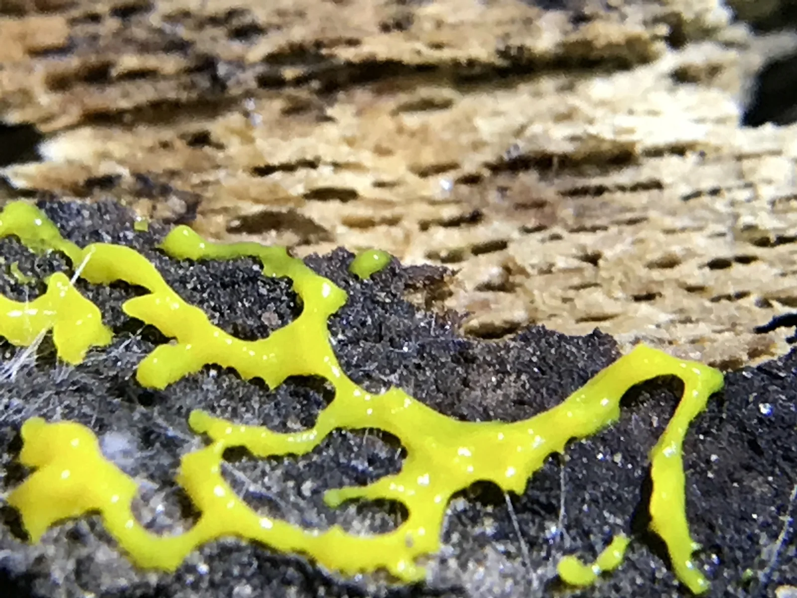
Physarum polycephalum plasmodium, a single cell that can spread from 0.5 cm to tens of centimetres [10−3–10−1 m]: yellow branches over dark, crumbly ground.
Color: native
"Physarum polycephalum 9986808" by jhubley — https://commons.wikimedia.org/wiki/File:Physarum_polycephalum_9986808.jpg — CC0 1.0 (https://creativecommons.org/publicdomain/zero/1.0/)

Is a slime mold a mold, or any kind of fungus?
No, although it was long filed as one. Myxomycetes, the plasmodial slime molds that include Physarum, were first thought to be fungi, Stephenson writes in a 2023 review: their fruiting bodies look like those of some small fungi, and both groups turn up in many of the same places. Up close the fruiting bodies are built very differently, and the two life cycles share little beyond making resistant spores. Myxomycetes were not universally recognized as a separate group until well into the twentieth century.
Genome work puts P. polycephalum in the Amoebozoa. Schaap and colleagues describe the Amoebozoa as the sister group of the opisthokonts, the branch that holds both fungi and animals (2016). The cellular slime mold Dictyostelium discoideum, the closest model organism with a genome sequenced before then, is an amoebozoan too. As of October 2026 the GBIF Backbone Taxonomy files P. polycephalum under kingdom Protozoa, phylum Mycetozoa, class Myxomycetes, and files baker's yeast under kingdom Fungi.
The slime is real. A plasmodium has no cell wall, but a thin layer of slime usually covers it and protects it against injury and drying out, by Stephenson's account; plasmodia range from colorless to black, and white and yellow are common. P. polycephalum grows on decaying plant material (Schaap and colleagues) and takes in bacteria, yeasts and small particles as food (Reid, 2023).
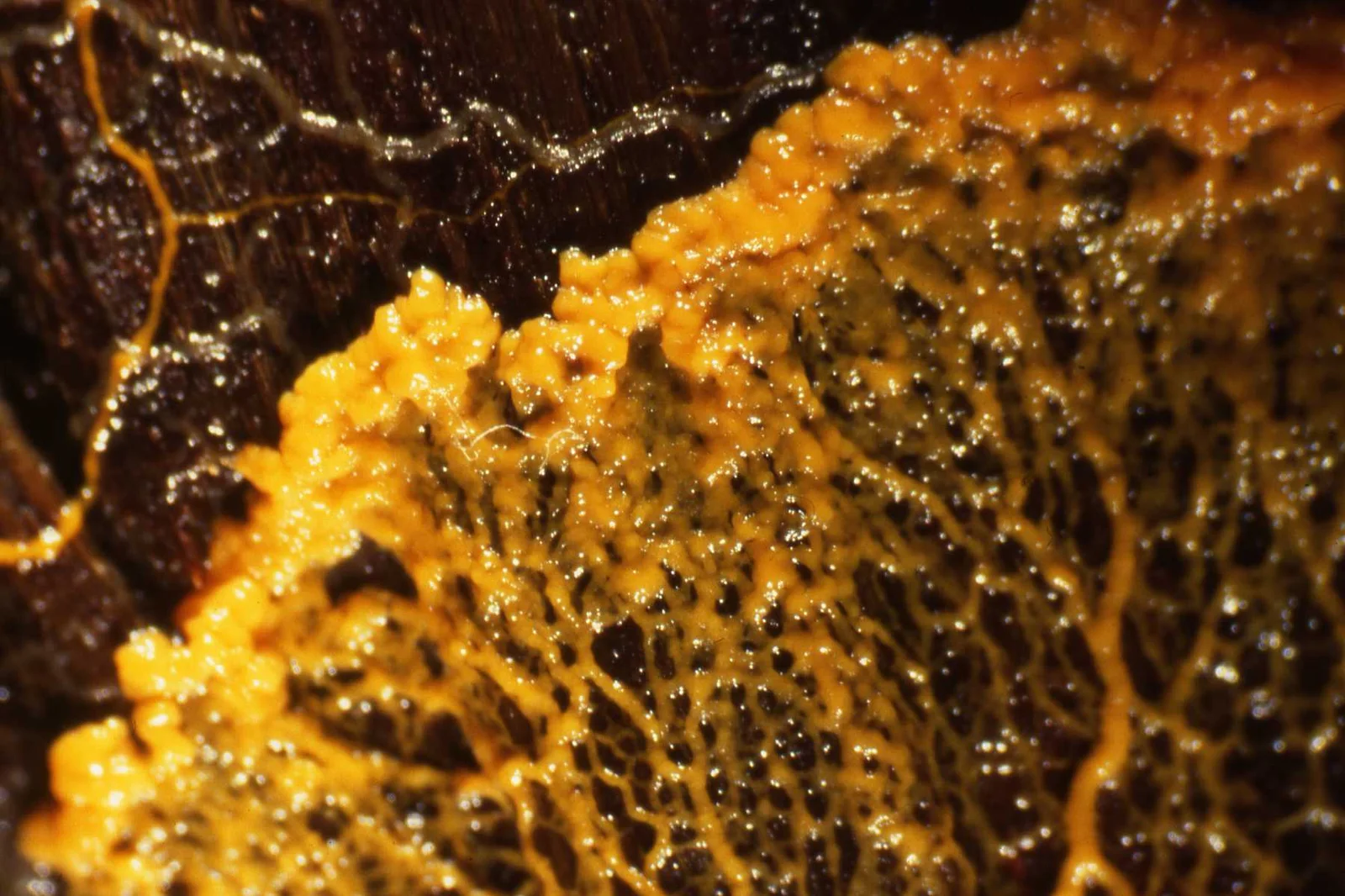
The advancing edge of a Physarum polycephalum plasmodium, part of one cell that can reach tens of centimetres [10−1 m]: a lumpy fan in front, a net of veins behind.
Color: native
"Physarum polycephalum" by HelenGinger — https://commons.wikimedia.org/wiki/File:Physarum_polycephalum.jpg — CC BY-SA 3.0 (https://creativecommons.org/licenses/by-sa/3.0/)

How can one cell hold millions of nuclei?
A plasmodium grows by dividing its nuclei without dividing the cell around them. The result is a syncytium, one mass of cytoplasm shared by many nuclei, Gerber and colleagues explain in a 2022 study. Citing earlier studies, they put the count at tens to millions of nuclei depending on the plasmodium's size, and in fixed, stained plasmodia grown on lab dishes they saw tens of thousands. Reid's review gives millions.
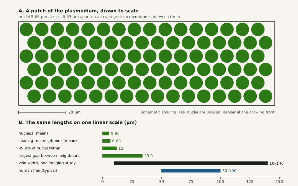
In Gerber's images the nuclei averaged an estimated 5.65 µm across and sat 6.63 µm apart on average, and 99.9% had a neighbour within 12 µm. They were packed more densely at the growing front than in the veins. Some sat trapped in the ectoplasm, which is made of cross-linked contractile fibres, while others rode the flow. By Schaap and colleagues' account, the nuclei of a plasmodium divide in step, keeping their nuclear envelopes intact, and a piece cut from it keeps that timing.
Slide the diagram sideways to read its labels, or open it full size.
Nuclei in a Physarum plasmodium, about 5.65 µm across and 6.63 µm apart on average [10−6 m], all in one cytoplasm.
Schematic: real nuclei are unevenly spread. Values from Gerber and colleagues (2022) and Marbach and colleagues (2023); hair range from this site's reference table. Data: slime-mold-measurements.csv (sources, version and license, JSON).
"One cell, many nuclei: a patch of Physarum plasmodium to scale" by Ants, Cells & Atoms, https://antscellsandatoms.com/microscopic-life/slime-mold/ — CC BY 4.0 (https://creativecommons.org/licenses/by/4.0/)
Data sources for this figure
Data: Nucleus size and spacing: Gerber T et al. 2022, eLife 11:e69745, CC BY 4.0, https://doi.org/10.7554/eLife.69745, retrieved 2026-10-25; Vein radii: Marbach S, Ziethen N, Bastin L, Bäuerle FK, Alim K 2023, eLife 12:e78100, CC BY 4.0, https://doi.org/10.7554/eLife.78100, retrieved 2026-10-25; Human hair 50-100 µm: Li C et al. 2023, Journal of Biomedical Optics 28(11):116501, CC BY 4.0, https://doi.org/10.1117/1.JBO.28.11.116501, as listed in this site's reference-objects.csv, retrieved 2026-10-25
Data sources for this figure
Data: Gerber T et al. 2022, eLife 11:e69745, CC BY 4.0, https://doi.org/10.7554/eLife.69745, retrieved 2026-10-25; Schaap P et al. 2016, Genome Biology and Evolution 8(1):109-125, CC BY 4.0, https://doi.org/10.1093/gbe/evv237, retrieved 2026-10-25; Reid CR 2023, Animal Cognition 26(6):1783-1797, CC BY 4.0, https://doi.org/10.1007/s10071-023-01782-1, retrieved 2026-10-25; Marbach S, Ziethen N, Bastin L, Bäuerle FK, Alim K 2023, eLife 12:e78100, CC BY 4.0, https://doi.org/10.7554/eLife.78100, retrieved 2026-10-25; Oettmeier C, Döbereiner HG 2019, PLOS ONE 14(4):e0215622, CC BY 4.0, https://doi.org/10.1371/journal.pone.0215622, retrieved 2026-10-25; Computed rows computed 2026-10-25
That makes the plasmodium an odd single cell. A Paramecium is also one cell, and P. caudatum is 140–311 µm long across the studies one database compiles (Velho Rodrigues and colleagues, 2021). Volvox goes the other way: a V. carteri sphere several hundred micrometres across is built from about 2,000 small cells and 12–16 large ones (Umen, 2020).
A plasmodium a few centimetres across would sit in the 0.2–10 cm macrozooplankton class, beside krill and comb jellies, by the size classes that one global ocean study uses for zooplankton, part of the plankton (Hatton and colleagues, 2021). Reid's review gives up to 900 cm2 under ideal conditions, and Gerber's team writes that a plasmodium can grow to cover square metres, near the top of the size range of cells.
Why does its inside flow back and forth?
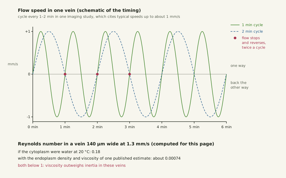
Behind the fan, the plasmodium is a network of veins. In networks that Marbach and colleagues imaged in 2023, the veins had radii of 5–70 µm, so the widest, at 140 µm, were wider than a human hair, typically 50–100 µm (Li and colleagues, 2023). The vein walls contract in a rhythm, once every 1–2 minutes in those measurements. The flow inside reverses within each cycle.
Other sources also give minutes. Reid's review says around 1–5 minutes, depending on the plasmodium's internal state, and Kishimoto's 1958 recordings (abstract) give 60 to 180 seconds for an electrical rhythm that matched the streaming.
For a cell, the flow is fast. Typical speeds reach about 1 mm/s, Marbach's team writes, citing earlier work, and Gerber's team gives up to 1.3 mm/s for nuclei carried along, among the fastest cytoplasmic flows ever measured. The whole plasmodium crawls at up to 5 cm an hour by Reid's account, so the fastest stated flow is roughly 90 times that, computed for this page.
Viscosity still wins in these veins. The Reynolds number — a ratio of inertia to stickiness — for 1.3 mm/s in a 140 µm vein is about 0.18 even if the cytoplasm were as thin as water at 20 °C, computed for this page. With the endoplasm density and viscosity that Oettmeier and Döbereiner use, 1,120 kg/m3 and 0.275 Pa·s, it drops to about 0.00074.
Slide the diagram sideways to read its labels, or open it full size.
Back-and-forth flow in a Physarum vein 10–140 µm wide [10−5–10−4 m]: one cycle every 1–2 minutes in one imaging study, which cites earlier work for typical speeds up to about 1 mm/s.
Smooth waves are a schematic of the timing, not a recording. Reynolds numbers computed for this page; inputs and sources in slime-mold-flow.csv (sources, version and license, JSON).
"The back-and-forth flow in a slime mold vein" by Ants, Cells & Atoms, https://antscellsandatoms.com/microscopic-life/slime-mold/ — CC BY 4.0 (https://creativecommons.org/licenses/by/4.0/)
Data sources for this figure
Data: Contraction period and flow speed: Marbach S, Ziethen N, Bastin L, Bäuerle FK, Alim K 2023, eLife 12:e78100, CC BY 4.0, https://doi.org/10.7554/eLife.78100, retrieved 2026-10-25; Fastest stated flow: Gerber T et al. 2022, eLife 11:e69745, CC BY 4.0, https://doi.org/10.7554/eLife.69745, retrieved 2026-10-25; Endoplasm density and viscosity as used by Oettmeier C, Döbereiner HG 2019, PLOS ONE 14(4):e0215622, CC BY 4.0, https://doi.org/10.1371/journal.pone.0215622, retrieved 2026-10-25; Water density and viscosity at 20 °C: International Association for the Properties of Water and Steam, IAPWS R6-95(2018) and IAPWS R12-08, as listed in this site's water-properties.csv; Reynolds numbers computed 2026-10-25
Data sources for this figure
Data: Gerber T et al. 2022, eLife 11:e69745, CC BY 4.0, https://doi.org/10.7554/eLife.69745, retrieved 2026-10-25; Marbach S, Ziethen N, Bastin L, Bäuerle FK, Alim K 2023, eLife 12:e78100, CC BY 4.0, https://doi.org/10.7554/eLife.78100, retrieved 2026-10-25; Oettmeier C, Döbereiner HG 2019, PLOS ONE 14(4):e0215622, CC BY 4.0, https://doi.org/10.1371/journal.pone.0215622, retrieved 2026-10-25; Reid CR 2023, Animal Cognition 26(6):1783-1797, CC BY 4.0, https://doi.org/10.1007/s10071-023-01782-1, retrieved 2026-10-25; Oxygen diffusion coefficient: Somerville GA, Proctor RA 2013, BMC Microbiology 13:9, CC BY 2.0, https://doi.org/10.1186/1471-2180-13-9, retrieved 2026-10-25; One-axis spread of the Gaussian solution: Vestergaard CL, Flyvbjerg H, Møller IM 2012, Frontiers in Plant Science 3:295, CC BY 3.0, https://doi.org/10.3389/fpls.2012.00295, retrieved 2026-10-25; Water density and viscosity at 20 °C: International Association for the Properties of Water and Steam, IAPWS R6-95(2018) and IAPWS R12-08, as listed in this site's water-properties.csv; values computed 2026-10-25
| Quantity | Value | Source |
|---|---|---|
| Cycle of the vein contractions | 1–2 min (measured) | Marbach and colleagues, 2023 |
| Period of the back-and-forth flow | about 1–5 min (review) | Reid, 2023 |
| Typical flow speed, upper end | up to about 1 mm/s | Marbach and colleagues, 2023, citing earlier work |
| Nuclei carried by the flow | up to 1.3 mm/s | Gerber and colleagues, 2022, citing earlier work |
| Re, 140 µm vein at 1.3 mm/s, water at 20 °C | about 0.18 (computed) | water values from IAPWS releases |
| Re, same vein, endoplasm values | about 0.00074 (computed) | inputs as used by Oettmeier and Döbereiner, 2019 |
| Time for oxygen to spread 1 cm in still water, 25 °C | about 2.4×10⁴ s (computed) | Somerville and Proctor, 2013; one-axis spread after Vestergaard and colleagues, 2012 |
Flow covers distance far faster than diffusion would. At 1 mm/s the flow carries material 1 cm in 10 seconds, while oxygen diffusing through still water at 25 °C would need about 24,000 seconds, some 6.6 hours, to spread that far along one axis, computed for this page. Reid's review says the flow moves biomass, nutrients and chemical signals around the cell. The same arithmetic of diffusion and distance comes up again in how insects breathe.
Can a slime mold solve a maze?
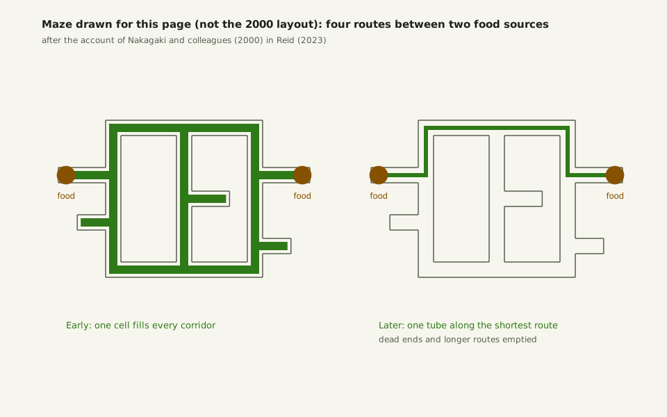
In the sense one experiment measured, yes: as a 2023 review describes it, a plasmodium spread through a maze ended up as one tube along the shortest route. The experiment appeared in Nature in 2000 under the title "Maze-solving by an amoeboid organism" (Nakagaki and colleagues, bibliographic record). In Reid's account, plasmodia were spread through a labyrinth linking two food sources, so that a single cell filled the maze. That maze had four possible solutions, and over time the cell pulled back from the dead ends and the longer paths until one tube remained.
The shortening, in Reid's account, runs through the flow itself. Tubes lined up with the waves of contraction get more of the flow and thicken, while tubes that lie across them get less and decay. The tubes that can carry the most flow are those on the shortest path, so feedback trims the network down to it. Reid also points out that these maze tests measured navigation, not learning: no plasmodium was given the same maze twice.
Slide the diagram sideways to read its labels, or open it full size.
Maze schematic: one Physarum plasmodium, a single cell that can reach tens of centimetres in other studies [10−1 m], fills every corridor, then keeps one tube along the shortest of four routes.
Drawn for this page from the text of Reid's 2023 review, not the layout or proportions of the 2000 maze; the four routes are 14, 18, 22 and 22 grid squares long.
"Maze drawn for this page: four routes, one tube left" by Ants, Cells & Atoms, https://antscellsandatoms.com/microscopic-life/slime-mold/ — CC BY 4.0 (https://creativecommons.org/licenses/by/4.0/)
Data: Reid CR 2023, Animal Cognition 26(6):1783-1797, CC BY 4.0, https://doi.org/10.1007/s10071-023-01782-1, retrieved 2026-10-25
A larger test followed. Tero and colleagues placed food on an agar plate shaped like the Tokyo area, at the positions of railway stations, and the plasmodium pruned its network to link them over about 26 hours, by Reid's account. The networks it built had efficiency, fault tolerance and cost comparable to the real Tokyo rail system, the study's abstract reports (2010).
How does a plasmodium turn into spores?
When food runs out, a plasmodium can turn into fruiting bodies. After several days without food, a P. polycephalum plasmodium becomes ready to sporulate, and in the lab a pulse of blue or far-red light, or a heat shock, sets it off, Barrantes and colleagues write (2010). The protoplasmic mass then forms multiple fruiting bodies, and each sporangium holds hundreds of spores made by meiosis, each with one nucleus (Schaap and colleagues). Among myxomycetes, Stephenson counts a fruiting body of about 1 mm as moderately sized.
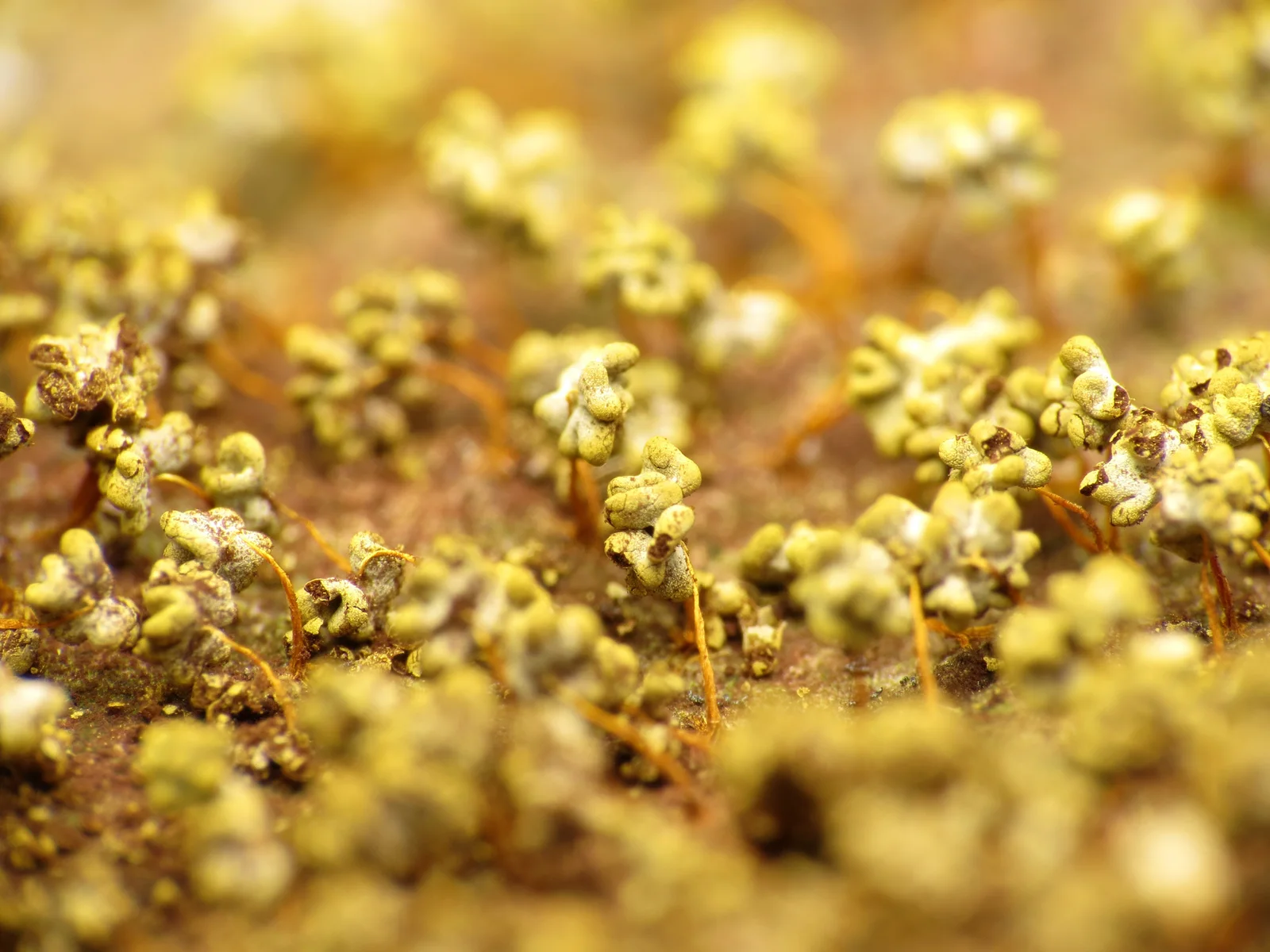
Sporangia of Physarum polycephalum on thin orange stalks; among myxomycetes a fruiting body of about 1 mm counts as moderately sized [10−3 m].
Color: native
"Physarum polycephalum 10744588" by Katja Schulz — https://commons.wikimedia.org/wiki/File:Physarum_polycephalum_10744588.jpg — CC BY 4.0 (https://creativecommons.org/licenses/by/4.0/)

One amoeba with a single nucleus hatches from each spore. In free water most such cells take a flagellated form, while in drier conditions most stay amoeboid, Stephenson writes of myxomycetes in general. Two amoebae of different mating types can fuse into a zygote, which grows into a new plasmodium by dividing its nucleus again and again (Schaap and colleagues; Gerber and colleagues).
So a new plasmodium starts from a cell with one nucleus. Gerber's team saw tens of thousands in plasmodia grown on lab dishes, and Reid's review gives millions, but none of the sources cited here counts the nuclei in a plasmodium of known size near the 900 cm2 that Reid gives as possible.
Sources
- Gerber T, Loureiro C, Schramma N, Chen S, Jain A, Weber A, Weigert A, Santel M, Alim K, Treutlein B, Camp JG. "Spatial transcriptomic and single-nucleus analysis reveals heterogeneity in a gigantic single-celled syncytium." eLife 11:e69745 (2022). https://doi.org/10.7554/eLife.69745 Read: full text. Text license: CC BY 4.0 (https://creativecommons.org/licenses/by/4.0/).
- Reid CR. "Thoughts from the forest floor: a review of cognition in the slime mould Physarum polycephalum." Animal Cognition 26(6):1783–1797 (2023). https://doi.org/10.1007/s10071-023-01782-1 Read: full text. Text license: CC BY 4.0 (https://creativecommons.org/licenses/by/4.0/).
- Schaap P, Barrantes I, Minx P, Sasaki N, Anderson RW, Bénard M, Biggar KK, Buchler NE, Bundschuh R, Chen X, Fronick C, Fulton L, Golderer G, Jahn N, Knoop V, Landweber LF, Maric C, Miller D, Noegel AA, Peace R, Pierron G, Sasaki T, Schallenberg-Rüdinger M, Schleicher M, Singh R, Spaller T, Storey KB, Suzuki T, Tomlinson C, Tyson JJ, Warren WC, Werner ER, Werner-Felmayer G, Wilson RK, Winckler T, Gott JM, Glöckner G, Marwan W. "The Physarum polycephalum Genome Reveals Extensive Use of Prokaryotic Two-Component and Metazoan-Type Tyrosine Kinase Signaling." Genome Biology and Evolution 8(1):109–125 (2016). https://doi.org/10.1093/gbe/evv237 Read: full text. Text license: CC BY 4.0 (https://creativecommons.org/licenses/by/4.0/).
- Stephenson SL. "Past and Ongoing Field-Based Studies of Myxomycetes." Microorganisms 11(9):2283 (2023). https://doi.org/10.3390/microorganisms11092283 Read: full text. Text license: CC BY 4.0 (https://creativecommons.org/licenses/by/4.0/).
- GBIF Secretariat. GBIF Backbone Taxonomy. Checklist dataset https://doi.org/10.15468/39omei, accessed 2026-10-25; Physarum polycephalum Schwein., 1822, usage key 3214606; Saccharomyces cerevisiae (Desm.) Meyen, usage key 7440012. Read: dataset record (species match and species records). Text license: CC BY 4.0 (https://creativecommons.org/licenses/by/4.0/).
- Velho Rodrigues MF, Lisicki M, Lauga E. "The bank of swimming organisms at the micron scale (BOSO-Micro)." PLOS ONE 16(6):e0252291 (2021). https://doi.org/10.1371/journal.pone.0252291 Read: full text. Text license: CC BY 4.0 (https://creativecommons.org/licenses/by/4.0/).
- Umen JG. "Volvox and volvocine green algae." EvoDevo 11:13 (2020). https://doi.org/10.1186/s13227-020-00158-7 Read: full text. Text license: CC BY 4.0 (https://creativecommons.org/licenses/by/4.0/).
- Hatton IA, Heneghan RF, Bar-On YM, Galbraith ED. "The global ocean size spectrum from bacteria to whales." Science Advances 7(46):eabh3732 (2021). https://doi.org/10.1126/sciadv.abh3732 Read: full text. Text license: CC BY 4.0 (https://creativecommons.org/licenses/by/4.0/).
- Marbach S, Ziethen N, Bastin L, Bäuerle FK, Alim K. "Vein fate determined by flow-based but time-delayed integration of network architecture." eLife 12:e78100 (2023). https://doi.org/10.7554/eLife.78100 Read: full text. Text license: CC BY 4.0 (https://creativecommons.org/licenses/by/4.0/).
- Li C, Du Y, Chen H, Han X, Wu W, Kong X, Zhang C, Yuan X. "Transmission infrared micro-spectroscopic study of individual human hair." Journal of Biomedical Optics 28(11):116501 (2023). https://doi.org/10.1117/1.JBO.28.11.116501 Read: full text. Text license: CC BY 4.0 (https://creativecommons.org/licenses/by/4.0/).
- Kishimoto U. "Rhythmicity in the protoplasmic streaming of a slime mold, Physarum polycephalum. I. A statistical analysis of the electric potential rhythm." The Journal of General Physiology 41(6):1205–1222 (1958). https://doi.org/10.1085/jgp.41.6.1205 Read: abstract.
- Oettmeier C, Döbereiner HG. "A lumped parameter model of endoplasm flow in Physarum polycephalum explains migration and polarization-induced asymmetry during the onset of locomotion." PLOS ONE 14(4):e0215622 (2019). https://doi.org/10.1371/journal.pone.0215622 Read: full text. Text license: CC BY 4.0 (https://creativecommons.org/licenses/by/4.0/).
- International Association for the Properties of Water and Steam. IAPWS R6-95(2018), Revised Release on the IAPWS Formulation 1995 for the Thermodynamic Properties of Ordinary Water Substance for General and Scientific Use (2018). https://iapws.org/technical-guidance/release/IAPWS-95 Read: values as computed from the release equations in this site's water-properties table. Text license: IAPWS: publication in whole or in part allowed with attribution (https://iapws.org/technical-guidance/release/IAPWS-95).
- International Association for the Properties of Water and Steam. IAPWS R12-08, Release on the IAPWS Formulation 2008 for the Viscosity of Ordinary Water Substance (2008). https://iapws.org/technical-guidance/release/viscosity Read: values as computed from the release equations in this site's water-properties table. Text license: IAPWS: publication in whole or in part allowed with attribution (https://iapws.org/technical-guidance/release/viscosity).
- Somerville GA, Proctor RA. "Cultivation conditions and the diffusion of oxygen into culture media: the rationale for the flask-to-medium ratio in microbiology." BMC Microbiology 13:9 (2013). https://doi.org/10.1186/1471-2180-13-9 Read: full text. Text license: CC BY 2.0 (https://creativecommons.org/licenses/by/2.0/).
- Vestergaard CL, Flyvbjerg H, Møller IM. "Intracellular signaling by diffusion: can waves of hydrogen peroxide transmit intracellular information in plant cells?" Frontiers in Plant Science 3:295 (2012). https://doi.org/10.3389/fpls.2012.00295 Read: full text. Text license: CC BY 3.0 (https://creativecommons.org/licenses/by/3.0/).
- Nakagaki T, Yamada H, Tóth Á. "Maze-solving by an amoeboid organism." Nature 407(6803):470 (2000). https://doi.org/10.1038/35035159 Read: bibliographic record only.
- Tero A, Takagi S, Saigusa T, Ito K, Bebber DP, Fricker MD, Yumiki K, Kobayashi R, Nakagaki T. "Rules for Biologically Inspired Adaptive Network Design." Science 327(5964):439–442 (2010). https://doi.org/10.1126/science.1177894 Read: abstract.
- Barrantes I, Glöckner G, Meyer S, Marwan W. "Transcriptomic changes arising during light-induced sporulation in Physarum polycephalum." BMC Genomics 11:115 (2010). https://doi.org/10.1186/1471-2164-11-115 Read: full text. Text license: CC BY 2.0 (https://creativecommons.org/licenses/by/2.0/).
Cite this page
Ants, Cells & Atoms. “Slime Mold: Not a Mold, and Its Body Can Be a Single Cell.” Published 2026-10-25. https://antscellsandatoms.com/microscopic-life/slime-mold/. Accessed (add the date you read it).
No DOI is issued for entries. Figures made here are CC BY 4.0; see each caption for the license of photographs.
Related entries
 Illustration
Diatoms: Why Most Glass-Walled Algae Shrink as They Multiply
Image: Illustration: Ants, Cells & Atoms, all rights reserved
Illustration: Ants, Cells & Atoms. Generated schematic illustration — not a photograph, micrograph or specimen record. © Ants, Cells & Atoms, all rights reserved.
Plankton: From Bacteria to Jellyfish, Sorted in Size Steps
Image: “Diatomea. - Schachtellinge” by Ernst Haeckel (artist) and Adolf Giltsch (lithographer) (1904) · Public domain
"Diatomea. - Schachtellinge" by Ernst Haeckel (artist) and Adolf Giltsch (lithographer) (1904) — https://commons.wikimedia.org/wiki/File:Diatomea._-_Schachtellinge_LCCN2015647735.jpg — Public domain (artist Ernst Haeckel died 1919 and lithographer Adolf Giltsch died 1911, so the plate is out of copyright in the EU (life plus 70 years) and in the US (published 1904, before 1930); the scan is from the Library of Congress Prints and Photographs division (lccn.loc.gov/2015647735), which lists no rights restriction)

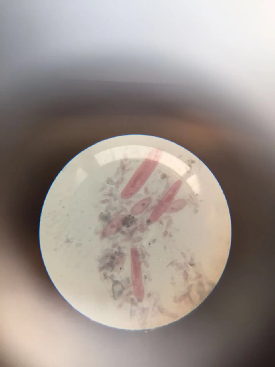
Paramecium: How a Nerve-Like Signal Sends One Cell Backward
Image: “Paramecium 1” by Nbnidhi (2017) · CC BY 4.0
"Paramecium 1" by Nbnidhi — https://commons.wikimedia.org/wiki/File:Paramecium_1.jpg — CC BY 4.0 (https://creativecommons.org/licenses/by/4.0/)
