How Long Is DNA? About 2 m per Diploid Human Nucleus

Slide the ladder sideways to read its labels, or open it full size.
DNA double helix, about 1.9–2.5 nm wide [10−9 m], the width set by where its edge is drawn; a typical hair, 50–100 µm, sits four to five rungs up.
Log scale from 10 cm to 0.1 nm; each rung is ten times smaller than the one to its left. Dashed lines: naked eye about 0.1 mm and light microscope about 0.2 µm, explained on the Zoom ladder page.
"Zoom ladder: how wide is DNA" by Ants, Cells & Atoms, https://antscellsandatoms.com/scale/dna-length/ — CC BY 4.0 (https://creativecommons.org/licenses/by/4.0/)
Data sources for this figure
Data: RCSB PDB 1BNA, CC0, coordinates from Drew HR, Wing RM, Takano T, Broka C, Tanaka S, Itakura K, Dickerson RE 1981, Proceedings of the National Academy of Sciences 78(4):2179–2183, https://doi.org/10.2210/pdb1bna/pdb (retrieved 2026-10-25); van der Waals radii C 170, N 155, O 152, P 180 pm from PubChem element records 6, 7, 8 and 15 (Los Alamos National Laboratory periodic table), PubChem 2025 update, https://doi.org/10.1093/nar/gkae1059 (retrieved 2026-10-25); Li C et al. 2023, Journal of Biomedical Optics 28(11):116501, CC BY 4.0, https://doi.org/10.1117/1.JBO.28.11.116501 (human hair, typically 50–100 µm; shared reference-objects.csv; retrieved 2026-10-25); subject band 1.9–2.5 nm from the article front matter zoom, same values as the helix geometry CSV; ladder computed 2026-10-25 for this page
Bench card
- Question
- How long is the DNA in one human cell, and in a whole body?
- Answer range
- about 2.0–2.4 m stretched out in one human cell with a nucleus and two chromosome sets [100 m]; about 6.1–7.1×10¹² m in the nucleated cells of a 70 kg reference man, 41–48 Earth–Sun distances; mature red blood cells, 84% of the human cells, have noneSource: dna-length-inputs.csv and dna-length-whole-body.csv on this page
- Method
- compiled measurements: base pairs in a complete human genome sequence times the rise per base pair computed from the PDB 1BNA crystal coordinates, cross-checked by DNA mass per nucleus; whole body = nucleated cells times DNA per cellSource: dna-length-inputs.csv on this page
- Inputs
- 7: base pairs in one genome set; sets per nucleus; rise per base pair (shared by both routes); DNA mass per nucleus; base pairs per picogram; nucleated cells per body; the astronomical unitSource: dna-length-inputs.csv and dna-length-whole-body.csv on this page
dna-length-bench.csv (CSV) · where each value comes from, and its license (JSON)
Data credits for this card
Data: Nurk S et al. 2022, Science 376(6588):44–53, CC BY 4.0, https://doi.org/10.1126/science.abj6987 (3,054,815,472 base pairs in the T2T-CHM13 set; 16,569 bp mitochondrial genome; retrieved 2026-10-25); RCSB PDB 1BNA, CC0, coordinates from Drew HR, Wing RM, Takano T, Broka C, Tanaka S, Itakura K, Dickerson RE 1981, Proceedings of the National Academy of Sciences 78(4):2179–2183, https://doi.org/10.2210/pdb1bna/pdb (retrieved 2026-10-25); Wyngaard GA et al. 2022, Scientific Reports 12:6616, CC BY 4.0, https://doi.org/10.1038/s41598-022-10585-2 (7.0 pg per nucleus assigned to male human white blood cells; 1 pg = 0.978 Gb, citing Doležel J et al. 2003; retrieved 2026-10-25); Sender R, Fuchs S, Milo R 2016, PLoS Biology 14(8):e1002533, CC BY 4.0, https://doi.org/10.1371/journal.pbio.1002533 (3.0e13 human cells, 84% red blood cells, about 0.3e13 nucleated; shared human-cell-counts.csv; retrieved 2026-10-25); BIPM, The International System of Units (SI Brochure), 9th edition, Table 8, CC BY 4.0, https://doi.org/10.59161/AUEZ1291 (1 au = 149,597,870,700 m; retrieved 2026-10-25); ranges computed 2026-10-25 for this page
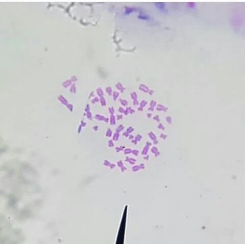
Female human chromosomes at metaphase, a stage of cell division; the largest about 8–10 µm long [10−6–10−5 m].
Color: stain (purple, not named by the photographer)
"Human Chromosomes" by SANCHITA20 — https://commons.wikimedia.org/wiki/File:Human_Chromosomes.jpg — CC BY 4.0 (https://creativecommons.org/licenses/by/4.0/)

This page works out the length twice, both times computed for this page. Route A counts base pairs, the matched bases that link the two strands, in a complete human genome sequence. Route B starts from the mass of DNA in one nucleus. The two routes share one input, the spacing between neighbouring base pairs, computed from the coordinates of a DNA crystal.
How wide is DNA, and how far apart are its base pairs?
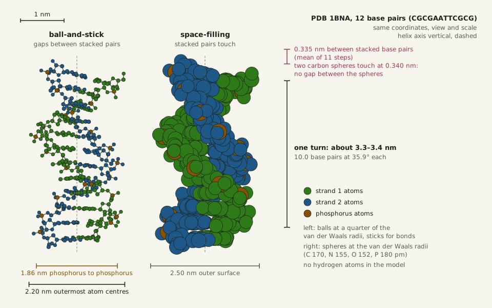
The crystal used here is short: twelve base pairs of the sequence CGCGAATTCGCG. Its two strands wind into a right-handed double helix. Drew and colleagues refined its structure in 1981 from X-ray data at 1.9 Å resolution (abstract). Its coordinates, open as entry 1BNA in the Protein Data Bank, place every atom except hydrogen.
Fitting a cylinder through the 16 phosphorus atoms of the central eight base pairs puts them 0.90–0.97 nm from the helix axis, so the backbone is about 1.9 nm across, computed for this page. The outermost atoms, phosphate oxygens, reach 1.1 nm out. Drawn as spheres of their van der Waals radius, which marks how close another atom can approach by Wikipedia's definition, the atoms make a surface 2.5 nm across. Still, 95% of the atoms in those eight pairs sit within 1.0 nm of the axis.
Slide the diagram sideways to read its labels, or open it full size.
PDB 1BNA, twelve base pairs with sticks and space-filling: about 1.9–2.5 nm wide [10−9 m], base pairs 0.335 nm apart on average.
Left, ball-and-stick; right, space-filling. Of the two, the space-filling drawing is closer to the electron cloud. Data: dna-length-helix-geometry.csv (sources, version and license, JSON), computed for this page from the 1BNA coordinates.
"The DNA double helix drawn two ways, ball-and-stick and space-filling: PDB 1BNA" by Ants, Cells & Atoms, https://antscellsandatoms.com/scale/dna-length/ — CC BY 4.0 (https://creativecommons.org/licenses/by/4.0/)
Data sources for this figure
Data: RCSB PDB 1BNA, CC0, coordinates from Drew HR, Wing RM, Takano T, Broka C, Tanaka S, Itakura K, Dickerson RE 1981, Proceedings of the National Academy of Sciences 78(4):2179–2183, https://doi.org/10.2210/pdb1bna/pdb (retrieved 2026-10-25); van der Waals radii C 170, N 155, O 152, P 180 pm from PubChem element records 6, 7, 8 and 15 (Los Alamos National Laboratory periodic table), PubChem 2025 update, https://doi.org/10.1093/nar/gkae1059 (retrieved 2026-10-25); Drew HR et al. 1981, Proceedings of the National Academy of Sciences 78(4):2179–2183, https://doi.org/10.1073/pnas.78.4.2179 (abstract: 36.9 degrees per step, 9.8 base pairs per turn in the central steps; retrieved 2026-10-25); widths, rise, twist and the drawing computed 2026-10-25 for this page
Data sources for this figure
Data: RCSB PDB 1BNA, CC0, coordinates from Drew HR, Wing RM, Takano T, Broka C, Tanaka S, Itakura K, Dickerson RE 1981, Proceedings of the National Academy of Sciences 78(4):2179–2183, https://doi.org/10.2210/pdb1bna/pdb (retrieved 2026-10-25); van der Waals radii C 170, N 155, O 152, P 180 pm from PubChem element records 6, 7, 8 and 15 (Los Alamos National Laboratory periodic table), PubChem 2025 update, https://doi.org/10.1093/nar/gkae1059 (retrieved 2026-10-25); Drew HR et al. 1981, Proceedings of the National Academy of Sciences 78(4):2179–2183, https://doi.org/10.1073/pnas.78.4.2179 (abstract: 36.9 degrees per step, 9.8 base pairs per turn in the central steps; retrieved 2026-10-25); all other values computed 2026-10-25 for this page
Along the axis, neighbouring base pairs sit 0.335 nm apart on average over the 11 steps, though single steps range from 0.29 to 0.42 nm; other ways of drawing the axis give 0.333 and 0.347 nm. Each step turns the helix by 35.9° on average, so a full turn takes 10.0 base pairs and runs about 3.4 nm, computed for this page. For the central steps, Drew and colleagues report a mean of 36.9° per step, or 9.8 base pairs per turn (abstract).
The left drawing puts sticks between the atoms, the habit this site calls the stick fiction. Its stacked pairs stand apart, with gaps that are not there. The right one draws every atom as a sphere of its van der Waals radius, so the stacked pairs touch: their 0.335 nm spacing is just under the 0.340 nm at which two carbon spheres meet, using PubChem's carbon radius. The acetylsalicylic acid page pairs the two kinds of drawing the same way.
Route A: how many base pairs does one nucleus hold?
The Telomere-to-Telomere consortium published a complete sequence of a human genome, T2T-CHM13, in 2022. Its 22 autosomes, the chromosomes other than X and Y, and its X chromosome hold 3,054,815,472 base pairs of nuclear DNA, plus a mitochondrial genome of 16,569 base pairs, Nurk and colleagues report. The CHM13 cells are homozygous, with matching copies of each chromosome, and have a 46,XX karyotype: 46 chromosomes, two of them X. So their two chromosome sets are nearly identical.
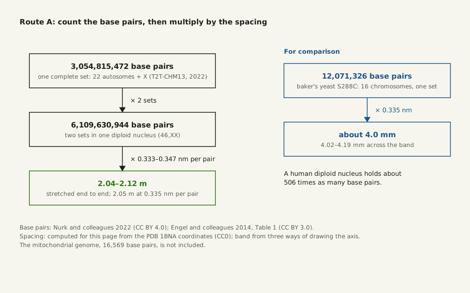
A diploid nucleus, one with two such sets as in a 46,XX cell, holds 6,109,630,944 base pairs. At 0.335 nm per pair they would stretch 2.05 m, and 2.04–2.12 m across the 0.333–0.347 nm band, computed for this page.
Slide the diagram sideways to read its labels, or open it full size.
Route A for one diploid nucleus: 6.11 billion base pairs × 0.335 nm = 2.05 m [100 m], 2.04–2.12 m across the rise band.
Data: dna-length-inputs.csv (sources, version and license, JSON), computed for this page.
"Route A: counting base pairs to the length of DNA in one nucleus" by Ants, Cells & Atoms, https://antscellsandatoms.com/scale/dna-length/ — CC BY 4.0 (https://creativecommons.org/licenses/by/4.0/)
Data sources for this figure
Data: Nurk S et al. 2022, Science 376(6588):44–53, CC BY 4.0, https://doi.org/10.1126/science.abj6987 (3,054,815,472 base pairs in the T2T-CHM13 set; 16,569 bp mitochondrial genome; retrieved 2026-10-25); Engel SR et al. 2014, G3 4(3):389–398, Table 1, CC BY 3.0, https://doi.org/10.1534/g3.113.008995 (16 chromosome lengths of S288C, summed for this page; retrieved 2026-10-25); RCSB PDB 1BNA, CC0, coordinates from Drew HR, Wing RM, Takano T, Broka C, Tanaka S, Itakura K, Dickerson RE 1981, Proceedings of the National Academy of Sciences 78(4):2179–2183, https://doi.org/10.2210/pdb1bna/pdb (retrieved 2026-10-25); products computed 2026-10-25 for this page
Data sources for this figure
Data: Nurk S et al. 2022, Science 376(6588):44–53, CC BY 4.0, https://doi.org/10.1126/science.abj6987 (3,054,815,472 base pairs in the T2T-CHM13 set; 16,569 bp mitochondrial genome; retrieved 2026-10-25); Wyngaard GA et al. 2022, Scientific Reports 12:6616, CC BY 4.0, https://doi.org/10.1038/s41598-022-10585-2 (7.0 pg per nucleus assigned to male human white blood cells; 1 pg = 0.978 Gb, citing Doležel J et al. 2003; retrieved 2026-10-25); Piovesan A et al. 2019, BMC Research Notes 12:106, CC BY 4.0, https://doi.org/10.1186/s13104-019-4137-z (diploid lengths 205.00 cm male and 208.23 cm female; whole-body 6.20e12 m; mitochondrial DNA 0.09–3.62 cm per cell; retrieved 2026-10-25); Engel SR et al. 2014, G3 4(3):389–398, Table 1, CC BY 3.0, https://doi.org/10.1534/g3.113.008995 (16 chromosome lengths of S288C, summed for this page; retrieved 2026-10-25); RCSB PDB 1BNA, CC0, coordinates from Drew HR, Wing RM, Takano T, Broka C, Tanaka S, Itakura K, Dickerson RE 1981, Proceedings of the National Academy of Sciences 78(4):2179–2183, https://doi.org/10.2210/pdb1bna/pdb (retrieved 2026-10-25); routes computed 2026-10-25 for this page
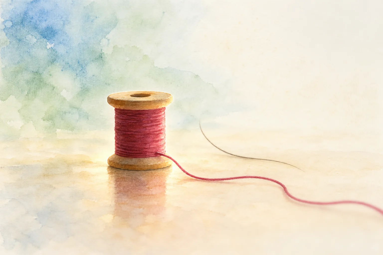 Illustration
Thread pulled off a sewing spool.
Illustration — not to scale
Illustration: Ants, Cells & Atoms. Generated schematic illustration — not a photograph, micrograph or specimen record. © Ants, Cells & Atoms, all rights reserved.
A male nucleus carries a Y chromosome in place of one X. In Piovesan and colleagues' 2019 figures from the older GRCh38 reference, one X holds 156 million base pairs and one Y 57 million, so a male diploid set comes to 205.00 cm against 208.23 cm for a female one. They estimate that people differ by about ±0.65 cm, from the roughly 20 million bases of variation in a typical diploid genome.
The same count for baker's yeast is far smaller. The 16 chromosomes of the laboratory strain S288C, which carries one set per cell, total 12,071,326 base pairs in Engel and colleagues' 2014 table: about 4.0 mm of DNA at the same spacing. A human diploid nucleus holds about 506 times as many base pairs, computed for this page.
Route B: weigh the DNA instead
DNA can also be weighed instead of read. In a 2022 genome-size study, Wyngaard and colleagues measured stained nuclei against reference cells and assigned male human white blood cells 7.0 pg of DNA per nucleus, a value they base on earlier work and the Animal Genome Size Database. They convert mass to length in base pairs with 1 pg = 0.978 billion base pairs, citing Doležel and colleagues (2003).
Those two figures give 6.846 billion base pairs in one male nucleus. At 0.335 nm per pair that is 2.29 m, and 2.28–2.38 m across the band, computed for this page: 12% longer than route A.
| Route | Base pairs in one nucleus | Rise per pair | Length |
|---|---|---|---|
| A: complete sequence, two sets (46,XX) | 6.11 billion | 0.333–0.347 nm | 2.04–2.12 m |
| B: 7.0 pg × 0.978 billion per pg (46,XY) | 6.85 billion | 0.333–0.347 nm | 2.28–2.38 m |
| Published: GRCh38, male / female | 6.27 / 6.37 billion | about 0.327 nm | 2.05 / 2.08 m |
The two routes share only the rise per base pair. Their base-pair counts come from different kinds of evidence: one from reading a sequence, one from the dye signal of stained nuclei. The 7.0 pg is a reference value assigned for calibration, not a measurement made in that study, and this page does not account for the 12% gap. Both routes still land near 2 m.
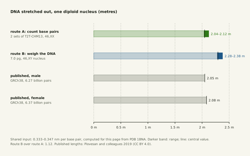
Slide the diagram sideways to read its labels, or open it full size.
DNA in one diploid human nucleus by two routes, 2.04–2.38 m [100 m]: counting base pairs gives 2.05 m, weighing the DNA 2.29 m.
Data: dna-length-inputs.csv (sources, version and license, JSON), computed for this page.
"DNA in one diploid human nucleus by two routes" by Ants, Cells & Atoms, https://antscellsandatoms.com/scale/dna-length/ — CC BY 4.0 (https://creativecommons.org/licenses/by/4.0/)
Data sources for this figure
Data: Nurk S et al. 2022, Science 376(6588):44–53, CC BY 4.0, https://doi.org/10.1126/science.abj6987 (3,054,815,472 base pairs in the T2T-CHM13 set; 16,569 bp mitochondrial genome; retrieved 2026-10-25); Wyngaard GA et al. 2022, Scientific Reports 12:6616, CC BY 4.0, https://doi.org/10.1038/s41598-022-10585-2 (7.0 pg per nucleus assigned to male human white blood cells; 1 pg = 0.978 Gb, citing Doležel J et al. 2003; retrieved 2026-10-25); Piovesan A et al. 2019, BMC Research Notes 12:106, CC BY 4.0, https://doi.org/10.1186/s13104-019-4137-z (diploid lengths 205.00 cm male and 208.23 cm female; whole-body 6.20e12 m; mitochondrial DNA 0.09–3.62 cm per cell; retrieved 2026-10-25); RCSB PDB 1BNA, CC0, coordinates from Drew HR, Wing RM, Takano T, Broka C, Tanaka S, Itakura K, Dickerson RE 1981, Proceedings of the National Academy of Sciences 78(4):2179–2183, https://doi.org/10.2210/pdb1bna/pdb (retrieved 2026-10-25); route lengths computed 2026-10-25 for this page
Data sources for this figure
Data: Nurk S et al. 2022, Science 376(6588):44–53, CC BY 4.0, https://doi.org/10.1126/science.abj6987 (3,054,815,472 base pairs in the T2T-CHM13 set; 16,569 bp mitochondrial genome; retrieved 2026-10-25); Wyngaard GA et al. 2022, Scientific Reports 12:6616, CC BY 4.0, https://doi.org/10.1038/s41598-022-10585-2 (7.0 pg per nucleus assigned to male human white blood cells; 1 pg = 0.978 Gb, citing Doležel J et al. 2003; retrieved 2026-10-25); Piovesan A et al. 2019, BMC Research Notes 12:106, CC BY 4.0, https://doi.org/10.1186/s13104-019-4137-z (diploid lengths 205.00 cm male and 208.23 cm female; whole-body 6.20e12 m; mitochondrial DNA 0.09–3.62 cm per cell; retrieved 2026-10-25); Engel SR et al. 2014, G3 4(3):389–398, Table 1, CC BY 3.0, https://doi.org/10.1534/g3.113.008995 (16 chromosome lengths of S288C, summed for this page; retrieved 2026-10-25); RCSB PDB 1BNA, CC0, coordinates from Drew HR, Wing RM, Takano T, Broka C, Tanaka S, Itakura K, Dickerson RE 1981, Proceedings of the National Academy of Sciences 78(4):2179–2183, https://doi.org/10.2210/pdb1bna/pdb (retrieved 2026-10-25); routes computed 2026-10-25 for this page
Piovesan and colleagues' published lengths fall between the two routes. Their 205.00 cm male set comes from 6.27 billion base pairs, an implied 0.327 nm per base pair, computed here from their numbers. The ranges they give with each length come from uncertainty in the number of base pairs per helical turn.
How does 2 m fit inside a nucleus?
Between divisions, those 2 m sit in the nucleus, and a typical nucleus is about 10 µm across, Paulson and colleagues write in a 2021 review. That is about 200,000 times the nucleus's width, computed for this page. The DNA is wound around proteins called core histones, forming a string of disk-shaped nucleosomes. High-resolution imaging of nuclei shows a disordered chain 5–24 nm thick rather than regular 30 nm fibres, by the review's account.
A bacteriophage packs its DNA into far less room. Phage T4 holds about 171,000 base pairs, some 57 µm at 0.335 nm per pair, in a head 120 nm long, by Rao and Black's 2010 figures; the length is computed for this page.
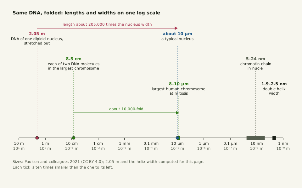
Slide the diagram sideways to read its labels, or open it full size.
From 2 m of DNA to a 10 µm nucleus [10−5 m]: each of the two DNA molecules in the largest human chromosome, 8.5 cm long, is packed into 8–10 µm, about 10,000-fold.
Data: dna-length-whole-body.csv (sources, version and license, JSON); sizes from Paulson and colleagues, 2021.
"From 2 metres of DNA to a 10 micrometre nucleus" by Ants, Cells & Atoms, https://antscellsandatoms.com/scale/dna-length/ — CC BY 4.0 (https://creativecommons.org/licenses/by/4.0/)
Data sources for this figure
Data: Paulson JR, Hudson DF, Cisneros-Soberanis F, Earnshaw WC 2021, Seminars in Cell and Developmental Biology 117:7–29, CC BY 4.0, https://doi.org/10.1016/j.semcdb.2021.03.014 (largest human mitotic chromosome 8–10 µm, with one DNA molecule 8.5 cm long in each of its two sister chromatids; nucleus about 10 µm; chain 5–24 nm; retrieved 2026-10-25); 2.05 m per nucleus and the 1.9–2.5 nm helix width computed 2026-10-25 for this page
Data sources for this figure
Data: Sender R, Fuchs S, Milo R 2016, PLoS Biology 14(8):e1002533, CC BY 4.0, https://doi.org/10.1371/journal.pbio.1002533 (3.0e13 human cells, 84% red blood cells, about 0.3e13 nucleated; shared human-cell-counts.csv; retrieved 2026-10-25); Moras M, Lefevre SD, Ostuni MA 2017, Frontiers in Physiology 8:1076, CC BY 4.0, https://doi.org/10.3389/fphys.2017.01076 (mature red cells lose the nucleus and mitochondria; retrieved 2026-10-25); Piovesan A et al. 2019, BMC Research Notes 12:106, CC BY 4.0, https://doi.org/10.1186/s13104-019-4137-z (diploid lengths 205.00 cm male and 208.23 cm female; whole-body 6.20e12 m; mitochondrial DNA 0.09–3.62 cm per cell; retrieved 2026-10-25); BIPM, The International System of Units (SI Brochure), 9th edition, Table 8, CC BY 4.0, https://doi.org/10.59161/AUEZ1291 (1 au = 149,597,870,700 m; retrieved 2026-10-25); Bajić D 2024, Entropy 26(5):430, CC BY 4.0, https://doi.org/10.3390/e26050430 (400 Earth–Sun distances; retrieved 2026-10-25); Darmasaputra GS et al. 2024, Journal of Cell Biology 223(8):e202403020, CC BY 4.0, https://doi.org/10.1083/jcb.202403020 (human liver cells 2N to 8N, up to 20% binucleated; retrieved 2026-10-25); Paulson JR, Hudson DF, Cisneros-Soberanis F, Earnshaw WC 2021, Seminars in Cell and Developmental Biology 117:7–29, CC BY 4.0, https://doi.org/10.1016/j.semcdb.2021.03.014 (largest human mitotic chromosome 8–10 µm, with one DNA molecule 8.5 cm long in each of its two sister chromatids; nucleus about 10 µm; chain 5–24 nm; retrieved 2026-10-25); Nurk S et al. 2022, Science 376(6588):44–53, CC BY 4.0, https://doi.org/10.1126/science.abj6987 (3,054,815,472 base pairs in the T2T-CHM13 set; 16,569 bp mitochondrial genome; retrieved 2026-10-25); totals and shares computed 2026-10-25 for this page
The largest human chromosome at mitosis, when a cell divides, is about 8–10 µm long, the same review notes. By then it consists of two sister chromatids, copies made when its DNA was replicated, and each holds one DNA molecule 8.5 cm long, packed about 10,000-fold lengthwise. Between divisions, that chromosome's DNA is compacted about 8,500-fold to fit into the nucleus, so the two states differ less in compaction than in looks.
The photograph near the top of this page shows female human chromosomes at metaphase, according to its description. Their purple comes from a stain, not from the chromosomes themselves: a case of borrowed color. No scale bar is printed on the photograph, so their size cannot be measured from it; the 8–10 µm in its caption is the review's figure for the largest human chromosome.
Most of your cells have none
Sender, Fuchs and Milo estimated in 2016 that a 70 kg reference man has about 3.0×10¹³ human cells (How Many Cells in the Human Body?). Red blood cells make up 84% of that count. They are among the smallest cell types in the body, with a volume of about 100 µm3, the authors note.
According to Piovesan and colleagues, human DNA is stored in the nucleus and the mitochondria. In a 2017 review, Moras and colleagues write that mammalian red blood cells expel their nucleus as they mature and then lose their other organelles, mitochondria included. So a mature red blood cell carries no DNA at all.
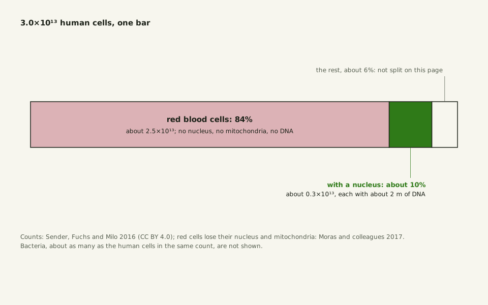
Slide the diagram sideways to read its labels, or open it full size.
Human cells in a 70 kg reference man, 3.0×10¹³ in all: 84% red blood cells with no DNA, about a tenth with a nucleus.
Data: dna-length-whole-body.csv (sources, version and license, JSON); counts from Sender and colleagues, 2016.
"Human cells in a 70 kg reference man, by whether they hold DNA" by Ants, Cells & Atoms, https://antscellsandatoms.com/scale/dna-length/ — CC BY 4.0 (https://creativecommons.org/licenses/by/4.0/)
Data: Sender R, Fuchs S, Milo R 2016, PLoS Biology 14(8):e1002533, CC BY 4.0, https://doi.org/10.1371/journal.pbio.1002533 (3.0e13 human cells, 84% red blood cells, about 0.3e13 nucleated; shared human-cell-counts.csv; retrieved 2026-10-25); Moras M, Lefevre SD, Ostuni MA 2017, Frontiers in Physiology 8:1076, CC BY 4.0, https://doi.org/10.3389/fphys.2017.01076 (mature red cells lose the nucleus and mitochondria; retrieved 2026-10-25); shares computed 2026-10-25 for this page
Data sources for this figure
Data: Sender R, Fuchs S, Milo R 2016, PLoS Biology 14(8):e1002533, CC BY 4.0, https://doi.org/10.1371/journal.pbio.1002533 (3.0e13 human cells, 84% red blood cells, about 0.3e13 nucleated; shared human-cell-counts.csv; retrieved 2026-10-25); Moras M, Lefevre SD, Ostuni MA 2017, Frontiers in Physiology 8:1076, CC BY 4.0, https://doi.org/10.3389/fphys.2017.01076 (mature red cells lose the nucleus and mitochondria; retrieved 2026-10-25); Piovesan A et al. 2019, BMC Research Notes 12:106, CC BY 4.0, https://doi.org/10.1186/s13104-019-4137-z (diploid lengths 205.00 cm male and 208.23 cm female; whole-body 6.20e12 m; mitochondrial DNA 0.09–3.62 cm per cell; retrieved 2026-10-25); BIPM, The International System of Units (SI Brochure), 9th edition, Table 8, CC BY 4.0, https://doi.org/10.59161/AUEZ1291 (1 au = 149,597,870,700 m; retrieved 2026-10-25); Bajić D 2024, Entropy 26(5):430, CC BY 4.0, https://doi.org/10.3390/e26050430 (400 Earth–Sun distances; retrieved 2026-10-25); Darmasaputra GS et al. 2024, Journal of Cell Biology 223(8):e202403020, CC BY 4.0, https://doi.org/10.1083/jcb.202403020 (human liver cells 2N to 8N, up to 20% binucleated; retrieved 2026-10-25); Paulson JR, Hudson DF, Cisneros-Soberanis F, Earnshaw WC 2021, Seminars in Cell and Developmental Biology 117:7–29, CC BY 4.0, https://doi.org/10.1016/j.semcdb.2021.03.014 (largest human mitotic chromosome 8–10 µm, with one DNA molecule 8.5 cm long in each of its two sister chromatids; nucleus about 10 µm; chain 5–24 nm; retrieved 2026-10-25); Nurk S et al. 2022, Science 376(6588):44–53, CC BY 4.0, https://doi.org/10.1126/science.abj6987 (3,054,815,472 base pairs in the T2T-CHM13 set; 16,569 bp mitochondrial genome; retrieved 2026-10-25); totals and shares computed 2026-10-25 for this page
Sender and colleagues put the number of nucleated human cells near 0.3×10¹³, about a tenth of the total. That number has one significant digit. It is the weakest input of the whole-body total below. This page counts human cells only: the bacteria in the same reference man, about as many as the human cells, are left out.
How much DNA is in a whole body?
Give each of those 0.3×10¹³ nucleated cells one diploid set of 2.04–2.38 m, the span of both routes, and one body's nuclear DNA comes to 6.1–7.1×10¹² m, computed for this page. The astronomical unit, conceived as the average Earth–Sun distance by Wikipedia's account, is exactly 149,597,870,700 m in the SI Brochure, so the total spans 41–48 astronomical units: to the Sun and back about 20–24 times.
Piovesan and colleagues ran a similar sum in 2019: 206.62 cm per diploid cell times 3×10¹² nucleated cells gives 6.20×10¹² m, more than 41 Earth–Sun distances.
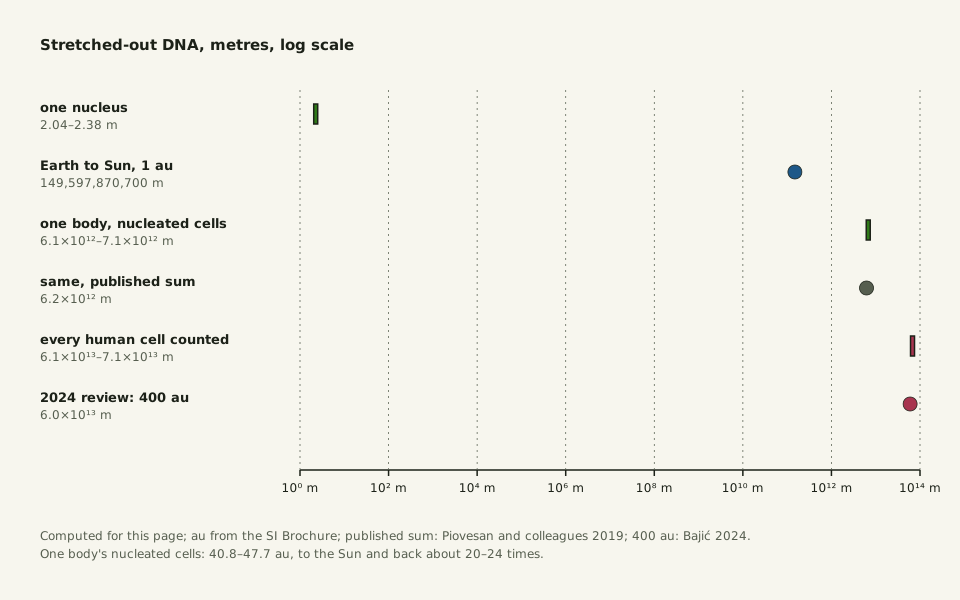
Slide the diagram sideways to read its labels, or open it full size.
One body's nuclear DNA, 6.1–7.1×10¹² m [1012 m]: 41–48 Earth–Sun distances, about a tenth of the figure that counts red blood cells too.
Data: dna-length-whole-body.csv (sources, version and license, JSON), computed for this page.
"One body’s nuclear DNA against the Earth–Sun distance" by Ants, Cells & Atoms, https://antscellsandatoms.com/scale/dna-length/ — CC BY 4.0 (https://creativecommons.org/licenses/by/4.0/)
Data sources for this figure
Data: Sender R, Fuchs S, Milo R 2016, PLoS Biology 14(8):e1002533, CC BY 4.0, https://doi.org/10.1371/journal.pbio.1002533 (3.0e13 human cells, 84% red blood cells, about 0.3e13 nucleated; shared human-cell-counts.csv; retrieved 2026-10-25); BIPM, The International System of Units (SI Brochure), 9th edition, Table 8, CC BY 4.0, https://doi.org/10.59161/AUEZ1291 (1 au = 149,597,870,700 m; retrieved 2026-10-25); Piovesan A et al. 2019, BMC Research Notes 12:106, CC BY 4.0, https://doi.org/10.1186/s13104-019-4137-z (diploid lengths 205.00 cm male and 208.23 cm female; whole-body 6.20e12 m; mitochondrial DNA 0.09–3.62 cm per cell; retrieved 2026-10-25); Bajić D 2024, Entropy 26(5):430, CC BY 4.0, https://doi.org/10.3390/e26050430 (400 Earth–Sun distances; retrieved 2026-10-25); totals computed 2026-10-25 for this page
Data sources for this figure
Data: Sender R, Fuchs S, Milo R 2016, PLoS Biology 14(8):e1002533, CC BY 4.0, https://doi.org/10.1371/journal.pbio.1002533 (3.0e13 human cells, 84% red blood cells, about 0.3e13 nucleated; shared human-cell-counts.csv; retrieved 2026-10-25); Moras M, Lefevre SD, Ostuni MA 2017, Frontiers in Physiology 8:1076, CC BY 4.0, https://doi.org/10.3389/fphys.2017.01076 (mature red cells lose the nucleus and mitochondria; retrieved 2026-10-25); Piovesan A et al. 2019, BMC Research Notes 12:106, CC BY 4.0, https://doi.org/10.1186/s13104-019-4137-z (diploid lengths 205.00 cm male and 208.23 cm female; whole-body 6.20e12 m; mitochondrial DNA 0.09–3.62 cm per cell; retrieved 2026-10-25); BIPM, The International System of Units (SI Brochure), 9th edition, Table 8, CC BY 4.0, https://doi.org/10.59161/AUEZ1291 (1 au = 149,597,870,700 m; retrieved 2026-10-25); Bajić D 2024, Entropy 26(5):430, CC BY 4.0, https://doi.org/10.3390/e26050430 (400 Earth–Sun distances; retrieved 2026-10-25); Darmasaputra GS et al. 2024, Journal of Cell Biology 223(8):e202403020, CC BY 4.0, https://doi.org/10.1083/jcb.202403020 (human liver cells 2N to 8N, up to 20% binucleated; retrieved 2026-10-25); Paulson JR, Hudson DF, Cisneros-Soberanis F, Earnshaw WC 2021, Seminars in Cell and Developmental Biology 117:7–29, CC BY 4.0, https://doi.org/10.1016/j.semcdb.2021.03.014 (largest human mitotic chromosome 8–10 µm, with one DNA molecule 8.5 cm long in each of its two sister chromatids; nucleus about 10 µm; chain 5–24 nm; retrieved 2026-10-25); Nurk S et al. 2022, Science 376(6588):44–53, CC BY 4.0, https://doi.org/10.1126/science.abj6987 (3,054,815,472 base pairs in the T2T-CHM13 set; 16,569 bp mitochondrial genome; retrieved 2026-10-25); totals and shares computed 2026-10-25 for this page
A 2024 review in the journal Entropy, citing a 2008 textbook, puts the total at 400 Earth–Sun distances. That is about what comes out if all 3.0×10¹³ human cells, red blood cells included, get about 2 m each: 408–477 astronomical units, computed for this page. Leaving out the cells without a nucleus cuts the total tenfold.
The total assumes one diploid set per nucleated cell. Some liver cells hold more: human liver cells range from two to eight chromosome sets, and up to 20% have two nuclei, Darmasaputra and colleagues write. Each mitochondrial genome adds a loop of 16,569 base pairs, about 5.6 µm if cut and stretched; Piovesan's range per cell, 0.09–3.62 cm of mitochondrial DNA, is under 2% of the nuclear 2 m, computed for this page.
The count of nucleated cells, given to one significant digit, still limits the total. Each nucleus about 10 µm across holds its own 2 m; at that count, together they would reach the Sun and back about twenty times.
Sources
- Sender R, Fuchs S, Milo R. "Revised Estimates for the Number of Human and Bacteria Cells in the Body." PLOS Biology 14(8):e1002533 (2016). https://doi.org/10.1371/journal.pbio.1002533 Read: full text. Text license: CC BY 4.0 (https://creativecommons.org/licenses/by/4.0/).
- Moras M, Lefevre SD, Ostuni MA. "From Erythroblasts to Mature Red Blood Cells: Organelle Clearance in Mammals." Frontiers in Physiology 8:1076 (2017). https://doi.org/10.3389/fphys.2017.01076 Read: full text. Text license: CC BY 4.0 (https://creativecommons.org/licenses/by/4.0/).
- Drew HR, Wing RM, Takano T, Broka C, Tanaka S, Itakura K, Dickerson RE. "Structure of a B-DNA dodecamer: conformation and dynamics." RCSB Protein Data Bank entry 1BNA (deposited 1981). https://doi.org/10.2210/pdb1bna/pdb Read: dataset record (atomic coordinates). Text license: CC0 1.0 (https://creativecommons.org/publicdomain/zero/1.0/).
- Drew HR, Wing RM, Takano T, Broka C, Tanaka S, Itakura K, Dickerson RE. "Structure of a B-DNA dodecamer: conformation and dynamics." Proceedings of the National Academy of Sciences of the USA 78(4):2179–2183 (1981). https://doi.org/10.1073/pnas.78.4.2179 Read: abstract.
- Wikipedia contributors. "Van der Waals radius." Wikipedia (read 2026-10-25). https://en.wikipedia.org/wiki/Van_der_Waals_radius Read: full text; paraphrased only. Text license: CC BY-SA 4.0 (https://creativecommons.org/licenses/by-sa/4.0/).
- PubChem, element records 6 (carbon), 7 (nitrogen), 8 (oxygen) and 15 (phosphorus), Atomic Radius; van der Waals radii from the Los Alamos National Laboratory periodic table. Database described in Kim S, et al. "PubChem 2025 update." Nucleic Acids Research 53(D1):D1516–D1525 (2025). https://doi.org/10.1093/nar/gkae1059 Read: full text of the Atomic Radius section of each element record.
- Nurk S, Koren S, Rhie A, Rautiainen M, et al. "The complete sequence of a human genome." Science 376(6588):44–53 (2022). https://doi.org/10.1126/science.abj6987 Read: full text. Text license: CC BY 4.0 (https://creativecommons.org/licenses/by/4.0/).
- Piovesan A, Pelleri MC, Antonaros F, Strippoli P, Caracausi M, Vitale L. "On the length, weight and GC content of the human genome." BMC Research Notes 12:106 (2019). https://doi.org/10.1186/s13104-019-4137-z Read: full text. Text license: CC BY 4.0 (https://creativecommons.org/licenses/by/4.0/).
- Engel SR, Dietrich FS, Fisk DG, Binkley G, Balakrishnan R, Costanzo MC, Dwight SS, Hitz BC, Karra K, Nash RS, Weng S, Wong ED, Lloyd P, Skrzypek MS, Miyasato SR, Simison M, Cherry JM. "The Reference Genome Sequence of Saccharomyces cerevisiae: Then and Now." G3: Genes, Genomes, Genetics 4(3):389–398 (2014). https://doi.org/10.1534/g3.113.008995 Read: full text. Text license: CC BY 3.0 (https://creativecommons.org/licenses/by/3.0/).
- Wyngaard GA, Skern-Mauritzen R, Malde K, Prendergast R, Peruzzi S. "The salmon louse genome may be much larger than sequencing suggests." Scientific Reports 12:6616 (2022). https://doi.org/10.1038/s41598-022-10585-2 Read: full text. Text license: CC BY 4.0 (https://creativecommons.org/licenses/by/4.0/).
- Doležel J, Bartoš J, Voglmayr H, Greilhuber J. "Nuclear DNA content and genome size of trout and human." Cytometry Part A 51A(2):127–128 (2003). https://doi.org/10.1002/cyto.a.10013 Read: bibliographic record only.
- Paulson JR, Hudson DF, Cisneros-Soberanis F, Earnshaw WC. "Mitotic chromosomes." Seminars in Cell & Developmental Biology 117:7–29 (2021). https://doi.org/10.1016/j.semcdb.2021.03.014 Read: full text. Text license: CC BY 4.0 (https://creativecommons.org/licenses/by/4.0/).
- Wikipedia contributors. "Astronomical unit." Wikipedia (read 2026-10-25). https://en.wikipedia.org/wiki/Astronomical_unit Read: full text; paraphrased only. Text license: CC BY-SA 4.0 (https://creativecommons.org/licenses/by-sa/4.0/).
- Bureau International des Poids et Mesures. The International System of Units (SI) (SI Brochure), 9th edition (2019; version 4.01, June 2026), Table 8. https://doi.org/10.59161/AUEZ1291 Read: Table 8 and its notes. Text license: CC BY 4.0 (https://creativecommons.org/licenses/by/4.0/).
- Bajić D. "Information Theory, Living Systems, and Communication Engineering." Entropy 26(5):430 (2024). https://doi.org/10.3390/e26050430 Read: full text. Text license: CC BY 4.0 (https://creativecommons.org/licenses/by/4.0/).
- Darmasaputra GS, Geerlings CC, Chuva de Sousa Lopes SM, Clevers H, Galli M. "Binucleated human hepatocytes arise through late cytokinetic regression during endomitosis M phase." Journal of Cell Biology 223(8):e202403020 (2024). https://doi.org/10.1083/jcb.202403020 Read: full text. Text license: CC BY 4.0 (https://creativecommons.org/licenses/by/4.0/).
- Rao VB, Black LW. "Structure and assembly of bacteriophage T4 head." Virology Journal 7:356 (2010). https://doi.org/10.1186/1743-422X-7-356 Read: full text. Text license: CC BY 2.0 (https://creativecommons.org/licenses/by/2.0/).
Cite this page
Ants, Cells & Atoms. “How Long Is DNA? About 2 m per Diploid Human Nucleus.” Published 2026-10-25. https://antscellsandatoms.com/scale/dna-length/. Accessed (add the date you read it).
No DOI is issued for entries. Figures made here are CC BY 4.0; see each caption for the license of photographs.
Related entries
How Many Cells in the Human Body? About Half Are Bacteria
Image: “E. coli Bacteria (7316101966)” by NIAID (2002) · Public domain
"E. coli Bacteria (7316101966)" by NIAID (2002) — https://commons.wikimedia.org/wiki/File:E._coli_Bacteria_(7316101966).jpg — Public domain (work of a United States federal agency (NIAID, part of NIH), public domain in the United States under 17 U.S.C. §105; Commons tag PD-USGov-NIH-NIAID; Flickr source page credits NIAID with no contractor note; dated 2002-11-14 in the file metadata; no copyright term applies in the EU for a US government work released as public domain by its author agency (used as the source's explicit PD mark))

Reynolds Number: Why a Bacterium Can't Coast and a Fish Can
Image: “Oczlik (Cyclops) - samica” by Mikron86 (2025) · CC BY 4.0
"Oczlik (Cyclops) - samica" by Mikron86 — https://commons.wikimedia.org/wiki/File:Oczlik_(Cyclops)_-_samica.jpg — CC BY 4.0 (https://creativecommons.org/licenses/by/4.0/)

How Big Is a Cell? Most Are a Hair's Width or Far Narrower
Image: “SEM blood cells” by Bruce Wetzel and Harry Schaefer, National Cancer Institute (1982) · Public domain
"SEM blood cells" by Bruce Wetzel and Harry Schaefer, National Cancer Institute (1982) — https://commons.wikimedia.org/wiki/File:SEM_blood_cells.jpg — Public domain (work of United States federal employees at the National Cancer Institute (NIH), public domain in the United States under 17 U.S.C. §105; Commons tag PD-USGov; the NCI Visuals Online source states that all images are in the public domain and may be reproduced without permission, with credit to the listed source or author; dated February 1982; used in the EU on the source's explicit public-domain statement)
